Dismantling The Viral Theory

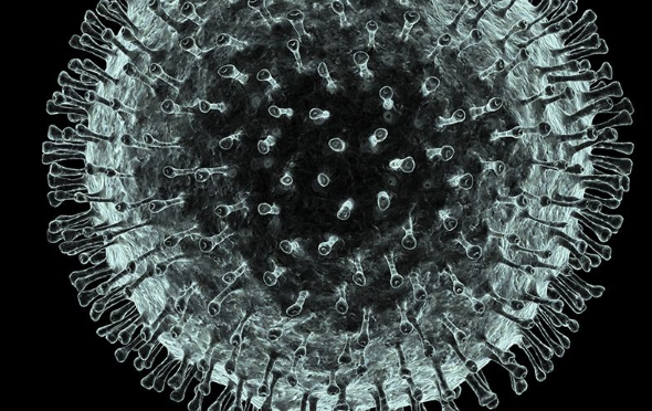
[Electron micrograph of the so-called Polio virus that has never been demonstrated scientifically to cause the symptoms of paralysis. Illustration has been colorized for effect]
The first isolation of a virus was achieved in 1892 by Russian bacteria hunter Dimitri Iwanowski, who gathered fluid from diseased tobacco plants. He passed this liquid through a filter fine enough to retain bacteria; yet to Iwanowski’s surprise, the bacteria-free filtrate easily made healthy plants sick. In 1898 a Dutch botanist, Martinus Willem Beijerinck, repeating the experiment, also recognized that there was an invisible cause and named the infectious agent “tobacco mosaic virus.” In the same year as Beijerinck’s report, two German scientists purified a liquid containing filterable viruses that caused foot-and-mouth disease in cattle (viruses were at one time called “filterable viruses,” but eventually the term “filterable” came to apply only to viruses, and was dropped). Walter Reed followed in 1901 with a filtrate responsible for yellow fever, and soon dozens of other disease-causing viruses were found.

In 1935 another American, Wendell M. Stanley, went back to the beginning and created pure crystals of tobacco mosaic virus from a filtered liquid solution. He affirmed that these crystals could easily infect plants, and concluded that a virus was not a living organism, since it could be crystallized like salt and yet remained infectious. Subsequently, bacteriologists all over the world began filtering for viruses, and a new area of biology was born-virology.
Historically, medical science has vacillated on the question of whether a virus is alive. Originally it was described as nonliving, but is currently said to be an extremely complex molecule or an extremely simple microorganism, and is usually referred to as a parasite having a cycle of life. (The term “killed” is applied to certain viral vaccines, thus implying an official conviction that viruses live.) Commonly composed of either DNA or RNA cores with protein coverings, and having no inherent reproductive ability, viruses depend upon the host for replication. They must utilize the nucleic acids of living cells they infect to reproduce their proteins (i.e., trick the host into producing them), which are then assembled into new viruses like cars on an assembly line. Theoretically, this is their only means of surviving and infecting new cells or hosts.
The Replicating Virus Theory
Then it was discovered that, when bacteria slowly begin to die, bacteria create tiny, apparently lifeless forms of survival, the so-called spores. It was then suspected that these spores were toxic and that they were the so-called pathogenic poisons. This was then refuted, since the spores are rapidly developing into bacteria when their vital resources are being restored. When scientists in the laboratory observed that the weak, highly inbred bacteria perished very quickly while turning into much smaller structures than the spores, it was first believed that the bacteria were being killed by the alleged pathogenic poisons, called viruses, and that the viruses were thereby replicating.
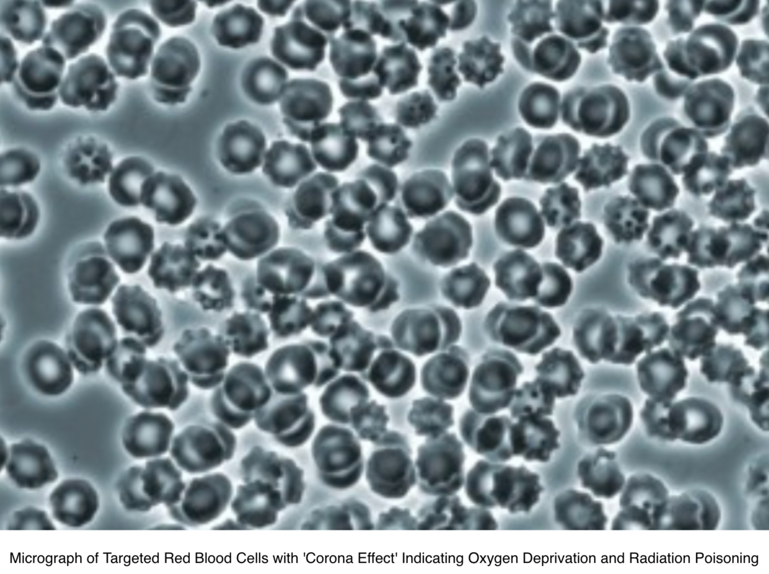
[The micrograph above was done using Dark Field Microscopy showing red blood cells and the evolution of bacterial pHages and bacterial spores (the white spots0 from red blood cell biological transformation]
The Replicating Virus Theory
Then it was discovered that, when bacteria slowly begin to die, bacteria create tiny, apparently lifeless forms of survival, the so-called spores. It was then suspected that these spores were toxic and that they were the so-called pathogenic poisons. This was then refuted, since the spores are rapidly developing into bacteria when their vital resources are being restored. When scientists in the laboratory observed that the weak, highly inbred bacteria perished very quickly while turning into much smaller structures than the spores, it was first believed that the bacteria were being killed by the alleged pathogenic poisons, called viruses, and that the viruses were thereby replicating.
The Invention of Bacterial Viruses
Due to the belief that these – at the time of their discovery still invisible- structures were killing the bacteria, they were called phages/bacteriophages, “eaters of bacteria”. Only later it was determined that merely highly inbred and therefore almost non-viable bacteria can be made to turn into phages, or bacteria which are being destroyed so fast that they do not have time to form spores.
The introduction of the electron microscopy led to the discovery of the structures resulting from the biological transformation or pleomorphism of bacteria when these were suddenly dying or when the metabolism of the highly inbred germs was overwhelmed by processes triggered by the adding of “phages”. It was also discovered that there are hundreds of types of different-looking “phages”. The discovery of phages, the so-called bacterial “viruses”, reinforced the wrong assumption and the belief that there were human and animal viruses that looked the same and had the same structure. This is not and cannot be the case, for several different reasons.
After introducing chemical examination techniques in biology, it was discovered that there are thousands of types of phages and that phages of one type always have the same structure. They consist of a particular molecule, made of nucleic acid, which is covered in a shell of proteins of a given number and composition. It was only later discovered that merely the bacteria which had been highly inbred in the test tube could turn into phages themselves, by contact with phages, but this never applied to natural bacteria or bacteria which had just been isolated from their natural environment. In this process, it was discovered that these “bacterial viruses” actually serve to provide other bacteria with important molecules and proteins, and that the bacteria themselves emerged from such structures.
Before it could be established that the “bacterial viruses” cannot kill natural bacteria, but they are instead helping them to live and that bacteria themselves emerge from such structures, these “phages” were already used as models for the alleged human and animal viruses. It was assumed that the human and animal viruses looked like the “phages”, were allegedly killing cells and thereby causing diseases, while at the same time producing new disease poisons and in this way transmitting the diseases. To date, many new or apparently new diseases have been attributed to viruses if their origin is unknown or not acknowledged. This reflex found an apparent confirmation in the discovery of the “bacterial viruses”.
It is important to note that the theories of fight and infection were accepted and highly praised by a majority of the specialists only if and when the countries or regions where they lived were also suffering from war and adversity. In times of peace, other concepts dominated the world of science.[272]
It is very important to note that the theory of infection – starting from Germany – has only been globalized through the third Reich, when the Jewish researchers, most of which had opposed and refuted the politically exploited theories of infection, were removed from their positions.[273]
The Detection of Phages and Biological Transformation
The existence of phages can be proved rapidly
[Bacterial pHage being born out of a blood and/or body cell. A biological process known as pleomorphism]
First step: their presence is confirmed through an effect, namely the transformation of bacteria into phages, and also through an electron micrograph of those phages. The control experiments show that phages do not appear if bacteria do not change or if bacteria randomly start decomposing due to extrinsic sudden annihilation, without forming phages.

[The micrographs (micrographs #1 through #6] above show the cellular transformation of red blood cells, using pHase contrast microscopy, into rod bacteria, cell-wall deficient bacteria, Y-form yeast and then bacteria pHages]
Second step: the liquid containing the phages is concentrated and applied on another liquid, which has a high concentration at the bottom of the test tube and a low concentration at the top of the test tube. The test tube with the phages is then powerfully spun (centrifuged) and all the particles gather according to their mass and weight to the place of their own density. The density is the ratio of weight (mass) per unit of volume, expressed as Kg/l or g/mg, respectively. That is why this concentration and purification step for particles with the same density is called density gradient centrifugation.
The layer where many particles of the same density gather becomes “cloudy”, which is called a “band.” This step is being documented, then the particles concentrated, purified and sedimented in a “band” are removed with a syringe needle. The extracted concentrated amount of particles is called an isolate. A fast and simple electron micrograph will confirm the presence of phages in the isolate, which at the same time is an indication for the purity of the isolate, if the micrograph shows no other particles but the phages. The appearance and the diameter of the phages will also be established with the help of this micrograph.
The control experiment performed for this step consists in treating and centrifuging the liquid from bacteria which did not form any phages, where no phages appear at the end of the procedure.
After the step of successfully isolating the phages, the decisive biochemical characterization of the phages follows. The biochemical characterization of their composition is essential for identifying the specific type of phage, since different types of phages often appear to be similar. The isolate obtained through the density gradient centrifugation is now divided in two parts. One part is used to determine the size, type and composition of the nucleic acid; in a separate procedure, the other part is used to determine the amount, size and morphology of the proteins of the phages. Since the 1970s, these tests have been simple standard techniques that are learned by every biology student in their first semesters.
These tests represent the biochemical characterization of the phages. In almost every case, these results have been and are being published in only one publication, since a phage has a very simple structure which is very easy to analyze. The control experiments for these tests use liquid from bacteria which do not form phages and thus cannot present any biochemical proof. The existence of approximately two thousand different types of phages have been scientifically demonstrated this way
The So-Called Pathogenic Viruses
The “bacteriophages,” correctly defined as incomplete mini spores and building blocks of the bacteria, have been scientifically isolated, while the so-called pathogenic viruses have never been observed in humans or animals or in their body fluids and have never been isolated and subsequently biochemically analyzed. To date, none of the researchers involved in virology research seems to have realized this very important point.
The use of electron microscopy and the biochemistry were very slowly returning to normal after 1945 and no one had realized that not one pathogenic virus had ever been isolated in humans or animals; thus, as of 1949 researchers started applying the same idea used for the (bacterio) phages, in order to replicate the human and animal “viruses.” John Franklin Enders, born in 1897 in the family of a rich financier, was active in various fraternities after having finished his studies, then he worked as a real estate agent and studied foreign languages for four years before turning to bacterial virology, which fascinated him. He then simply transferred the ideas and concepts that he learned in this area of research to the supposed pathogenic viruses in humans.
UnScientific Experiments and Interpretations Gave Birth to Virology
With his unscientific experiments and interpretations that he had never confirmed through negative controls, Enders brought the entire “viral” infectious medicine to a dead end. It is important to note at this point that Enders, like many infectious diseases specialists, worked for the U.S. military, which had always been and remains to date a huge victim of the fear of contagions. It was mainly the U.S. military which spread its erroneous belief that besides chemical weapons there were also biological weapons in the form of bacteria and viruses.
In 1949, Enders announced that he had managed to cultivate and grow the alleged polio virus in vitro on various tissues. The American expert opinion believed everything immediately. What Enders did was to add fluids from patients with poliomyelitis to tissue cultures which he claimed to have had sterilized, then he alleged that the cells were dying because of the virus, that the virus was replicating in this way and that a vaccine could be harvested from the respective culture. At that time, summer polio epidemics (polio = flaccid paralysis) were very frequent during summer and they were believed to be caused by the polio virus. A vaccine was to help eradicate the alleged virus. After the polio vaccine was introduced, the symptoms were then re-diagnosed among other things as multiple sclerosis, flaccid acute paralysis, aseptic meningitis etc. and later polio was claimed to have been eradicated. During his experiments, Enders et al. sterilized the tissue cultures in order to exclude the possibility of bacteria killing the cells. What he didn’t take into consideration was that the sterilization and the treatment of the cell culture when preparing it for the alleged infection was exactly what was destroying and killing the cells. Instead, he interpreted the cytopathic effects as the existence and the action of a so-called polio virus, without ever having isolated a single virus and describing its biochemistry. The necessary negative control experiments, which would have shown that the sterilization and the treatment of the cells prior to the “infection” in the test tube was killing the cells, have never been performed. However, for this “performance” Enders received the Nobel prize in 1954.
The Invention of the Polio Virus and ‘YES” the Measles Virus Too!

[Measles virus or a bacterial pHage?]
1954 is also the year in which Enders applied and introduced the same technique in order to allegedly replicate the measles virus. As he had been awarded the Nobel prize for the alleged polio virus the same year, all researchers believed his technique to be scientifically valid. Thus, to date, the entire concept of polio and measles has been based upon this unscientific technique and fraud.
Thus, the polio and measles vaccines do not contain viruses, but particles of dead monkey kidney tissue or human cancerous body cells. To date, no negative control experiments have been done with respect to the so-called polio and measles viruses either, which would have shown that it was the laboratory procedures that lead to the cytopathic effects on the cells.
Additionally, all claims and experiments made by Enders et al. and subsequent researchers lead to the only objective conclusion, that in fact they were observing and analyzing the cellular particles or fragments and the activity thereof in the test tube, misinterpreting these as particles and characteristics of the alleged polio and/or measles viruses.
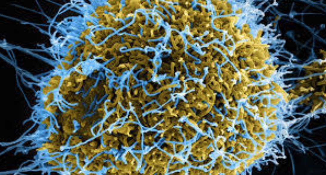
ALL Viruses from HIV, EBV, CMV, Hepatitis C, West Nile Virus, Ebola, Measles, Zika, etc., are ALL Phantom Viruses
Viral Existence Has NEVER Been Scientifically Demonstrated and Never Proven!
The following explanations applies to all the so-called (human or animal) “pathogenic viruses”. The six papers provided by Dr. Bardens in the course of the “measles trial” as proof for the existence of the measles virus described in a didactically ideal way the various steps of the chain of misinterpretations up to the belief in the existence of a measles virus.
The first paper was published in 1954 by Enders et al.: “Propagation in tissue cultures of cytopathogenic agents from patients with measles” (Proc Soc Exp Biol Med. 1954 Jun; 86 (2): 277–286).
This publication can be found on the internet, like all the other publications presented at the measles trial. In that experiment, Enders et al. cut down dramatically on the nutrient solution and added cell-destroying antibiotics to the cell culture before introducing the allegedly infected fluid. The subsequent dying of the cells was then misinterpreted as presence and also isolation of the measles virus. No control experiments were performed to exclude the possibility that it was the deprivation of nutrients as well as the antibiotics which led to the cytopathic effects.
Enders’ and his colleagues’ blindness can be explained by the fact that he truly wanted to help people, while the ‘virus hysteria’ was intensifying after the war and during the cold war. It can also be explained by the fact that Enders and many of his colleagues had no idea about medicine or biochemistry and they were competing with the Soviet Union for the development of the first measles vaccine. Such a pressure for success can also explain why Enders and his colleagues ignored their own reservations and cautions expressed in 1954, when they had observed and noted that many cells also died after being treated normally (i.e. without being “infected”), which they thought to have been caused by unknown viruses and other factors. All these facts and cautions were subsequently disregarded.
The second paper presented by the claimant in the ‘measles trial’ was published in 1959[274] and, for the reasons presented above, the authors concluded that the technique introduced by Enders was not appropriate for the isolation of ANY virus. This rebuttal is not only NOT being discussed by ALL the other researchers, but it is being ignored completely!
The ‘Viral Dogma’ of Pathogenic Viruses is Still Being Promoted Today!
In a third paper[275], the authors photographed typical cellular particles inside the cells and misinterpreted these as measles virus. They did not isolate any virus. For unexplained reasons, they failed to determine and describe the biochemical structure of what they were presenting as a virus in a separate experiment. In the short description of the methods used, one can read that the authors did not apply the standard isolation technique for viruses, i.e. the density gradient centrifugation. They simply centrifuged fragments of dead cells at the bottom of a test tube and then, without describing their biochemical structure, they misinterpreted the cellular debris as viruses.
From the way the experiments were performed, one can only conclude that cellular particles were misinterpreted as viruses. We find the same situation in the fourth[276] and the sixth[277] publication put forward by the claimant as proof of the existence of a measles virus. The fifth publication[278] is a review describing the consensus process as to which nucleic acid molecules from the dead cells would represent the so-called genome of the polio or measles virus. The result is that dozens of research teams work with short pieces of cell-specific molecules, after which -following a given model – they put all the pieces together on paper. However, this jigsaw puzzle made of so many pieces was never scientifically proven to exist as a whole and was never isolated from a virus, for a polio, measles, HIV or Hepatitis C, Ebola or Zika viruses have never been seen, neither in humans nor in a test tube. Referring to this publication, the court-appointed expert stated that it described the gold standard, i.e. the entire virus genome. It is obvious that the expert did not read this paper, whose authors stated that the exact molecular composition and functions of the measles virus genome will have to be the object of further research, which is why they had to rely on other virus models in order to achieve a consensus on the structure and functions of ANY virus genome. The easiest thing for anyone to notice is that in all of these publications, as well as in all other publications on the “measles virus” and other pathogenic viruses, including HIV, EBV, CMV, Ebola and Zika, no control experiments have ever been performed. No researchers used the density gradient centrifugation technique; instead, they only centrifuged cellular debris at the bottom of a test tube. This technique, used to collect all the particles from a fluid, is called pelletizing. From a logical and scientific perspective, it can be said that in all publications on the so-called “pathogenic viruses”, the researchers demonstrated in fact only particles and characteristics of cells. I would also like to point out that the so-called giant viruses[279] , i.e. an enwrapped nucleic acid can be found everywhere in the sea and in basic organisms. Like all bacterial phages, not only are they harmless, but they have beneficial functions. They can be also isolated by using the density gradient centrifugation, which proves their existence (see the graphic above).
I also recommend Prof. Lüdtke’s relevant review (1999).[280] He noted that at the early beginnings of virology, the majority of virologists always concluded that the structures they had mistaken for viruses turned out to be components of the cells and thus, they were only the result of the experiment and not the cause of the changes observed.
After the discovery and characterization of the phages and after introducing the dogma that the nucleic acid was the genome of all cells and viruses, the consensus was born, according to which such viruses must exist in humans and animals as well. In 1992, the dogma stating that the nucleic acid is the genotype of all cells was retracted in the scientific community. The ‘viral dogma’ of pathogenic viruses, however, is still being promoted today to the harm of billions of people. – for what?
The Bottom Line Concerning Phantom Viruses and the Polio and Measles Virus
[An Electron micrograph of the so-called Polio virus that has never been demonstrated scientifically to cause the symptoms of paralysis. Illustration has been colorized for effect]
My bottom line still holds the truth that the terrain or internal environment is everything and the germ or so-called virus is NOTHING! The germ or so-called virus can only be a symptom of cellular breakdown due to an imbalance of the delicate alkaline pH balance of the body fluids and NOT the cause of that breakdown. That is why years ago I offered any scientist in the World a finders fee of 5 million US dollars if they could prove the existence of the HIV virus using Koch’s postulates. It has now been over 20 years and I am still waiting even though currently I no longer have the funds to pay the prize due to political assassination! It is unfortunate that a former 5 million US dollar prize offered 20 years ago was not enough money to change the current medical viral dogma that is currently paying out trillions of dollars to guess who?[281]

Click here to read more: http://medcraveonline.com/IJVV/IJVV-02-00032.php
To order your copy of Second Thoughts About Viruses, Vaccines and the HIV/AIDS Hypothesis go to: https://www.amazon.com/…/ref=dbs_a_def_rwt_hsch_vapi_taft_p…
Lecture in Dubai – The 2nd Annual Conference on Bacterial, Viral and Infectious Diseases
http://www.drrobertyoung.com/events.html
Join Robert O Young PhD and Galina Migalko MD in Dubai on June 18th and 19th, 2019 for the Annual Conference on Bacterial, Viral and Infectious Diseases. They will be Key Note Speakers and doing a workshop on the New Biology.

For more information and to register go to: https://bacterialdiseases.infectiousconferences.com/organiz…

The following is the abstract for Dr. Young’s lecture:
The Dismantling of the Viral Theory
Robert O Young CPT, MSc, DSc, PhD, Naturopathic Practitioner
Abstract
There is now over 100 years of documented history and research on the Polio virus and whether or not its treatment by inoculation has been successful in eradicating Polio. I am suggesting in this article and in my lecture that there are significant findings based on historical and past and current research, including my own that the viral theory of Polio and possibly other modern-day diseases, such as Post-Polio Syndrome, Polio Vaccine-Induced Paralysis, Legionnaires, CNS disease, Cancer, HIV/AIDS and now Zika may be caused by acidic chemical poisoning from DDT (dichloro-diphenyl-trichloroethane) and other related DDT pesticides, acidic vaccinations, and other factors including lifestyle and dietary factors rather than from a lone infectious virus. I will present ten historical graphs outlining the history of Polio, the production of DDT, BHC, Lead, Arsenic, Polio vaccinations and the author’s theory that chemical poisoning, vaccination, and lifestyle and dietary choices are a more likely causes for the symptoms of Polio, neurological diseases, Cancer, HIV/AIDS and now Zika.
https://www.linkedin.com/…/lecture-dubai-annual-conference…/
https://bacterialdiseases.infectiousconferences.com/organiz…
References:
[1] Morton S. Biskind, MD. “Public Health Aspects of the New Insecticides”. American Journal of Digestive Diseases, New York, 1953, v 20, p331.
[2] Handbook of Pesticide Toxicology, edited by Wayland J. Hayes, Jr. and Edward R. Laws, Academic Press Inc., Harcourt Brace Jovanovich, Publishers, San Diego, 1991, p769
[3] Toxicological Profile: for DDT, DDE, and DDE. Agency for Toxic Substances and Disease Registry, September 2002.
[4] U. Beck, E. Löser “Chlorinated Benzenes and other Nucleus-Chlorinated Aromatic Hydrocarbons” Ullmann’s Encyclopedia of Industrial Chemistry, 2012, Wiley-VCH, Weinheim.
[5] Chlorobenzene”. Immediately Dangerous to Life and Health. National Institute for Occupational Safety and Health (NIOSH)
[6] U.S. Vital Statistics, U.S. Government Printing Office, Washington, D.C.
[7] Historical Statistics of the U.S., The U.S. Government Printing Office, Washington, D.C.
[8] Van Nostrand’s Encyclopedia of Science and Engineering (1995), vol. 5, p1725. The phrase “Pesticides As A Panacea: 1942-1962” is a subtitle found in Encyclopedia Britannica, Macropaedia (1986).
[9] Thomas, Robert E. (1955), Salt & Water, Power & People: A Short History of Hooker Electrochemical Co. Niagara Falls, NY: Hooker Chemical Co.
[10] Booth, Gerald (2000), “Ullmann’s Encyclopedia of Industrial Chemistry – Nitro Compounds, Aromatic”. doi:10.1002/14356007.a17_411. ISBN 3527306730
[11] Weber, Manfred; Weber, Markus; Kleine-Boymann, Michael (2004). “Ullmann’s Encyclopedia of Industrial Chemistry – Phenol”. doi:10.1002/14356007.a19_299.pub2. ISBN 3527306730.
[12] Haller, H. L., Bartlett, P. D., Drake, N. L., and others: The Chemical Composition of Technical DDT, American Chemical Society, Journal, volume 67, pages 1591- 1602, 1945.
[13] Jo-Yu Chin, Christopher Godwin, Chunrong Jia, Thomas Robins, Toby Lewis, Edith Parker, Paul Max, and Stuart Batterman, “Concentrations and Risks of p-Dichlorobenzene in Indoor and Outdoor Air,” Indoor Air, 2013 Feb; 23(1): 40–49, Published online 2012 Jul 18. doi: 10.1111/j.1600-0668.2012.00796.x.
[14] Duesberg, PH, “Inventing the AIDS Virus,” Regnery, (1996). ISBN 0-89526-399-8. [15] Icon Group International (Author), Chlorobenzene: Webster’s Timeline History, 1851 – 2007 May 17, 2010
[16] Ibid [17] Ibid
[18] Risse, GB (1988). Fee E, Fox DM, eds. Epidemics and History: Ecological Perspectives. in AIDS: The Burden of History. University of California Press, Berkeley. ISBN 0-520-06396-1.
[19] A Disease of Cleanliness: Polio in New York City, 1900-1990, in David Rosner, ed., Hives of Sickness: Public Health and Epidemics in New York City Rutgers University Press, 1995, pp. 115-130.
[20] McDonough, F., The Origins of the First and Second World Wars (Cambridge Perspectives in History), Cambridge University Press, August 28, 1997.
[21] Goel, A, Aggarwal, P, “Pesticide Poisoning,” Natl Med J India. 2007 Jul-Aug; 20(4):182-91.
[22] Ibid.
[23] Biskind, MS (1953) “Public Health Aspects of the New Insecticides,” American Journal of Digestive Diseases 20: 331-341.
[24] TIME Magazine, U.S. Edition, March 14, 1994 Vol. 143 No. 11. [25] Baily, J. W.: J. Am. Vet. M. A. 113: 251, Sept. 1948.
[26] Biden-Steele, K. and Stuckey, R. E.: “Poisoning by DDT Emulsion: Report of a Fatal Case”, Lancet, 2: 235-236, Aug. 17, 1946.
[27] Biskind, M. S.: “DDT Poisoning and X Disease in Cattle”, J. Am. Vet. M. A. 114: 20, Jan. 1949.
[28] Biskind, M. S.: “DDT Poisoning a Serious Public Health Hazard”, Am. J. Dig. Dis. 16: 73, Feb. 1949.
[29] Biskind, M. S.: “DDT Poisoning and the Elusive ‘Virus X’: A New Cause for Gastro- Enteritis”, Am. J. Dig. Dis. 16: 79, March 1949.
[30] Boyd, C. L.: “A Report on “XX Disease in Texas”, J. Am. Vet. M. A. 113: 463, Nov. 1948.
[31] Cameron, C. R., and Burgess, F.: “The Toxicity of DDT”, Brit. M. J. 1: 865-871, June 23, 1945.
[32] Carte; R. H., Hubanks, P. E., et al: “Effect of Cooking on the DDT Content of Beef”, Science, 107: 347, April 2, 1948.
[33] Case, R. A. M.: Toxic Effects of DDT in Man”, Brit. M. J., 2: 842-845, Dec. 15, 1945.
[34] Council on Pharmacy and Chemistry, A. M. A.: “Health Hazards of Pesticides”, J. A. M. A. 137: 1603, Aug. 28, 1948.
[35] Crescitelli, F., and Gillman, A.: “Electrical Manifestations of Cerebellum and Cerebral Cortex Following DDT Administration to Cats and Monkeys”, Am. J. Physiol., 147: 127- 137, Sept. 1946.
[36] Deederer, C.: “DDT Toxicity”, M.Rec. 161: 216-220, April 1948
[37] Domenici, T. J.: “Hepatitis without Jaundice and without Hepatomegaly”, N. Eng. J. Med. 240: 88, Jan. 20, 1949
[38] Dunn, J. E., Dunn, J. C., and Smith, R. S.: “Skin Sensitising Properties of DDT for 31
Guinea Pig”, Pub. Health Rep. 61: 1614-1620, 1949.
[39] Editorial: Pesticides: “Chemical Contaminants of Foods”, J.A.M.A. 137: 1604, Aug. 28, 1948.
[40] Fitzhugh, O. G., and Nelson, A. A.: “The Chronic Oral Toxicity of DDT”, J. Pharm.acol. and Exper. Therap. 89: 18-30, Jan. 1947.
[41] Gamier, G.: “Treatment of Scabies with DDT”, .Presse Med. 56: 458, June 23, 1948. [42] Garett, ii. M., “Toxicity of DDT for Man”, Alabama St. M. A. J., 17: 74, Aug. 1947.
[43] Globus, J. H.: “DDT Poisoning; Histopathologic Observations on the Central Nervous System in So-Treated Monkeys, Dogs, Cats and Rats”, J. Neuropath. 7: 418-431, Oct. 1948.
[44] Haymaker, W., Ginzler, A. M., and Ferguson, J. L.: “Toxic Effects of Prolonged Ingestion of DDT on Dogs, with Special Reference to Lesions in Brain”, Am. J. M. Sc. 212: 423, Oct. 1946.
[45] Hill, K. R., and Daniiani, C. R.: “Death Following Exposure to DDT, Report of a Case”, New Eng. J. Med., 235: 897-899, Dec. 19, 1946.
[46] Hill, K. 3. and Robinson, G.: “A Fatal Case of DDT Poisoning in a Child, with an Account of Two Accidental Deaths in Dogs”. Brit. M. J. 2: 845-847, Dee. 15, 1945.
[47] Ingle, L.: “Toxicity of Chlordane to White Rats”, J. Econ. Entomol. 40: 264-268, 1947.
[48] Jandorf, B. J;. Sanett, H. P., and Bodansky, Oscar: “Effect of Oral Administration of DDT on Metabolism of Glucose and Pyruvie Acid in Rat Tissues”, J. Pharmaeol. and Exper. Therap. 88: 333-337, Dec. 1946.
[49] Jenkins, D. W.: “A Review of the Insecticide Hexachloro-cyclohexane (‘666’)”, Office of Technical Services, U. S. Dept of Commerce, Washington, D • C., No. PB 4034, Med. Div. Rept. No. 56, Sept. 26, 1945.
[50] Kempe, H. E.: “Progress Report on Benzene Hexachloride for the Destruction of Sheep Scab Mites”, Vet. Med., Feb. 1948, pp. 76-79.
[51] Kirk, H.: Vet. Red. 58: 43, 1946.
[52] Kirk, H.: “DDT in Canine Practice”, Vet. Med. Feb. 1947, PP. 76-78.
[53] Lawhon, G. J., Jr.: “X Disease in South Carolina”, N. Am. Vet. 29: 643, Oct. 1948.
[54] Leider, M.: “Allergenic Eczematous Contact-Type Dermatitis Caused by DDT”, J. Invest. Dermatol. 8: 125-126., March 1947.
[55] Lillie, R. D., Smith, M. I., and Stohlman, E. F.: Pathologic Action of DDT and Certain of its Analogs and Derivatives”, Arch. Path. 43: 127-142, Feb. 1947.
[56] Mackerras, I. M., and West, R. F. K.: “DDT Poisoning in Man”, M. J. Australia, 1: 400-401, March 23, 1946.
[57] Mobbs, J. F.:” Toxicity of Hexaehloroeyclohexane in Scabies, J.A.M.A. 138: 1253, Dec. 25, 1948. Personal Communication.
[58] Morrill, C. C.: “Hyperkeratosi.s or X Disease”, N. Am. Vet. 29: 642, Oct. 1948.
[59] Neal, P. A., Sweeney, T. B., Spicer, S. S., and von Oettingen, W. F.: “The Excretion of DDT in Man, Together with Clinical Observations”, Pub. Health Rep., 61: 403, March 22, 1946.
[60] Neal, P. A., von Oettingen, W. F., Smith, W. W., et al: Toxicology and Potential Dangers of Aerosols, Mists and Dusting Powders Containing DDT”, Pub. Health Rep. Suppl. 177, 1944.
[61] Neal, P. A., von Oettingeu, W. F., Dunn, R. C., and Sharpless, N. E.: “Toxicology and Potential Dangers of Aerosols and Residues from Aerosols Containing 3 Percent of DDT. Second Report, ibid., Suppl. 183, 1945.
[62] Nelson, A. A., Draize, 3. H., Woodard, G., et al: “Histopathological Changes Following Administration of DDT to Several Species of Animals”, U. S. Pub. Health Rep. 59: 1009, Aug. 4, 1944.
[63] Neve, Helen: “Toxic Effects of DDT on a Cat”, Vet. Rec. 58: 43, 1946. Vet. Med., Feb. 1947, p. 78.
[64] Niedelman, M. L.: “Contact Dermatitis Due to DDT”, Occup. Med. 1: 391-395, April 1946.
[65] Radeleff, R. D.: “DDT Spray Outmodes Dipping Vat”, Vet. Med. Oct. 1947, pp. 372- 373.
[66] Radeleff, R. D.: “Chlordane Poisoning: Symptomatology and Pathology, Vet. Med. Aug. 1948, pp. 342-347.
[67] Robinson, J. H.: “Harvest Analysis of DDT Residues”, Food Packer, 29: 50-53, 1948.
[68] Riker, W. F., Jr., Huebner, Virginia, R., Raska, S. B., and Cattell, McKeen: “Studies on DDT, Effects on Oxidative Metabolism”, J. Pharmacol. and, Exper. Therap., 88: 327- 332, Dec. 1946.
[69] Sarrett, H. P., and Jandorf, B. J.: “Effects of Chronic DDT Intoxication in Rats on Lipids and Other Constituents of Liver”, ibid., 91: 340-344, Dec. 1947.
[70] Smith, M. I.: “Accidental Ingestion of DDT, with a Note on its Metabolism in Man”, J.A.M.A., 131: 519-520, Juno 8, 1946.
[71] Smith, M. I., and Stohlnian, E. F.: “Pharmacologic Action of 2, 2 his (p-Chlorophenyl) 1,1,1-Trichloroethane and its Estimation in the Tissues and Body Fluid”, Pub. Health Rep., 59: 984, July 28, 1944.
[72] SmIth, M. I., and Stohlman, E. F.: “Further Studies on the Pharmacologic Action of DDT”, ibid., 60: 289, March 16, 1945.
[73] Smith, N. 3.: “Death Following Accidental Ingestion of DDT”, J.A.M.A., 136: 469- 471, Feb. 14, 1948.
[74] Smith, R. F., Fullmes, O. H., and Messenger, P. S.: “DDT Residues on Alfalfa Hay and Seed Chaff”, J. Econ. Entomol. 41: 755-8, 1948.
[75] Strycker, G. V., and Godfroy, B.: “Dermatitis Resulting from Exposure to DDT”, J. Missouri St. M. A., 43: 384-386, June 1948.
[76] Taylor, E. L.: “Danger of Ununction with DDT”, Lancet, 2: 320, Sept. 8, 1945.
[77] Telford, H. S., and Guthrie, J. E.: “Transmission of the Toxicity of DDT Through the Milk of White Rats and Goats”, Science, 102: 647, Dec. 21, 1945.
[78] Thoungh, TI. C.: “Poisonous Effects of DDT on Humans”, Indian M. Ga:. 81: 432, Oct. 1946.
[79] U. S. Dept. Agriculture, “Bureau of Entomology and Plant Quarantine: Now Insecticides in Grasshopper Control”, Bull. E-722, May 1947. Bull. EC.1, March 1948.
[80] U. S. Dept. Agriculture, Bureau of Entomology and Plant Quarantine: “New Insecticides for Controlling External Parasites of Livestock”, Bull. E. 762, Dec. 1948.
[81] Westerfteld, C.: “The Use of DDT in Medicine-A Review”, Vet. Med., Oct. 1946, pp. 355-360.
[82] Wigglesworth, V. D.: “A Case of DDT Poisoning in Man”, Brit M. J. 1: 517, April 14, 1945.
[83] Wilson, J. B.: Are Pesticides Making Your Food Unsafer? Hygiea, Jan. 1949. p. 44.
[84] Woodard, G., Ofner, Ruth B., and Montgomery, C. M.: “Accumulation of DDT in the Body Fat and its Appearance in the Milk of Dogs”, Science, 102: 177-178, Aug. 17, 1945.
[85] Wright, C. S., Doan, C. A., and Haynie, H. C.: “Agranulocytosis Occurring after Exposure to DDT Pyrethrum Aerosol Bomb”, Am. J. Med., 1: 562-567, Nov. 1946.
[86] The Pesticide Residues Amendment of 1954, Pub. L. No. 83-518, ch. 559, 68 Stat. 511 [codified at 21 USC § 346a (1981)]; and the Food Additives Amendments of 1958, Pub. L. No. 85-529, Ch. 4.72 Stat. 1785 [codified at 21 USC § 348 (1981)], respectively.
[87] 20 Fed. Reg. 750 (1955) [codified until repealed at 21 CFR § 120. 1(f) (1956). [88] DDT Regulatory History: A Brief Survey (to 1975). United States Environmental
Protection Agency (EPA).
[89] Ibid.
[90] TIME Magazine, U.S. Edition, March 14, 1994 Vol. 143 No. 11.
(91] Handbook of Pesticide Toxicology, edited by Wayland J. Hayes, Jr. and Edward R. Laws, Academic Press Inc., Harcourt Brace Jovanovich, Publishers, San Diego, 1991.
[92] Peter Duesberg and Brian J. Ellison, Inventing the AIDS Virus, Regnery Pub.,1996. [93] Ibid.
[94] Biskind, MS (1953) “Public Health Aspects of the New Insecticides,” American Journal of Digestive Diseases 20: 331-341.
[95] Peter Duesberg and Brian J. Ellison, Inventing the AIDS Virus, Regnery Pub.,1996. [96] DDT Regulatory History: A Brief Survey (to 1975). United States Environmental
Protection Agency (EPA).
[97] Poliomyelitis: Fact sheet N°114″. World Health Organization. Sep 2016. Retrieved 14 Sep 2016.
[98] Ibid.
[99] DDT Regulatory History: A Brief Survey (to 1975). United States Environmental
Protection Agency (EPA).
[100] Ibid.
[101] Handbook of Pesticide Toxicology, edited by Wayland J. Hayes, Jr. and Edward R.
Laws, Academic Press Inc., Harcourt Brace Jovanovich, Publishers, San Diego, 1991.
[102] Rea WJ, Johnson AR, Fenyves E, Butler J. Related Articles: The environmental aspects of the post-polio syndrome. Birth Defects Orig Artic Ser. 1987;23(4):173-81. No abstract available. Pub Med ID: 3620615; UI: 87299998.
[103] Ibid.
[104] Casarett and Doull’s Toxicology (1996).
[105) Rea WJ, Johnson AR, Fenyves E, Butler J. Related Articles: The environmental aspects of the post-polio syndrome. Birth Defects Orig Artic Ser. 1987;23(4):173-81. No abstract available. Pub Med ID: 3620615; UI: 87299998.
[106] PubMed ID: 7611631, UI: 95336052 (London, May, 1995)
[107] Pub Med ID: 7611630, UI: 95336051 (Bethesda, MA, May, 1995)
[108] Pub Med ID: 8818905, UI: 96415998 (Lyon, France, Aug., 1996)
[109] Alfredo Morabia (1 January 2004). A History of Epidemiologic Methods and Concepts. Springer. pp. 133–4. ISBN 978-3-7643-6818-0. Retrieved 22 June 2013.
[110] Ibid.
[111] Morton S. Biskind, MD. “Public Health Aspects of the New Insecticides”. American
Journal of Digestive Diseases, New York, 1953, v 20, p331. [112] Ibid.
[113] Young RO (2016) Second Thoughts Concerning Viruses, Vaccines and the HIV/AIDS Hypothesis – Part 2. Int J Vaccines Vaccin 2(3): 00034. DOI: 10.15406/ijvv.2016.02.00034
[114] Dirt and Disease: Polio before FDR Rutgers University Press, 1992. [115] Ibid.
[116] Menkes, John H., Child Neurology, pg. 420, (1995).
[117] A Paralyzing Fear: The Story of Polio in America. Produced by Paul Wagner, Nina Gilden Seavey. Directed, written by Nina Gilden Seavey. Narration written by Stephen Chodorov. With: Narrator: Olympia Dukakis. Camera (Colorlab color), Allen Moore, Reuben Aaronson; editor, Catherine Shields; music, Paul Christianson; associate producers, Tom Wentworth, Malvina Anderson Martin. Reviewed on videocassette, N.Y., March 3, 1998. Running time: 90 min.
[118] FILM REVIEW; Once a Fear Beyond Fear Itself, by STEPHEN HOLDEN, Published: March 4, 1998, New York Times.
[119] Ibid.
[120] Duesberg, Peter and Ellison, Brian J., Inventing the AIDS Virus, Regnery Pub.,1996.
[121] Ibid.
[122] Ibid.
[123] Ibid.
[124] Ibid.
[125] Rose DR (2004). “Fact Sheet—Polio Vaccine Field Trial of 1954.” March of Dimes Archives. (2004).
[126] Ibid.
[127] American Journal of Digestive Diseases, 1953 20:330 [128] Ibid.
[129] Ibid.
[130] Jenkins, D. W.: “A Review of the Insecticide Hexachloro-cyclohexane (‘666’)”, Office of Technical Services, U. S. Department of Commerce, Washington, D.C., No. PB 4034, Med. Div. Rept. No. 56, Sept. 26, 1945.
[131] Biskind, M., “DDT Poisoning and the Elusive ‘Virus X’.” A New Cause for Gastroenteritis.” Am. J. Dig., Vol. 16, Num 3, pg. 79-84, (1949).
[132] Biskind, MS, Bieber, I, “DDT Poisoning A New Syndrome With Neuropsychiatric Manifestations,” American Journal of Psychotherapy, p261, (1949).
[133] Presented before the Select Committee to Investigate the Use of Chemicals in Food Products, United States House of Representatives, U.S. December 12, 1950 Westport, Conn.
[134] “Salk and Sabin: poliomyelitis immunisation”. J Neurol Neurosurg Psychiatry. 75 (11): 1552. doi:10.1136/jnnp.2003.028530. PMC 1738787. PMID 15489385.
[135] H. Rept. No. 2356, 82d Cong., 2d sess. 1 (1952), reprinted in A Legislative History of the Federal Food, Drug and Cosmetic Act and Its Amendments 499 (hereinafter Legislative History)
[136] Scobey, RR, “Is The Public Health Law Responsible For The Poliomyelitis Mystery?” Syracuse, N.Y., Archive of Pediatrics (May, 1951).
[137] White, Mark; Sharon M. McDonnell; Denise H.Werker; Victor M. Cardenas; Stephen B. Thacker (2001). “Partnerships in International Applied Epidemiology Training and Service,”. American Journal of Epidemiology 154 (11): 993–999. doi:10.1093/aje/154.11.993.
[138] Van Nostrand’s Encyclopedia of Science and Engineering, Van Nostrand Reinhold 1995, v 5, p1775
[139] “Salk and Sabin: poliomyelitis immunisation”. J Neurol Neurosurg Psychiatry. 75 (11): 1552. doi:10.1136/jnnp.2003.028530. PMC 1738787. PMID 15489385.
[140] Ralph R. Scobey, MD. “The Poison Cause of Poliomyelitis and Obstructions to Its Investigation.” Archive of Pediatrics, April 1952.
[141] The National Adipose Tissue Survey, reported in Handbook of Pesticide Toxicology, edited by Wayland J. Hayes, Jr. and Edward R. Laws, Academic Press Inc., Harcourt Brace Jovanovich, Publishers, San Diego, 1991, pg. 303.
[142] The National Adipose Tissue Survey, reported in Handbook of Pesticide Toxicology, edited by Wayland J. Hayes, Jr. and Edward R. Laws, Academic Press Inc., Harcourt Brace Jovanovich, Publishers, San Diego, 1991, pg. 303.
[143] Van Nostrand’s Encyclopedia of Science and Engineering (1995), vol. 5, pg.1725. [144] Offit, Paul A. (2007). The Cutter Incident: How America’s First Polio Vaccine Led to
the Growing Vaccine Crisis. Yale University Press. p. 38. ISBN 0-300-12605-0. [145] Albert Sabin to Henry Kumm, Sabin Papers, UC, Pittsburgh Press, 1954. [146] American Journal of Digestive Diseases, 1953 20:330.
[147] Trevelyan, B., Smallman-Raynor, M. and Cliff, A.D., The Spatial Dynamics of Poliomyelitis in the United States: From Epidemic Emergence to Vaccine-Induced Retreat, 1910–1971, Ann Assoc Am Geogr. 2005 Jun; 95(2): 269–293.
[148] Baicus, A., History of Polio Vaccination, World J Virol. 2012 Aug 12; 1(4): 108–114. Published online 2012 Aug 12. doi: 10.5501/wjv.v1.i4.108.
[149] Ibid.
[150] Women’s History Month: “Oveta Culp Hobby” by Senator Kay Bailey Hutchison
Humanities Texas, March 2012.
[151] Harry M. Marks, “The 1954 Salk Poliomyelitis Vaccine Field Trial,” Institute of the History of Medicine, Johns Hopkins University, Baltimore, MD: 2008.
152[ National Museum of American History, “Whatever Happened to Polio?” Time line, http://americanhistory.si.edu/polio/timeline/index.htm (accessed March 28,, 2012).
[153] Abid.
[154] Norrby E., Prusiner S.B., Polio and Nobel Prizes: looking vack 50 years. Ann Neurol.
2007 May;61(5):385-95.
[155] Eloise Batic, You Are There 1955: Ending Polio exhibit text (2012).
[156] Boston Herald newspaper, April 18, 1955, “Drug Companies Expecting Big Profit on
Salk Vaccine”,
[157] Washington Bureau of the Detroit Free Press reports, June 3, 1955.
[158] Michigan University. Poliomyelitis Evaluation Center (1955), An evaluation mof the 1954 poliomyelitis vaccine trials; summary report. Ann Arbor: n.p. , pp. 17-18 as quoted in Marks, Harry M. “The 1954 Salk Poliomyelitis Vaccine Field Trial.” Institute of the History of Medicine, Johns Hopkins University. Baltimore: 2008, p. 20.
[160] McBean E. The Poisoned Needle. Mokelumne Hill, California: Health Research,1957:1
[161] McBean E. The Poisoned Needle. Mokelumne Hill, California: Health Research, 1957:119.
[162] McBean E. The Poisoned Needle. Mokelumne Hill, California: Health Research,1957:1
[163] Offit, Paul A. The Cutter Incident: How America’s First Polio Vaccine Led to the Growing Vaccine Crisis, Yale University Press, 2005, pp. 100, 116–19, 133. ISBN 0-300- 10864-8
[164] Ibid.
[165] Smith, JS, “Patenting the Sun: Polio and the Salk Vaccine,” 1st Edition, William
Morrow & Co; 1st edition (April 1990).
[166] Offit PA (2005), “The Cutter incident, 50 years later” (PDF). N. Engl. J. Med. 352 (14): 1411–1412. doi:10.1056/NEJMp048180. PMID 15814877
[167] McBean E., The Poisoned Needle. Mokelumne Hill, California: Health Research,1957:1.
[168] Harris RJ et al Contaminant viruses in two live vaccines produced in chick cells. J Hyg (London) 1966 Mar:64(1) : 1-7
[169] McBean E. The Poisoned Needle. Mokelumne Hill, California: Health Research,1957:1
[170] Ibid.
[171] Ibid.
[172] Ibid.
[173] Ii. Results. American journal of public health and the nation’s health. 1955;45:15–48. [PMC free article] [PubMed]
[174] Harper’s Magazine. “’Who is responsible, and why, for the chaotic confusion over the polio inoculations?’ A noted medical journalist disentangles the essential facts.” August, 1955.
[175] Ibid.
[176] Ibid.
[177] American Cancer Society, Volume 8, Issue 1, Pages 1–218, (1955).
[178] Paul JR. A history of poliomyelitis. New Haven, CT: Yale University Press; 1971.
[179] Ibid.
[180] Ibid.
[181] Ibid.
[182] Rogers N. Dirt and disease: Polio before fdr. New Brunswick, NJ: Rutgers University Press; 1992.
[183] Ibid.
[184] Smith, Derek R; Leggat Peter A (2005). “Pioneering figures in medicine: Albert Bruce Sabin–inventor of the oral polio vaccine”. The Kurume medical journal. 52 (3): 111–6. doi:10.2739/kurumemedj.52.111. PMID 16422178
[185] Rose, David, March of Dimes Archives, August 26, 2010. http://www.marchofdimes.org/mission/a-history-of-the-march-of-dimes.aspx
[186] American Journal of Public Health and the Nations Health: May 1956, Vol. 46, No. 5: 547–562. Citation | PDF (2177 KB) | PDF Plus (744 KB)
[187]
[188] Sweet BH, Hilleman MR. The Vacuolating Virus: SV-40. As cited in The polio vaccine and simian virus 40 by Moriarty, T.J. http://www.chronicillnet.org/online/bensweet.html
[189] O’Hern M. Profiles: Pioneer Women Scientists. Bethesda, MD: National Institutes of Health.
[190] Curtis T, Manson P. Scientist’s Polio Fear Unheeded: How U.S. Researcher’s Warning Was Silenced. The Houston Post 1992:A1 and A12.
[191] Sweet BH, Hilleman MR. The Vacuolating Virus: SV-40. As cited in The polio vaccine and simian virus 40 by Moriarty, T.J. http://www.chronicillnet.org/online/bensweet.html
[192] Moriarty T.J. The polio vaccine and simian virus 40. Online News Index.
http://www.chronicillnet.org/online/bensweet.html
[193] Shah K, Nathanson N. Human exposure to SV40. American Journal of Epidemiology, 1976;103:1-12.
[194] Curtis T. The origin of AIDS: A startling new theory attempts to answer the question, “Was it an act of God or an act of man”, Rolling Stone, March 19,1992:57.
[195] Bookchin D, Schumaker J. Tainted Polio Vaccine Still Carries Its Threat 40 Years Later. The Boston Globe, January 26, 1997.
[196] Innis MD. Oncogenesis and poliomyelitis vaccine. Nature, 1968;219:972–3. [197] Soriano F, et al. Simian virus 40 in a human cancer. Nature, 1974; 249:421–4.
[198] Weiss AF, et al. Simian virus 40-related antigens in three human meningiomas with defined chromosome loss. Proceedings of the National Academy of Science, 1975;72(2):609–13.
[199] Scherneck S, et al. Isolation of a SV-40-like papovavirus from a human glioblastoma. International Journal of Cancer, 1979;24:523–31.
[200] Stoian M, et al. Possible relation between viruses and oromaxillofacial tumors. II. Research on the presence of SV40 antigen and specific antibodies in patients with oromaxillofacial tumors. Virologie, 1987;38:35–40.
[201] Stoian M, et al. Possible relation between viruses and oromaxillofacial tumors. II. Detection of SV40 antigen and of anti-SV40 antibodies in patients with parotid gland tumors. Virologie, 1987;38:41–6.
[202] Bravo MP, et al. Association between the occurrence of antibodies to simian vacuolating virus 40 and bladder cancer in male smokers. Neoplasma, 1988;35:285–8.
[203] O’Connell K, et al. Endothelial cells transformed by SV40 T-antigen causeKaposi’s sarcoma-like tumors in nude mice. American Journal of Pathology, 1991;139(4):743–9.
[204] Weiner LP, et al. Isolation of virus related to SV40 from patients with progressive multifocal leukoencephalopathy. New England Journal of Medicine, 1972;286:385–90.
[205] Tabuchi K. Screening of human brain tumors for SV-40-related T-antigen. International Journal of Cancer 1978;21:12–7.
[206] Meinke W, et al. Simian virus 40-related DNA sequences in a human brain tumor. Neurology 1979;29:1590–4.
[207] Krieg P, et al. Episomal simian virus 40 genomes in human brain tumors. Proceedings of the National Academy of Science 1981; 78:6446-50.
[208] Krieg P, et al. Cloning of SV40 genomes from human brain tumors. Virology 1984;138:336–40.
[209] Geissler E. SV40 in human intracranial tumors: passenger virus or oncogenic >hit- and-run= agent? Z Klin Med, 1986;41:493–5.
[210] Geissler E. SV40 and human brain tumors. Progress in Medical Virology, 1990;37:211–22.
[211] Bergsagel DJ, et al. DNA sequences similar to those of simian virus 40 in ependymomas and choroid plexus tumors of childhood. New England Journal of Medicine, 1992;326:988–93.
[212] Martini, M., et al. Human brain tumors and simian virus 40. Journal of the National Cancer Institute, 1995;87(17):1331.
[213] Lednicky JA, et al. Natural Simian Virus 40 Strains are Present in Human Choroid Plexus and Ependymoma Tumors. Virology, 1995;212(2):710–7.
[214] Tognon M, et al. Large T Antigen Coding Sequence of Two DNA Tumor Viruses, BK and SV-40, and Nonrandom Chromosome Changes in Two Gioblastoma Cell Lines. Cancer Genetics and Cytogenics, 1996;90(1): 17–23.
[215] Vilchez RA, et al. Association between simian virus 40 and non-hodgkin lymphoma. Lancet, (March 9, 2002), 359: 817–23.
[216] Carbone, M., et al. SV-40 Like Sequences in Human Bone Tumors. Oncogene, 1996;13(3):527–35.
[217] Pass, HI, Carbone, M., et al. Evidence For and Implications of SV-40 Like Sequences in Human Mesotheliomas. Important Advances in Oncology, 1996:89-108.
[218] Rock, Andrea. The Lethal Dangers of the Billion Dollar Vaccine Business, Money, December 1996:161.
[219] Carlsen, W. Rogue virus in the vaccine: Early polio vaccine harbored virus now feared to cause cancer in humans. San Francisco Chronicle, July 15,2001:7. Research by Susan Fisher, epidemiologist, Loyola UniversityMedical Center.
[220] National Institutes of Health. Zones of Contamination: Globe Staff Graphic.
[221] Bookchin D, Schumacher J. Tainted polio vaccine still carries its threat 40 years later. The Boston Globe, January 26, 1997.
[222] SV-40 Contamination of Polio Vaccine. Well Within Online, (February 3,2001, updated). http://www.nccn.net/~wwithin/polio.htm
[223] Rosa FW, et al. Absence of antibody response to simian virus 40 afterinoculation with killed-poliovirus vaccine of mothers offspring with neurological tumors. New England Journal of Medicine, 1988;318:1469.
[224] Rosa FW, et al. Response to: Neurological tumors in offspring after inoculation of mothers with killed poliovirus vaccine. New England Journal of Medicine, 1988, 319:1226.
[225] Martini F, et al. SV-40 Early Region and Large T Antigen in Human Brain Tumors, Peripheral Blood Cells, and Sperm Fluids from Healthy Individuals. Cancer Research, 1996;56(20):4820–5.
[226] Fisher, Barbara. Vaccine safety consumer group cites conflict of interest in government report on cancer and contaminated polio vaccine link. National Vaccine Information Center (NVIC); Press Release, January 27, 1998.
[227] National Cancer Institute (June 2001).
[228] The Landsteiner and Popper study, first published in Germany, was reported in Robert W Lovett, MD. The Occurrence of Infantile Paralysis in Massachusetts in 1908. Boston Medical and Surgical Journal, pg. 112, July 22, 1909.
[229] Young, RO (2016) Second Thoughts about Viruses, Vaccines, and the HIV/AIDS Hypothesis – Part 1. Int J Vaccines Vaccin 2(3): 00032. DOI: 10.15406/ijvv.2016.02.00032
[230] Young, RO (2016) Second Thoughts Concerning Viruses, Vaccines and the HIV/AIDS Hypothesis – Part 2. Int J Vaccines Vaccin 2(3): 00034. DOI: 10.15406/ijvv.2016.02.00034
[231] Young RO (2016) Second Thoughts Concerning Viruses, Vaccines and the HIV/AIDS Hypothesis – Part 3 HIV/AIDS and the Monomorphic Disease Model. Int J Vaccines Vaccin 2(3): 00035. DOI: 10.15406/ijvv.2016.02.00035
[232] Young RO (2016) Who Had Their Finger on the Magic of Life – Antoine Bechamp or Louis Pasteur?. Int J Vaccines Vaccin 2(5): 00047. DOI: 10.15406/ijvv.2016.02.00047
[233] Peter Duesberg and Brian J. Ellison, Inventing the AIDS Virus, Regnery Pub., 1996. [234] Gerald L. Geison, The Private Science Of Louis Pasteur, Princeton University Press, 1995.
[235] The Landsteiner and Popper study, first published in Germany, was reported in Robert W Lovett, MD. The Occurrence of Infantile Paralysis in Massachusetts in 1908. Boston Medical and Surgical Journal, pg. 112, July 22, 1909.
[236] Shaw D. Unintended casualties in war on polio. Philadelphia Inquirer June 6, 1993:A1.
[237] Moriarty T.J. The polio vaccine and simian virus 40. Online News Index. http://www.chronicillnet.org/online/bensweet.html
[238] Koprowksi H. Tin anniversary of the development of live virus vaccine. Journal of the American Medical Association 1960;174:972–6.
[239] Hayflick L, Koprowski H, et al. Preparation of poliovirus vaccines in a human fetal diploid cell strain. American J Hyg 1962;75:240–58.
[240] Hayflick L, Koprowski H, et al. Preparation of poliovirus vaccines in a human fetal diploid cell strain. American J Hyg 1962;75:240–58.
[241] Koprowski H. In a letter sent to the Congressional Health and Safety Subcommittee, April 14, 1961.
[242] Rock, Andrea. The Lethal Dangers of the Billion Dollar Vaccine Business, Money, December 1996:161.
[243] Scheibner V. Vaccination: 100 Years of Orthodox Research Shows that Vaccines represent a Medical Assault on the Immune System. Blackheath, NSW, Australia: Scheibner Publications, 1993153.
[244] Curtis T. Expert says test vaccine: backs check of polio stocks for AIDS virus. The Houston Post, March 22, 1992:A-21.
[245] Carlsen, W. Rogue virus in the vaccine: Early polio vaccine harbored virus now feared to cause cancer in humans. San Francisco Chronicle, July 15,2001:7. Research by Susan Fisher, epidemiologist, Loyola UniversityMedical Center.
[246] Neustaedter R. The Vaccine Guide. Berkeley, California: North Atlantic Books, 1996:107–8
[247] Curtis T. Expert says test vaccine: backs check of polio stocks for AIDS virus. The Houston Post, March 22, 1992:A-21.
[248] Essex M, et al. The origin of the AIDS virus. Scientific American, 1988; 259:64–71. [249] Karpas A. Origin and Spread of AIDS. Nature, 1990; 348:578.
[250] Kyle WS. Simian retroviruses, poliovaccine, and origin of AIDS. Lancet, 1992; 339:600–1.
[251] Elswood BF, Stricker RB. Polio vaccines and the origin of AIDS. Medical Hypothesis, 1994:42:347–54.
[252] Myers G, et al. The emergence of simian/human immunodeficiency viruses. AIDS Res Human Retro 1992:8:373–86.
[253] Curtis T. The origin of AIDS: A startling new theory attempts to answer the question “Was it an act of God or an act of man”, Rolling Stone, March 19,1992:57.
[254] O’Hern M. Profiles: Pioneer Women Scientists. Bethesda, MD: National Institutes of Health.
[255] Curtis T. Expert says test vaccine: backs check of polio stocks for AIDS virus. The Houston Post, March 22, 1992:A-21.
[256] Curtis T. Expert says test vaccine: backs check of polio stocks for AIDS virus. The Houston Post, March 22, 1992:A-21.
[257] Essex M, et al. The origin of the AIDS virus. Scientific American, 1988; 259:64–71. [258] Karpas A. Origin and Spread of AIDS. Nature, 1990; 348:578.
[259] Kyle WS. Simian retroviruses, poliovaccine, and origin of AIDS. Lancet, 1992; 339:600–1.
[260] Elswood BF, Stricker RB. Polio vaccines and the origin of AIDS. Medical Hypothesis, 1994:42:347–54.
[261] Workshop on Simian Virus-40 (SV-40): A Possible Human Polyomavirus. National Vaccine Information Center, January 27-28, 1997. http://www.909shot.com/polio197.htm (Includes a summary of evidence presented at the Eighth Annual Houston Conference on AIDS.)
[262] Martin B. Polio vaccines and the origin of AIDS: The career of a threatening idea. Townsend Letter for Doctors, January 1994:97–100.
[263] Curtis T. Did a polio vaccine experiment unleash AIDS in Africa? The Washington Post, April 5, 1992:C3+.
[264] Myers G, et al. The emergence of simian/human immunodeficiency viruses. AIDS Res Human Retro 1992:8:373–86.
[265] World Health Organization. T-lymphotropic retroviruses of nonhuman primates. WHO informal meeting. Weekly Epidemiology Records, 1985; 30:269–70.
[266] Ibid.
[267] Elswood BF, Stricker RB. Polio vaccines and the origin of AIDS. Medical
Hypothesis, 1994:42:347–54.
[268] Ohta Y, et al. No evidence for the contamination of live oral poliomyelitis vaccines with simian immunodeficiency virus. AIDS, 1989; 3:183–5.
[269] Huet T, et al. Genetic organization of a chimpanzee lentivirus related to HIV-1. Nature, 1990; 345:356–9.
[270] Desrosiers RC. HIV-1 origins: A finger on the missing link. Nature, 1990;345:288– 9.
[271] Sabin AB. Properties and behavior of orally administered attenuated poliovirus vaccine. Journal of the American Medical Association, 1957; 164:1216–23.
[272] Siehe Ausführungen zu Virchows Leben und Wirkung in WissenschafftPlus Nr. 5/2015 und Nr. 6/2015. 2 Anticontagionism between 1821 and 1867.
[273] Aufsatz von Erwin H. Ackerknecht in der Zeitschrift Bulletin of the History of Medicine, Volume XXII, The Johns Hopkins Press, 1948.
[274] Bech V, Magnus Pv. Studies on measles virus in monkey kidney tissue cultures. Acta Pathol Microbiol Scand. 1959; 42 (1): 75–85.
[275] Nakai M, Imagawa DT. Electron microscopy of measels virus replication. J. Virol. 1969 Feb; 3v (2): 187–97.
[276] Lund GA, Tyrell, DL, Bradley RD, Scraba DG. The molecular length of measles virus RNA and the structural organization of measles nucleocapsids. J. Gen. Virol. 1984 Sep;65 (Pt 9): 1535–42.
[277] Daikoku E, Morita C, Kohno T, Sano K. Analysis of Morphology and Infectivity of Measles Virus Particles. Bulletin of the Osaka Medical College. 2007; 53 (2): 107–14.
[278] Horikami SM, Moyer SA. Structure, Transcription, and Replication of Measles Virus. Curr Top Microbiol Immunol. 1995; 191: 35–50.
[279] Siehe WissenschafftPlus Nr. 1/2014.
[280] Zur Geschichte der frühen Virusforschung. Übersichtsarbeit von Prof. Karlheinz Lüdtke. Reprint 125 des MAX-PLANCK-INSTITUT FÜR WISSENSCHAFTSGESCHICHTE, 89 Seiten, 1999.
[281) The government of the United States of America holds patents on the following viruses: Ebola, Patent number #CA2741523A1, Swine Flu, Patent number 8124101, HIV, Patent number #5676977, the cure for cancer, Patent number #6630507.


You must be logged in to post a comment.