Category Archives: Before and After

The Efficacy of Sodium Bicarbonate in the Treatment of Medically Diagnosed Breast Cancer

Micrographs Indicating Breast Cancer Using Thermography (Left) and UltraSound with Doppler (Right) Showing a 14.2 cm Tumour
Micrographs Indicating Breast Cancer Using Thermography (Left) and UltraSound with Doppler (Right) Showing a 14.2 cm Tumor

PubMed

US National Library of MedicineNational Institutes of Health

Br J Cancer. 1999 Jun;80(7):1005-11.

Enhancement of chemotherapy by manipulation of tumour pH.

Raghunand N1, He Xvan Sluis RMahoney BBaggett BTaylor CWPaine-Murrieta GRoe DBhujwalla ZMGillies RJ.

Author information

Abstract

The extracellular (interstitial) pH (pHe) of solid tumours is significantly more acidiccompared to normal tissues. In-vitro, low pH reduces the uptake of weakly basic chemotherapeutic drugs and, hence, reduces their cytotoxicity. This phenomenon has been postulated to contribute to a ‘physiological’ resistance to weakly basic drugs in vivo. Doxorubicin is a weak base chemotherapeutic agent that is commonly used in combination chemotherapy to clinically treat breast cancers. This report demonstrates that MCF-7 human breast cancer cells in vitro are more susceptible to doxorubicin toxicity at pH 7.4, compared to pH 6.8. Furthermore 31P-magnetic resonance spectroscopy (MRS) has shown that the pHe of MCF-7 human breast cancer xenografts can be effectively and significantly raised with sodium bicarbonate in drinking water. The bicarbonate-induced extracellular alkalinization leads to significant improvements in the therapeutic effectiveness of doxorubicin against MCF-7 xenografts in vivo. Although physiological resistance to weakly basic chemotherapeutics is well-documented in vitro and in theory, these data represent the first in vivo demonstration of this important phenomenon.

PMID: 10362108 PMCID: PMC2363059 DOI: 10.1038/sj.bjc.6690455

Ariel Green reversed her medically diagnosed breast cancer with 3 cancerous tumors living the pH alkaline diet!

One of the 3 golf ball sized lumps in my breast that disappeared after changing to a pH alkaline diet. 

The following is Ariel Green’s personal story of reversing her cancerous breast condition involving 3 tumors without surgery, chemotherapy and radiation!

“Do you have a health condition you think is incurable? Do you want to lose weight and keep it off permanently? Do you want to reverse aging? Do you do everything you can to be healthy but still don’t feel quite right? The alkaline diet could cure all this and more; but is it too good to be true?”

“The alkaline diet is quickly becoming popular with backing of celebrates like Kate Moss, Gwyneth Paltrow, Jennifer Aniston, Linda Gray, Bill Clinton, Larry Hagman, and Kirsten Dunst. In 2003 Cris Carr, former Budweiser girl, made a move documentary on her battle with cancer and how she reversed the cancer with an alkaline diet. You may have heard about the alkaline diet on the news or in one of several interviews on the Oprah Winery show. You can find testimonies of people all over the internet that completely reversed every day illnesses as well as cancer, HIV MS, diabetes type1&2, and other chronic diseases.”

“How does it work? The alkaline diet works on the premise that our bodies are self healing. In order for the body to heal itself it needs the right tools one being the correct pH, others being sufficient nutrients, water, and exercise. The main thing that affects our pH is our diets. By eating alkalizing foods and minimizing acidic foods our bodies can begin to heal, prevent sickness, and help protect from external acid factors like stress and radiation. To maintain a good pH in our bodies we need to eat at least 70% alkaline foods and no more than 30% mildly acidic foods. Alkaline foods include most cooked and raw vegetables, some beans, and few fruits, grains, & nuts. Acidic foods include meat, dairy, sugar, processed foods, coffee, and most fruits, grains, and nuts.”

“Sound too hard? Well, you don’t have to jump right in. Most people have better results by making slow gradual changes to their diet. Some people only need to make a couple of small changes to start seeing results. There are also many tasty alkaline versions of acidic foods; so don’t worry about felling deprived.”

“So does the alkaline lifestyle and diet really work? Apparently it does from all the testimonies on the internet. I tried it myself in 2006 when I found out I had three breast tumors that my doctor told me had to be surgically removed. Within six months the cancerous tumors were gone, and so were my allergies, chronic knee & back pain, and my problem with vertigo that my doctors could not explain or treat. I also have more energy and I don’t get colds anymore. I have been on the pH alkaline diet since 2006 and continue to maintain excellent health. I have met many people that have completely reversed their health problems with the pH alkaline diet. I also know a couple of people that it did not work completely for but it did drastically improve their health. Many people give up on alkalizing before it has a chance to work because they feel deprived. They think they can only eat salad; but this is not true.”

“Supplementation is also important as there are some vitamins and minerals than can be hard to get on an alkaline diet. There are also many supplements that can make alkalizing quicker and easier. The pH alkaline diet can be hard and take a long time to get results if you don’t know enough about it. So it is best to read up on it and get a good pH coach. There is very little clinical research on the pH alkaline diet and its effects on specific disease conditions. However, an article published in PubMed says there supporting research that shows the pH alkaline diet can support health and reverse disease but more research is needed http://www.ncbi.nlm.nih.gov/pubmed/22013455.”

“It will be many years before clinical research can be done on the pH alkaline diet with every health problem. So it is best to consult a health professional before changing your diet especially if you have a chronic disease.”

“Some health problems with supporting clinical studies on the alkaline diet & treatments include cancer, low back pain, bone loss, and increased lean tissue mass in older adults:”

“In a study published in PubMed a high pH treatment was tested on over 30 humans with cancer. In each case the cancer disappeared. http://www.ncbi.nlm.nih.gov/pubmed?term=6522424

Supplementation with alkaline minerals reduces symptoms in patients with chronic lower back pain. http://www.ncbi.nlm.nih.gov/pmc/articles/PMC3195546/?tool=pubmed

“Increasing the alkaline content of the diet may slow bone loss in healthy older adults. http://www.ncbi.nlm.nih.gov/pmc/articles/PMC2630872/

“Alkaline diets favor lean tissue mass in older adults. http://www.ncbi.nlm.nih.gov/pmc/articles/PMC2597402/


To learn more read and share, The pH Miracle book 1, The pH Miracle revised and updated book 2, The pH Miracle for Cancer and the newly released book The Cancer Solution by Robert O. Young C PT, MSc, DSc, PhD, Naturopathic Practitioner

 http://www.phoreveryoung.comhttp://www.amazon.comhttp://www.phmiracle.com

Sing Your Way To a Long and Healthy Life!

I Love To Sing -  It Makes My Heart Happy!
I Love To Sing – It Makes My Heart Happy! 

Singing Daily Reduces Stress, Clears Sinuses, and Helps YOU Live Longer


https://www.youtube.com/watch?v=cODGupi1ir8&t=14s

Music makes everything better! It can relieve pain, reduces stress, makes you work harder, and helps you relax. Music is one of life’s most beautiful gifts.

Jimi Hendrix had this to say about music, “music doesn’t lie”. If there is something to be changed in this world, then it can happen through music.


One of the best ways to capture the benefits of music is through singing. It allows you to truly feel the song with your mind, body, and soul.

Research has shown singing can improve your health, increase happiness and even extend your life!

https://www.frontiersin.org/articles/10.3389/fpsyg.2013.00334/full

No matter who or where you are, you can reap the many benefits of music by singing along to some tunes! Sing wherever you are.

“Singing is good for your brain and can make you feel high. All I know is it makes me happy and gives me a greater sense of gratitude and love for God, family and friends”. Dr. Robert O. Young


https://m.starmakerstudios.com/share?recording_id=4785074259088880&share_type=fb&app=sm

It releases endorphins, hormones that produce pleasure, simultaneous to oxytocin, hormones that diminish stress and anxiety.

Oxytocin also decreases feelings of depression and loneliness, making us feel more connected with the world, which is precisely why singing with other people feels even better!

A study done by scientists at the University of Gothenburg in Sweden found people who sing together become so connected they exhibit synchronized heartbeats.

https://www.telegraph.co.uk/news/health/10168914/All-together-now-singing-is-good-for-your-body-and-soul.html


Anyone who has ever been in choir can attest to this. When the magical sound of several people singing together is created, there is an unexplained unity between those singing.

Singing also requires deep concentration on breathing, which works major muscle groups in the upper body and is great for both lung and cardiovascular health.

Björn Vickhoff, the leader of the study, stated:

“Song is a form of regular, controlled breathing, since breathing out occurs on the song phrases and inhaling takes place between these. It gives you pretty much the same effect as yoga breathing. It helps you relax, and there are indications that it does provide a heart benefit.”

Therefore, one could make the argument that singing is better for you than doing yoga!

Research has also proven that singing produces lower levels of cortisol, reducing stress while improving our immune systems.

Lastly, a joint study from Harvard and Yale Universities in 2008 found singing increases life expectancy. If you want to feel less stressed, become happier, and live longer: Start singing!


Do you want to stress less, sleep better, and feel abundantly happier… without drugs or anything crazy?

Start Singing!

http://www.drrobertyoung.com

http://www.phmiracleretreat.com

References

Bernardi, L., Gabutti, A., Porta, C., and Spicuzza, L. (2001a). Slow breathing reduces chemoreflex response to hypoxia and hypercapnia, and increases baroreflex sensitivity. J. Hypertens. 19, 2221–2229. doi: 10.1097/00004872-200112000-00016

Pubmed Abstract | Pubmed Full Text | CrossRef Full Text

Bernardi, L., Porta, C., Gabutti, A., Spicuzza, L., and Sleight, P. (2001b). Modulatory effects of respiration. Auton. Neurosci. 90, 47–56.

Pubmed Abstract | Pubmed Full Text | CrossRef Full Text

Bernardi, L., Sleight, P., Bandinelli, G., Cencetti, S., Fattorini, L., Wdowczyc-Szulc, J., et al. (2001c). Effect of rosary prayer and yoga mantras on autonomic cardiovascular rhythms: comparative study. BMJ 323, 1446–1449. doi: 10.1136/bmj.323.7327.1446

Pubmed Abstract | Pubmed Full Text | CrossRef Full Text

Bernardi, L., Keller, F., Sanders, M., Reddy, P. S., Griffith, B., Meno, F., et al. (1989). Respiratory sinus arrhythmia in the denervated human heart. J. Appl. Physiol. 67, 1447–1455.

Pubmed Abstract | Pubmed Full Text

Bernardi, L., Spadacini, G., Bellwon, J., Hajric, R., Roskamm, H., and Frey, A. W. (1998). Effect of breathing rate on oxygen saturation and exercise performance in chronic heart failure. Lancet 351, 1308–1311. doi: 10.1016/S0140-6736(97)10341-5

Pubmed Abstract | Pubmed Full Text | CrossRef Full Text

Brillinger, D. (2001). Time Series: Data analysis and Theory. San Francisco, CA: Society for Industrial and Applied Mathematics (SIAM). Unabridged republication of same title published by Holden Day, 1981.

Csikszentmihalyi, M. (1997). Finding Flow: The Psychology of Engagement with Everyday Life, New York, NY, BasicBooks.

David-Barrett, T., and Dunbar, R. I. (2012). Cooperation, behavioural synchrony and status in social networks. J. Theor. Biol. 308, 88–95. doi: 10.1016/j.jtbi.2012.05.007

Pubmed Abstract | Pubmed Full Text | CrossRef Full Text

De Manzano, O., Theorell, T., Harmat, L., and Ullen, F. (2010). The psychophysiology of flow during piano playing. Emotion 10, 301–311. doi: 10.1037/a0018432

Pubmed Abstract | Pubmed Full Text | CrossRef Full Text

Degiorgio, C. M., Miller, P., Meymandi, S., Chin, A., Epps, J., Gordon, S., et al. (2010). RMSSD, a measure of vagus-mediated heart rate variability, is associated with risk factors for SUDEP: the SUDEP-7 Inventory. Epilepsy Behav. 19, 78–81. doi: 10.1016/j.yebeh.2010.06.011

Pubmed Abstract | Pubmed Full Text | CrossRef Full Text

Friedman, E. H., and Coats, A. J. (2000). Neurobiology of exaggerated heart oscillations during two meditative techniques. Int. J. Cardiol. 73, 199. doi: 10.1016/S0167-5273(00)00214-X

Pubmed Abstract | Pubmed Full Text | CrossRef Full Text

Grape, C., Sandgren, M., Hansson, L. O., Ericson, M., and Theorell, T. (2003). Does singing promote well-being?: an empirical study of professional and amateur singers during a singing lesson. Integr. Physiol. Behav. Sci. 38, 65–74.

Pubmed Abstract | Pubmed Full Text

Hayano, J., Sakakibara, Y., Yamada, A., Yamada, M., Mukai, S., Fujinami, T., et al. (1991). Accuracy of assessment of cardiac vagal tone by heart rate variability in normal subjects. Am. J. Cardiol. 67, 199–204. doi: 10.1016/0002-9149(91)90445-Q

Pubmed Abstract | Pubmed Full Text | CrossRef Full Text

Huron, D. (2006). Is music an evolutionary adaptation? Ann. N.Y. Acad. Sci. 930, 43–61. doi: 10.1111/j.1749-6632.2001.tb05724.x

Pubmed Abstract | Pubmed Full Text | CrossRef Full Text

La Rovere, M. T., Bigger, J. T., Jr., Marcus, F. I., Mortara, A., and Schwartz, P. J. (1998). Baroreflex sensitivity and heart-rate variability in prediction of total cardiac mortality after myocardial infarction. ATRAMI (Autonomic Tone and Reflexes After Myocardial Infarction) Investigators. Lancet 351, 478–484. doi: 10.1016/S0140-6736(97)11144-8

Pubmed Abstract | Pubmed Full Text | CrossRef Full Text

Ludwig, C. (1847). Beiträge zur Kenntnis des Einflusses der Respirationsbewegungen auf den Blutumlauf im Aortensystem. Arch. Anat. Physiol. 13, 242–302.

Malik, M. Task Force of the European Society of Cardiology the North American Society. (1996). Heart rate variability: standards of measurement, physiological interpretation and clinical use. Circulation 93, 1043–1065. doi: 10.1161/01.CIR.93.5.1043

Pubmed Abstract | Pubmed Full Text | CrossRef Full Text

Marti, T. (2012). Wirkungen des Chorsingens auf die cardiorespiratorische Koordination und das subjektive Befinden bei Erwachsenen. Res. Steiner Edu. 3, 146–168.

Muller, V., and Lindenberger, U. (2011). Cardiac and respiratory patterns synchronize between persons during choir singing. PLoS ONE6:e24893. doi: 10.1371/journal.pone.0024893

Pubmed Abstract | Pubmed Full Text | CrossRef Full Text

Nesterov, S. V., Nesterov, V. P., and Burdygin, A. I. (2005). The effect of respiratory frequency on heart rate variability. Dokl. Biol. Sci. 400, 25–27. doi: 10.1007/s10630-005-0033-x

CrossRef Full Text

Ostborn, P., Aberg, S., and Ohlen, G. (2003). Phase transitions towards frequency entrainment in large oscillator lattices. Phys. Rev. E68:015104. doi: 10.1103/PhysRevE.68.015104

Pubmed Abstract | Pubmed Full Text | CrossRef Full Text

Pacherie, E. (2011). Framing joint action. Rev. Philos. Psychol. 2, 173–192. doi: 10.1007/s13164-011-0052-5

CrossRef Full Text

Paton, J. F., Boscan, P., Pickering, A. E., and Nalivaiko, E. (2005). The yin and yang of cardiac autonomic control: vago-sympathetic interactions revisited. Brain Res. Brain Res. Rev. 49, 555–565. doi: 10.1016/j.brainresrev.2005.02.005

Pubmed Abstract | Pubmed Full Text | CrossRef Full Text

Porges, S. W. (1995). Orienting in a defensive world: mammalian modifications of our evolutionary heritage. A Polyvagal Theory. Psychophysiology 32, 301–318. doi: 10.1111/j.1469-8986.1995.tb01213.x

Pubmed Abstract | Pubmed Full Text | CrossRef Full Text

Porges, S. W. (2003). The Polyvagal Theory: phylogenetic contributions to social behavior. Physiology Behav. 79, 503–513. doi: 10.1016/S0031-9384(03)00156-2

Pubmed Abstract | Pubmed Full Text | CrossRef Full Text

Porges, S. W. (2011). The Polyvagal Theory. New York, NY: W.W. Norton and Company.

Pramanik, T., Sharma, H. O., Mishra, S., Mishra, A., Prajapati, R., and Singh, S. (2009). Immediate effect of slow pace bhastrika pranayama on blood pressure and heart rate. J. Altern. Complement. Med. 15, 293–295. doi: 10.1089/acm.2008.0440

Pubmed Abstract | Pubmed Full Text | CrossRef Full Text

Richter, D. W., and Spyer, K. M. (1990). “Cardiorespiratory control,” in Central Regulation of Autonomic Function. eds A. D. Loewly and K. M. Spyer (New York, NY: Oxford University Press), 189–207.

Schafer, C., Rosenblum, M. G., Kurths, J., and Abel, H. H. (1998). Heartbeat synchronized with ventilation. Nature 392, 239–240. doi: 10.1038/32567

Pubmed Abstract | Pubmed Full Text | CrossRef Full Text

Sebanz, N., Bekkering, H., and Knoblich, G. (2006). Joint action: bodies and minds moving together. Trends Cogn. Sci. 10, 70–76. doi: 10.1016/j.tics.2005.12.009

Pubmed Abstract | Pubmed Full Text | CrossRef Full Text

Sharma, M., Frishman, W. H., and Gandhi, K. (2011). RESPeRATE: nonpharmacological treatment of hypertension. Cardiol. Rev. 19, 47–51. doi: 10.1097/CRD.0b013e3181fc1ae6

Pubmed Abstract | Pubmed Full Text | CrossRef Full Text

Sleight, P., La Rovere, M. T., Mortara, A., Pinna, G., Maestri, R., Leuzzi, S., et al. (1995). Physiology and pathophysiology of heart rate and blood pressure variability in humans: is power spectral analysis largely an index of baroreflex gain? Clin. Sci. 88, 103–109.

Pubmed Abstract | Pubmed Full Text

Snyder, C. R., and Lopez, S. J. (2009). Oxford Handbook of Positive Psychology. Oxford; New York: Oxford University Press.

Song, H. S., and Lehrer, P. M. (2003). The effects of specific respiratory rates on heart rate and heart rate variability. Appl. Psychophysiol. Biofeedback 28, 13–23. doi: 10.1023/A:1022312815649

Pubmed Abstract | Pubmed Full Text | CrossRef Full Text

Sukhova, G. S., and Mazurov, M. E. (2007). Mechanical-electrical processes involved in the synchronization of the sinus node from atria. Dokl. Biochem. Biophys. 412, 22–24. doi: 10.1134/S1607672907010073

Pubmed Abstract | Pubmed Full Text | CrossRef Full Text

Taha, B. H., Simon, P. M., Dempsey, J. A., Skatrud, J. B., and Iber, C. (1995). Respiratory sinus arrhythmia in humans: an obligatory role for vagal feedback from the lungs. J. Appl. Physiol. 78, 638–645.

Pubmed Abstract | Pubmed Full Text

Vickhoff, B. (2008). A Perspective Theory of Music Perception and Emotion, Gothenburg: University of Gothenburg.

Wiltermuth, S. S., and Heath, C. (2009). Synchrony and cooperation. Psychol. Sci. 20, 1–5. doi: 10.1111/j.1467-9280.2008.02253.x


Colloids and Colloidal Systems in Human Health and Nutrition

images-48

Abstract

Colloids and colloidal systems are essential to life. They are extremely useful, even indispensable, in many commercial and industrial situations as well. They function in every body cell, in the blood, and in all body fluids, especially the intercellular fluids, formerly known as “humors.” Therefore, increased understanding of colloids and their attendant phenomena, as well as the application of their operating principles, to enhance human health considerably is discussed in this article. Colloidal science is relatively young, however, and the number of qualified experts are few compared to other areas of science. In addition, most study in colloids has been confined to industrial processes. Having found use in agriculture, the concrete industry, horticulture, the floral industry, mining, electroplating and cosmetics, to name a few, colloids may have specific application in almost every form of human endeavor. Each of the few general areas just mentioned can be broken down into extensive subdivisions as discussed in this article.

[Key words: Colloids, Colloidal Chemistry, Colloidal Silver, Health, Nutritional supplements]

General Information

Definition of Terms

A material system in which one kind of matter, usually in the form of fine particles, is distributed more or less evenly throughout another is called a dispersion. The term colloid is usually applied either to a particle of matter falling within a specified size range, or to a colloidal system, which is a combination of particles and a containing medium, i.e., a dispersion. In this essay, colloid will refer to particles only, and colloidal system to the dispersion. A colloidal system may consist of one kind of colloid or a combination of solid, liquid or gas colloids dispersed in the medium. Essentially, particle size distinguishes colloidal systems from other material systems, such as suspensions and solutions (suspensions have larger particles and solutions have smaller.)

Classification of Dispersions

In 1925, H. Freundlich classified dispersions into three basic categories: true solutions, colloidal solutions, and emulsions/suspensions. There were four parameters for categorizing a dispersion: (1) particle size; (2) Brownian movement (movement of suspended particles resulting from the impact of molecules of the medium); (3) ability to pass through ordinary filter paper; and (4) level of solubility. Freundlich’s success in categorizing dispersions was somewhat limited due to the laboratory equipment available at that time. As a result, he and his contemporaries had to calculate the size of the particles. The table below identifies these categories of solutions.

Half a century later, the Russian scientist S.S. Voyutsky wrote his book on colloidal chemistry, and his findings.

is-matter-around-us-pure-chapter-2-class10-ncert-27-638

(Figure 1: The differences between suspensions, solutions and colloidal solutions)

Particle size was still of major importance in distinguishing systems from one another, but although the range of particle sizes in the diagram is the same as that originally calculated, the difference is that the emulsion/suspension level is divided into the microheterogeneous level (next up from colloidal) and the coarsely dispersed system. Voyutsky’s term for a true solution is the more accurate molecularly dispersed system, in which particles tend to be below 1 nanometer (nm). Though particle size is critical in making distinctions among systems, exact points of transition between the various degrees of dispersion cannot be established. Rather, there is a continuity much like the color transitions in a rainbow. Even so, colloidal systems are quite different from micro-heterogeneous ones, which do not have the very large total surface area of colloids.

In certain concentrations, micro heterogeneous systems display many of the same characteristics as colloids because a lot of the particles are within the colloidal range. However, due to the fact that a critical percentage of the particles are larger than 100 nm, a significant number of them will tend to settle. Micro-heterogeneous systems are a different color than a corresponding colloidal system. The color will tend more toward black because light is blocked by the coarser particles, and this will increase as concentration is raised. (See “Testing a Colloidal System” below.) Colloids may also be found in microheterogeneous systems and coarse suspensions. Relatively speaking, the total area of coarsely dispersed systems is quite small.

Colloidal Characteristics

Colloid size ranges from 0.001 to 0.1 micron (1 to 100 nm) in diameter. Since a micron is one-millionth of a meter, and a meter is about 40 inches, a micron is four one-hundred- thousandths of an inch. Thus, a colloid measures about four-millionths of an inch to about four one-hundred-millionths of an inch, or 10 angstroms at the smaller end of the range. This puts the size of the smallest colloids at about 10 times the size of a hydrogen atom.

Colloids do not settle, and are fdterable by ordinary techniques in the same sense as fdterable bacteria, whereas coarser particles in the dispersion size range are retained. They differ from “particles” in molecularly dispersed systems in that dispersed colloids cannot pass through the fine pores of passive membranes. Because of their size, colloids diffuse slowly.

In addition to particle size, a colloidal system must have the following three properties in order to be differentiated from other dispersions:

  1.  It must be heterogeneous, i.e., consist of dissimilar constituents, for example silver and water.

2.  It must be multiphasic, i.e., solid/liquid, gas/liquid, etc.

3.  The particles must be insoluble in the solution or suspension.

Each of these characteristics interacts with the others, giving a colloidal system its unique qualities. One fascinating thing is that even though particle size and concentration may vary, as long as most particles are in the proper range, the system will retain its colloidal properties, even though it may not be ideal.

Colloidal systems may be further identified according to the phases of their constituents. A solid dispersed in a liquid is called a sol, a solid or semi-solid colloidal system is a gel. An emulsion consists of one liquid dispersed in another. An aerosol, such as smoke or mist, consists of a solid or liquid dispersed in a gas. Some alloys are solid-in-solid colloids. The most common system, especially where human physiology is concerned, is the sol, or solid-in-liquid dispersion.

Sol Interaction

There are three main classifications of sols based upon the interaction of the particles and the medium:

(1) lyophobic (solvent-hating);

(2) lyophilic (solvent-loving); and

(3) association colloids.

Lyophobic colloids have little attraction for the medium and are therefore not highly stable (not able to remain dispersed). Metallic sols in water are examples of lyophobic colloids, that is, they lack the affinity for that medium. But they are atypical lyophobics due to the exceptional ability of metals to hold a charge. In lyophilic systems the particles are attracted to the medium. These systems are inherently more stable than lyophobic ones because the attraction of the colloid for the medium resists settling. Both types of colloidal systems are found in plant and animal fluids.

The molecules of association colloids have a hydrocarbon chain with a hydrophilic “head” group and a hydrophobic “tail” group. When dissolved in water the molecules form clusters called micelles. These keep the head groups in contact with the water while protecting the tails within the micelle from water. The interior of the micelle corrals oily material and keeps it stable within the system. Detergents and soaps are common examples of association colloids.

Surface Charge

The most important characteristic of colloidal systems is surface charge on the particles. Keep in mind that a “particle” is a group of bonded atoms or molecules. Charged particles repel each other, overcoming the tendency to aggregate (the attraction force) and remaining dispersed.  Particle size plays a major role in the capacity to bear a charge, and the colloidal size range is set by this capacity. In manufactured systems, the charge can be greatly increased over what might occur naturally. Within the range, the smaller the particle, the greater the surface and the greater the charge that can be applied in manufacture.  Only heterogeneous, highly dispersed colloidal systems, containing the smallest possible particles, have a well-developed surface area. Given a constant voltage applied to the system, particle charge is not automatically increased as the substance is made finer, but total charge in the system will increase.

Already coarse particles will tend to fall out even if they have received an electrical charge like the smaller particles, because gravity will have a greater influence than the electrical forces which maintain the dispersion. Metallic particles have a great affinity for each other at the atomic level. They are magnetically attracted to each other and want to bond. But the magnetism of metals does not create an added difficulty of attraction against maintaining a colloidal system because of the superior capacity of metals to hold a charge.

Given a constant particle size, the higher the concentration in a solution, the more likely the attraction force will overcome the repelling charge, creating larger masses. At some point, the mass will precipitate out due to gravitation. At lesser concentrations, the attraction force is insufficient for precipitative particle bonding, and groups are light enough that gravitation will not pull them out of solution. This is an ideal colloidal system.

Manufacturing Colloids and Systems

At least five methods were used to manufacture colloids before 1938, including: (1) Grind, (2) Wave, (3) Liquid, (4) Chemical, (5) Electrical. For medical or health purposes, the FDA now allows both the grind and electrical manufacturing techniques to be used. Of these two methods, however, the electro-colloidal process is generally considered to be far superior. (The chemical method, described below, is restricted to industrial or commercial applications.) With the grind method, the inorganic or organic particles are usually no finer than four one-hundred-thousandths of an inch, or about one micron, which is outside the upper end of the ideal size range by a factor of 10. Such particles may or may not be electrically charged. Even if a charge is present, the size of the particles may be great enough that the repelling forces are unable to overcome the pull of gravity. Thus, particles will tend to settle to the bottom of the solution, and much of the effectiveness of the colloidal system will be lost. While some sols owe their stability to particle size, charge and high dispersion, others employ a mechanical stabilizer added to the medium. Such stabilizers include gelatin, glycoproteins, and starch, among other things, which increase solution viscosity and cause the particles to settle much more slowly. The downside to this is that stabilizers tend to block the effects of the colloids, and the particles will still eventually settle if the solution is allowed to stand long enough. If the inorganic or organic particles are within the size range of 1 to 100 nm and are uniformly charged, no stabilizer is required to maintain suspension indefinitely in deionized water, as long as no disruptive influence intrudes. Thus, the integrity and power of a colloidal system is a factor of the interplay among size, charge, concentration, and interaction between particle and medium. It should be mentioned that shape is also a factor.

In recent years, the chemical process has been widely employed to replace the inferior grind method, because it provides a convenient shortcut to the more difficult electro- colloidal process. But it also has drawbacks, one of which is the difficulty in getting the chemicals (acids) back out of the colloidal solution. Consequently, traces of the chemicals are frequently left in solution, which can cause unwanted effects, especially in nutritional/medical applications. After studying the health benefits of various forms of colloidal silver, Dr. Leonard Keene Hirschberg, A.M.M.D. (Johns Hopkins) concluded, “There are two principal ways of producing metallic colloids, viz., chemical and physical (electrical). The two methods yield widely different results, and from a therapeutic point of view I need only deal with the electric colloid metals, since only these present the necessary homogeneity, minuteness of granules, purity, and stability.”

A simple illustration will suggest the immense power potential of a colloidal system. The total surface of a one-inch cube of iron is six square inches. By colloidal chemistry, the cube can be divided into particles having a total surface area in the range of 800,000,000 square inches, all expressing electrical energy. The total surface area of the particles in a quarter teaspoon is greater than that of a football field.

The Ultimate Colloid

The highest quality colloidal systems are produced by the electro-colloidal method, meaning the inorganic or organic particles and (usually) water have been completely “colloided.” This is simultaneous dispersion and bonding by a current sent through the combination. This is the only method that will create a true colloidal system by manufacture. Products that are simple mixtures of metal and liquid cannot possess nearly the potential of electrocolloids, and are therefore of questionable value. The proper electrical process allows inorganic or organic particles that are well within the colloidal size range to be drawn off an ingot. Animated by Brownian movement, they are able to remain in suspension in a liquid medium almost indefinitely. (Because many nutrients are best transported through the body in water, the best medium to use for ingested nutritional products is pure, de-ionized water.)

All other things being equal, the number of particles varies inversely according to the cube of the size change, so if size is reduced 50 percent, overall number is multiplied by eight. This is a mathematical proof, and is determined by actual count using an electron microscope and by atomic absorption. Obviously, ideal size is element dependent. Size is controlled by frequency, amperage and micro-meshes, among other things.

The ultimate colloidal sol contains ultra-fine and ultra-light particles in the range of 0.015-0.005 microns in diameter, and they will remain suspended in de-ionized water without need of any other ingredient. There is no visible accumulation of inorganic or organic particles either in the solution or settled on the bottom. Products that show visible particles in the solution or at the bottom of the container indicate that the particles are either too large or have not received the proper electrical charge.

The metallic particles in a sol may vary in concentration, but more is not necessarily better, unless we have correspondingly smaller particles. In fact, the reverse is usually true- less is better, and in essence, less is more, functionally speaking, because as noted earlier, the higher the concentration in a solution, the more likely the attraction force will overcome the repelling charge. But even before this happens, effectiveness is reduced. The highest quality colloid will have a certain maximum number of particles. They will be of the minimum possible size, and ideally no more than a “handful” of atoms hooked together per molecule of water utilized, and in a negatively charged state. This will prevent further aggregation at that size.

Testing a Colloidal System

A quick way to see if a solution contains colloids is by observing the Tyndall effect. A clear colloidal dispersion will appear turbid when a sharp and intense beam of light is passed through. The scattered light also takes on a cone shape within the solution. A simple way to observe this is to shine a very bright flashlight through a test tube of the dispersion in a dark room.

images-46


[Figure 2; Viewing the colloidal particle size of silver using a laser beam]

As noted earlier, a critical indicator of a colloidal system’s quality is its color. The ideal form of colloidal silver, for example, will have a golden yellow color. As the size of each particle increases, the color of the suspension proceeds from yellow to brown to red to gray to black. Therefore, the color range could also be read as “best to fair to mediocre to inferior.” In all cases, systems produced using the electro-colloidal method are a different color than those from other methods except where an artificial dye is used to imitate the proper color. Additionally, color varies with concentration, use of a stabilizer, and the presence or absence of other trace elements.

To confirm that a product is a true colloidal, examine the ingredients. If it contains an ingredient other than the designated colloidal particles, the product may not be suitable. If no additional ingredient is listed, but the product requires refrigeration, it means there is an ingredient in it that might spoil at room temperature. Properly prepared using the electro-colloidal method, a colloidal system requires no such ingredient. Needless to say, a product with instructions to shake before using is also quite suspect.

Colloids and the Living System

Humoral medicine died in 1863 as a result of the influence of the Virchow school. Cellular pathology tells only part of the story of disease. The colloidal integrity of the intercellular fluids, or humoral milieu, is the sine qua non of life itself. The state of health is present when there is unhindered flow of life- force through every part of the humoral system. The real function of body cells is to maintain the milieu in a state of functional efficiency.

-O.C. Gruner, M.D.

Colloid Pioneers

At the present time, colloidal chemistry plays a major role in over 7,000 industries, and, as noted earlier, most study in colloids has been applied to industrial processes. In the last few decades of the 19th century, and even well into the 20th, there was considerable awareness of the importance of colloids to human health. But with the rise and sway of monomorphic, allopathic, cell-oriented medicine, this area of study fell by the wayside. Therefore, there is currently comparatively little awareness of, or focus on, colloids in the living system. Professor Wolfgang Ostwald has noted, “All life processes take place in a colloidal system, and that is true both of the normal fluids and secretions of the organism, and of the bacterial toxins, as well as, in large measure, of the reactions which confer immunity.” Based on that premise, Alfred Searle wrote:

Fortunately, the recognition of bacteria and their products as essentially colloidal in character has greatly facilitated the study of disinfection. It is now realized that-disregarding the fact that bacteria are alive-they may, owing to their colloidal character and that of the toxins and some other substances they produce-be destroyed by substances which bear an electrical charge opposite to that of the bacteria or their colloidal products.  ” . . . The great advantage of dealing with germs as colloids lies in the fact that the agents used for their coagulation and consequent destruction are not necessarily poisonous . . ..”

Colloidal silver, for example, has no recorded side effects. Its power to “disinfect” stems from the frequencies of vibration presented on the surface of the particles; i.e., they are very highly charged oppositely to microorganisms, and very active. Yet they are completely safe and natural. It is a situation opposite to the antibiotic one, which is “toxichemical,” not only to the target microform but to the host as well, and which sets more improperly charged toxins free in the system rather than catalytic, energetically harmonious colloids. Moreover, the antibiotic cannot really alter the condition which supported the evolution of the target form, and may not even kill it but only instigate an evasive pleomorphosis and potentiate a worse scenario later. This is not to mention the negative effect on the health and vitality of the intestinal villi. (The intestinal villi are safe from colloidal silver because the silver is absorbed and/or used to buffer dietary acids in the mouth and stomach before reaching the small intestines and the intestinal villi.)

Alfred Searle:

Metal solids have the further therapeutic advantage of acting most rapidly in faintly alkaline solutions, so that when properly prepared they are not affected adversely by normal blood. Before a therapeutic substance can exert its full effect it must be converted into the ionized or into the colloidal state. . . . The drugs employed to combat disease should be in the colloidal state, i.e. in a form in which they may be isomorphic and isotonic with the elements of the body. Only so can they be expected to exert their full potency.

The task of thus bringing their remedial virtue to its highest point is not an easy one, for colloidal substances, unless prepared with consummate skill and meticulous care, lack stability, and are prone to precipitation when brought into contact with the electrolytes normally present in the body tissues and fluids.

Though Searle was speaking in terms of pharmaceuticals, the information also applies to food supplements.

Colloidal Behavior

Since surfaces present, and interact through, electrical and magnetic energies, the electrical characteristics of colloids take on fundamental importance. For example, sick, dead and broken-down cells are attracted to colloids by electromagnetic force, as iron filings are attracted to a magnet. The resulting complexes are carried into the lymph, which recycles what it can, while the rest are carried to the bloodstream to be eliminated. The recycling/breakdown process is carried on by a normal level of fermentation, and it is highly likely that (colloidal) microzymas provide the enzymes as theorized by French scientist and medical doctor,  Antione Bechamp.

The surface energies of colloids have powerful effects on physical and chemical activity. It is well known that like charges repel and opposite ones attract, thus surface charges on colloids maintain an energetic system that resists coagulation. Often behaving like enzymes in life processes, certain colloids act as catalysts in chemical reactions. The high surface energies that may accompany them account for the action and sensitivity of colloids in the living system. The electrical potential on the surface of a colloid is known as the zeta potential. Another colloid pioneer, Thomas Riddick, believed zeta potential to be a basic law of nature, or an expression of a universal law-the Law of Balance, or the tension of opposites. Nowhere is this law better represented than in positive vs. negative charge, the fundamental vibrations of elements. Thus, it becomes obvious that zeta potential is basic to life. But each substance, being uniquely configured, will present its own quality of influence.

Gotto-rouleau-300x225

(Figure 3; Red blood cell clotting know as Rouleau due to lost surface charge)

Blood Clumping (Rouleau)

These photographs show the blood of a healthy person after eating a large meal with a high fat content. The  picture in Figure 3 shows the red blood cells immobile and aggregated due to loss of surface charge or zeta potential. At this point the person felt tired and listless. Figure 4 picture is the same person’s blood 15 minutes after drinking a glass of alkaline water with 5 drops of liquid colloidal alkalizing minerals. (pH Miracle’s LiquidLightning Siver DepHense Minerals™ were used.)

mycoplasma-300x225

[Figure 4: The normal profile of red blood cells is to be separate residing in their own space]

These highly negatively charged particles impart a vibrational energy which breaks the cells apart and restores the zeta potential-what I call the negative spark of life. Blood cells are once again discrete and highly mobile, regaining their efficiency as carriers of oxygen. The subject reported feeling bright and energetic.

The Colloidal Computer

The human body is literally a gigantic liquid crystal!

-Robert O. Becker, MD

We know today that all living organisms are composed of highly structured colloidal systems and that these form the basis of a gigantic colloidal computer. Every cell has an internal colloidal system arranged in patterns to create specific functions. The cells surrounding nerve fibers-glial cells in the brain, Schwann cells in the rest of the body-are made up of high-zeta-potential colloids arranged in a structured matrix. (The reader may be aware that the difference between Einstein’s brain and ordinary brains is that his brain had more glial cells.)

Dr. Robert O. Becker, formerly of the Upstate New York Veterans’ Hospital, is considered the world’s foremost authority on electrically accelerated healing of tissues. In his book The Body Electric, he describes his discovery of an analog computer-type healing system in the body. (Until he made this discovery, it was thought that the nervous system was a digital information transfer system.) In an analog computer, information is transmitted by voltage levels or by current intensity. Becker found that glial and Schwann cells form the basis of a direct-current, analog healing system for broken bones. The body produces a negative electrical current across the break, which mediates the healing process-by attracting nutrient ions to the area, for one thing.

He also found that glial cells and Schwann cells are semiconductors just like transistors and that there is a direct correspondence between these cells and the acupuncture energy system mapped out by the ancient Chinese. (Despite his brilliance, however, Becker joins other pioneers in being ignored and shunned by establishment savants because his work threatened vested interests and carefully guarded concepts on several fronts. Thus, for one thing, a marvelous development of his-a safe, effective method of regenerative, scarless healing of severe injury-has been denied to the world.)

The body has a special mechanism-called, for convenience, the blood-brain barrier-that somehow prevents many substances from entering the brain. It is very likely an electro­chemical condition in which the balance of electrolytes in the brain and nervous system is critical. When the electrolyte balance is disturbed and the crystalline structure of this vital liquid changes, unwanted materials may pass the barrier. Since a colloidal system supports intercellular fluid, the same situation may exist here, so that if the colloidal system is compromised, the electrolytes lose their ability to reject unwanted substances.

Dr. Becker has shown that the “permeability” of the barrier is altered by external, pulsed electrical and magnetic fields of extra-low frequencies (ELF, or 1 to 100 Hz). ELF signals are transmitted by power lines and hundreds of industrial and household products. Everything from hair dryers, TV sets, computers, washing machines, clothes dryers, even electric wrist watches emit these low frequency waves. Dr. Becker believes the health and possibly the existence of the human race is threatened by these signals, as they alter the chemistry of life (an area in which he threatened money interests). Experiments have shown that colloidal dispersion is threatened with collapse by ELF signals when the zeta potential of the colloidal system is low. At this point it takes very little stress to trigger flocculation in the system. This occurs with most colloids at a zeta potential of around 1 millivolt.

Dead Zones

It is possible that the increase in zeta potential achieved by adding high energy colloids and anionic electrolytes to the body will protect us from these harmful energies. As noted, colloids and electrolytes relate to and support one another by their electrical bio-energy. Health is vitally characterized by organized matter that carries a high negative surface charge. Death/disease in the terrain is the neutralization of a colloidal system in an area of the body by the positively charged influence of toxins, residues from acid-forming food, and resulting morbidly evolved microforms and their acids. I refer to such areas as dead zones. They exist in the blood as rouleau, as fused cells in tissues, and in the intercellular fluid itself (see below). Toxins and debris in the system promote loss of negative surface charge and act as mortar to glue cells together, reducing function and efficiency. Given the chance, the body will deal with these areas in an electrical manner somewhat analogous to Dr. Becker’s revelations about broken bones. Here, however, the electricity is not a current, but is static. Highly negatively charged colloidal mineral particles will be attracted to these zones, exert their influence and re-establish balance, allowing blood cells to dissociate and opening up cellular communication in tissue. They do this by providing the “negative spark of life.”

Intercellular Fluid

A most crucial area for appreciating the signifi of cance colloids and their flocculation, is the intercellular fluid, or body humors. As described by O.C. Gruner, M.D., the flow of life is maintained in the space between cells, where the living system has enclosed part of the ocean, so to speak. This humoral milieu, or “lake” as Gruner refers to it, has inflow and outflow. The specialized cells of the “tissue spongework” rely upon this substance that bathes them, maintains pH, brings in nutrients, carries away wastes, and shields them from toxins. The flow moves from the capillaries through the interstitial spaces, then, after exchanges with the cells, back into the lymphatic channels.

Some remarks from Gruner:

“Unless the outlet of the pool is patent, and the canaliculi likewise, chemical changes take place in the stagnant fluid. Moreover, the parts beyond cease to receive the materials needed. . . . Should this movement be arrested in any part of the body, however small be the site, and however short be the time-what we call “disease” begins. … It may be some time before the signs of obstruction become evident. . . for the surplus capacity of the “lake” is so great. . . . We have this principle then: there is only one disease. All the 414 (or so) diseases described in textbooks of medicine are fundamentally forms of one and the same disorder …. The problem to be solved in every case of sickness is, “for what reason has the flow of intercellular fluid ceased, and in what site has it ceased?” But the final reason is the presence in the fluid of the “pathogen,” whose makeup is chemical [emphasis added].”

The pathogen is of course not a microform but a toxin. Nutritional deficiency, the other major pathogenic factor, must also be kept in mind.

Colloids and Inner Light

In emphasizing the importance of the dynamics of the lake, Gruner describes an “. . . .intangible circulation, . . . namely the flow of ‘vitality,’ life-force, bioenergy, along the neuro-endocrine system, following the physiological processes of absorption, assimilation, and elimination. As long as this flow is unhindered, the individual is in a state of health.” It is the extraordinary surface area of the colloidal system which makes it capable of carrying a high level of charge and thus vibrational energy, or bioenergy. The neuro-endocrine system, the neuro-lymphatic system and the muscular system are all tied together via the meridians identified by the Chinese over 5,000 years ago and used in acupuncture ever since. And the operating principle of acupuncture recalls the yogic understanding of prana (life force) and the chakras. The Law of Balance is again manifest as the yin and yang aspects of life force. Thus, in addition to food, the body ingests, assimilates and excretes subtle vibratory energies, including normally invisible light frequencies.

An interesting aspect is that Gruner sees both the lake and the lymphatics as vital sites of parenteral digestion. In the lymphatics: “There is a real digestion of the entering fluid carried on by the ferments [emphasis added] supplied by their cell population.” And in the intercellular fluid: “The ‘lake’ is the site of parenteral digestion. This is the real digestion. It is the site of constant building up and tearing down, through enzyme activity.” As indicated, poisonous waste products from dietary improprieties find their way into the fluid and interfere with the flow because they cannot be perfectly digested. They are “acid decomposition products,” which create a condition of anoxia in the fluid:

Besides this anoxia . . . the lack of ferment [emphasis added] needed to break up the pathological substances itself causes interference with the onward flow. . . . Failing the breakdown of the materials in question, deposition will occur. As Lumiere pointed out, it is not the chemistry per se that accounts for health, it is the due retention of the colloidal state [emphasis added]. When flocculation (precipitation, deposition) occurs, the signs of disease appear. … To sum up, the effects of interference are evident to the clinician in all the manifold forms which disease assumes. . . . Their real significance is appreciated in terms of biocolloidology.

What structure could be responsible for most or all of the critical fermentations and anabolic enzyme activity carried on in this inner sanctum? Our fundamental anatomical elements, the colloidal microzymas of the brilliant Antoine Bechamp? It is the electrical, or bioenergetic nature of the fluid that supports the flow, and it is primarily a colloidal system, in concert with (molecular) electrolytes, that constitutes its electrical nature. A fascinating point to be made with respect to anabolic activity is that Gruner believes the lake to be the birthplace of cells, which must also be the work of the tireless and indestructible microzymas .

With regard to the presence of light in living systems, recent discoveries indicate that colloids play a critical role in the vital function of intercellular communication.  Discoveries by Dr. Fritz Albert Popp of Germany indicate that the DNA molecule transmits its blueprint information to other cells by means of encoded bursts of ultraviolet laser light. The optical pathways for this information are “light pipes” consisting of highly structured cellular water {Fusion, Sept./Oct. 1985). Popp indicates that the structure of cellular water is created by minute quantities of highly charged colloidal minerals.

Applications in Health

In the billions of cells comprising tissues and organs, energy is obtained from certain metals, among them iron, iodine, manganese and copper. There are some 32 minerals in the body, with traces of at least as many others. Colloidal nutritional chemistry is the science which converts those elements into particles so minute they can be utilized directly by cells and the intercellular fluid.

Ideally, a nutrient should be administered in such a form that its essential constituent will travel through the body until it reaches the part where it is required, and that it arrives at that organ or tissue in such a state as to be used to the greatest advantage. To administer a substance in chemical form because it has been isolated from brain matter, for example, may be to misunderstand the chemical and physical changes which take place in assimilation, and thus to supply the material in a form from which it has to be converted. The fact that this conversion occurs shows how marvelous an alchemist the human organism is; but it is no reason for the administration of agents in less than ideal form, especially when the physiology is weakened. The treatment of a disease condition by the administration of various compounds is much more completely understood when it is realized that the reactions deal largely with colloidal materials and systems. Nutritional and remedial treatments become most efficient when they can be based entirely upon this principle or can include it, eliminating or minimizing further disturbance to the system.

Before the 1940s, even though certain colloids had seen substantial success-colloidal silver for example-the full health potential of colloids in general had not been demonstrated. This is due to a number of factors, of which the two most important are: (1) the difficulty of studying the special properties of substances in the colloidal state with the technology at hand in the first three decades of the 20th century; (2) failure of early investigators to realize the necessity of stabilizing the remedial colloid in terms of rendering it isosmotic with the blood serum, thus preventing precipitation. Then, in 1941 and later, antibiotics and other “miracle drugs” came on the scene, and mainstream interest turned away from the great potential of colloids. Thus, their potential has waited to outlive the devastation of health by drugs and has become more fully realized only in the last decade.

Stabilization

It is now quite evident that the application of colloidal sols to the disease condition of the human body is distinctly encouraging. As suggested, a supply of improperly prepared and unstable colloidal systems has been one of the most serious sources of drawbacks and discouragement. In sols, the presence of very small quantities of certain salts, the ions or ultimate particles of which have an electrical charge opposite to that of the active colloid, will coagulate the latter unless it is protected by some means. In this case, coagulation or precipitation can occur only when the protective agent has been destroyed or removed.

In the 1970s, upon recognition of the colloidal nature of the chief body fluids, German investigators saw the enormous possibilities for the application of colloidal disinfectants and medicines. Subsequently (1980s), several colloidal products were placed on the market and their therapeutic properties carefully and vigorously “hyped.” Most of these systems rapidly deteriorated in value, however. Some were so unstable they contained no active colloid at the time of use, and others, not being isosmotic with body fluids, coagulated immediately upon administration.

On the other hand, if a substance is converted to the colloidal state in the presence of certain other colloids and of certain salts, the activity of the desired colloid will not be impaired and the system will be quite stable, mixing easily with blood or other body fluids without being rendered inactive by them. A potential drawback is that, while the additional colloids and salts enable use under otherwise impossible circumstances, the action of the protective agents becomes a factor. Usually it appears to be negligible, but occasionally it has been observed to be of considerable importance.

Practicality

A great advantage which colloidal sols of elements possess over compounds is the facility with which their action may be studied. If a salt or other compound is administered, there is always the chance of it undergoing hydrolysis or ionization in the alimentary canal or bloodstream, thereby setting up a complex situation in which elements other than the one under investigation become involved. For example, iron may be administrated in the form of a carbonate which is converted  in the stomach into chloride and this, on dilution, is hydrolyzed so that eventually there are formed both hydroxide and chloride of iron. If the iron were administered as a colloidal element, these complications would be avoided and the investigator would be much more certain in drawing conclusions.

Also, an element in the ionized state is always associated with its corresponding ion(s). Thus, ionized potassium chloride separates into potassium ions and chlorine ions, and the net charge of the system is neutralized. When the element is administered in the colloidal state, however, it is introduced alone as an active agent, the charge in the particles is quite definite, and activity is correspondingly great. Also, the extreme toxicity of many combined or non-colloidal elements is avoided when administered in the form of colloidal sols, while usefulness is brought to the fore. The remarkable fact that colloidal silver and iodine do not stain the skin, whereas pharmaceutical preparations of silver and iodine do so strongly, is a further indication of the striking difference between colloidal sols and ordinary solutions.

The effect of administering elements in a colloidal state to persons suffering from certain symptomologies is extremely interesting, partly because of the progress of the recovery, and partly because of the absence of complications such as occur when the same element is administered in another form. Consequently, there is much truth in the statement that a substance to be fully efficient must be in a colloidal state, or very easily convertible to it in the body of the subject.

Power

Many metallic colloids serve as powerful catalysts. For example, colloidal silver, titanium, gold, platinum, rhodium, iridium, osmium and palladium are effective catalysts in the despecialization and respecialization of cells. I have observed that these highly negatively charged metal sols can cause red or white blood cells to dedifferentiate to their embryonic state and then redifferentiate into cells needed due to emotional or physical stresses, e.g.,. bone, muscle, nerve or skin cells. This phenomenon was seen in a patient with third degree burns on his hands and face. As silver and titanium metal sols were administered topically, new skin grew rapidly through the respecialization of blood cells. Even where there were skin grafts, new skin was created underneath them, where the metal sols were administered. In The Body Electric, Dr. Becker also reports observations of the healing power of the negative charge.

The electrically charged particles of some metal sols have activity and catalytic power so great as to be barely conceivable. They can induce chemical reactions which would otherwise require conditions unattainable in the human subject. Dr. Hirschberg found that they cause strong chemical actions “out of all proportion with the quantity of matter brought to bear.” Marked catalytic properties are evident when only 0.0000002 grain of a platinum sol is present, for example. This intense power of promoting reactions and of being themselves left free at the end of the reaction results in very small quantities of metal sols being capable of effecting changes highly disproportionate to the amount of sol present. Changes which are extremely complex or which would otherwise require a long time if relying on a series of successive reactions may be readily effected by the presence of an elemental sol. These considerations throw light on the importance of trace elements in nutrition. Metal sols have the further therapeutic advantage of acting most rapidly in slightly alkaline solutions, so that when properly prepared they are not affected adversely by normal blood.

Of interest is the fact that substances (e.g., mercuric chloride or arsenic acid) that block catalysis by metallic colloids, have a like effect on biological systems-that is, they block biochemical reactions and hence can be lethal.

Conclusion

The use of specific colloids in health is not a universal cure-all, but just as the administration of extracts or isolates marks an advantage in many cases over the use of the cruder source, so the employment of colloidal elements marks a clear step forward in many cases over traditional supplements or remedies. The ability to energize blood and body fluids alone offers major support to health and a means of addressing a fundamental physiological dysfunction. From this standpoint, the positive effect of colloidal supplements on a host of symptomologies could be anticipated, and has been observed clinically.

General References

  1. Alexander, J. Colloid Chemistry. New York: Van Nostrand Co., 1924.

2.  Becker, Robert O., M.D. and Selden, Gary. The Body Electric. Electromagnetism and the Foundation of Life. New York: QuillAVilliam Morrow, 1985.

3.  Becker, R.O. and Spadaro, J.A. Treatment of orthopedic infection with electrically generated silver ions. Journal of Bone and Joint Surgery, January 1978; 60-A: 871-81.

4.  Becker, R.O. and Spadaro, J.A.. Experience with low current silver electrode treatment of nonunion. In: (Grighton, Carl T., Black, Jonathan, and Pollack, Solomon, eds.). Electrical Properties of Bone and Cartilage. New York: Grune and Stratton, 1979, pp. 631-38.

5.  Freundlich, H. Colloid and Capillary Chemistry (H.S. Hatfield, translator). New York: E.P. Dutton and Company, Inc., 1922.

6.  Freundlich, H. The Elements of Colloidal Chemistry (G. Barger, translator). London: Methuen & Co. Ltd., 1925.

7.  Goddard, E.D. Colloid. In: The World Book Encyclopedia, Vol. 4. Chicago: World Book, Inc., 1985.

8.  Gruner, M.D. and Cameron, O. An Interpretation of Cancer. Montreal, 1947. Reprinted Pomeroy, Washington: Health Research, 1973.

9.  Higher Education Library Publishers (H.E.L.P.). Colloidal Silver-A closer look. H.E.L.P.fulNews, Vol. 9, p. 11.

10.  Hirschberg, Dr. Leonard Keene. Electrical Colloids, from an article out of Johns Hopkins University Hospital.

11. Searle, A.B. The Use of Colloids in Health and Disease. New York: E.P. Dutton & Co.,1919.

12. R.O.  Young, Sick and Tired, Reclaim Your Inner Terrain. Woodland Publishing, Orem, Utah, 1999.

13.  R.O. Young, S.R. Young, The pH Miracle revised and updated.  Hachette Publishing and Grand Central Publishing, New York, New York, 2010.

14.  Voyutsky, S. Colloid Chemistry (N. Bobrov, translator). Moscow: Mir Publishers, 1978. Webster, D.A., Spadaro, J.A., Becker, R.O., and Cramer, S. Silver anode treatment of chronic osteomyelitis. Clinical Orthopedic and Related Research, 1981; 161: 104-14.

Pathological Blood Coagulation and the Mycotoxic Oxidative Stress Test

 Robert Young PhD

Naturopathic Practitioner – The pH Miracle Ti Sana Detox Medical Spa and Universal Medical Imaging Group

Abstract

Historical analysis suggests that conventional understandings of Disseminated Intravascular Coagulation (DIC) may be misguided; further examination may be necessary.  Here, a theoretical analysis provides an alternative explanation for DIC pathology; it is suggested that the cause and mechanics of DIC are largely due to the proliferation of several intravascular microforms and their associated metabolic toxic acidic waste products — Mycrozymian Acidic Toxins (MAT) and Exotoxic-Mycotoxic-Producing Microorganisms (EMPO).  The Mycotoxic Oxidative Stress Test (MOST) is presented here as an easy, inexpensive and non-invasive alternative to conventional measurements for the detection of intravascular  acidic toxins, DIC  and oxidative stress.

Introduction and Historical Perspective

More than 150 years ago, British physician T. W. Jones asked the question, “Why does the blood circulating in the vessels not coagulate?”[1]  though a general answer to this question is now obvious, the biochemical mechanisms involved in how the blood coagulates (clots) are complex and varied, and all the intricacies have not yet been explained. A. Trousseau, recognized that the blood of cancer patients is in a hyper-coagulable state in the process of coagulation, even while confined in the blood vessels.[2]  The name given to this discovery is still in use today, as “Trousseau’s Syndrome.”[2]  Early in his career, Rudolph Virchow, the Father of Pathology, was interested in thrombosis and embolism.  He speculated that intravascular blood could be altered so it would clot as a result of a stimulus too weak to clot normal blood.[3]  In 1856 Virchow delivered a lecture setting forth this concept.

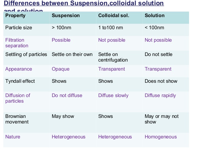
Although the concept of partial clotting within vessels reaches back to the beginnings of modern medicine, much of the discovery of its biochemical mechanisms – the activation of clotting factors – has been left to chance.  The admission of a patient to the hospital with an unceplained bleeding disorder challenged researchers to discover the cause of hemorrhaging.  Analysis of blood from normal persons helped in the study of the patient with the blood disorder. A new clotting factor was hereby discovered which was missing from the  patient’s blood.  For this reason, several clotting factors have been named after the individuals in which they were missing: e.g., Christmas factor (factor IX)[4], Hageman factor (factor XII)[4].

In this article, the causes of pathological (intravascular) clotting will be described, as will various methods of detecting this condition, especially a blood test I call the Mycotoxin Oxidative Stress Test (MOST).

The Mechanics of Blood Coagulation

Blood clotting is a highly detailed chemical-mechanism involving many distinct components.  The problem for the hematologist hs been to understand it at the biochemical level.  Undoubtedly, efforts to fully understand blood clotting will continue for many more years.

Recalling Antione Bechamp’s[8] and Gunther Enderlein’s[9] research into the sub cellular living elements and combining this with what is known of colloidal flocculation[6], it is suggested that the clotting of blood begins with the end-linking (polymerizing) of the fundamental protein unit called by Bechamp the microzyma[8].  A chain of these living units constitutes fibrinogen, which is still dispersed 9micro-hetergenous0 in the blood, and it may or may not be further processed.  If processing continues, it will be either by continued end-linking or by cross-linking.  End-linked fibrinogen is referred to here as fibrin monomer, which I have suggested is a repair protein also dispersed in the blood. Due to a number of blood clotting factors, the process may continue until the excess fibrin monomer and/or until fibrin becomes excessively end-linked.

Cross-linking the polymerized strands to form a three-dimensional network results in what is called the hard clot (fibrin – the major protein of clotting blood).  Factor XIII, which instigates the forming of these blood networks. is always present but latent in the blood, and must be activated before the formation can occur.  Persons who are producing fibrin monomer or excessively linked fibrinogen are said to be in a hyper-coagulable state, while those having diminished  ability to form clots are in a hypo-coagulated state.  It is the activation of the colloidal clotting factors which is so complex.  Blood clotting may occur through many pathways and be initiated by many different stimuli.  Regardless of initiation factors, the process is a sequence of events in which the activation of one factor triggers another, until, after a series of discrete steps, fibrin is formed.

When blood is clotted prematurely, and the factors involved are consumed (incorporated into) the body recognizes a deficiency of clotting agents and generates more.  Thus, people with a tendency to clot excessively will alternate between a hyper coagulable state and a hypo-coagulatable state.  When in the hypo coagulated state, such people hemorrhage until the deficient clotting factors are replaced.[4]  When only fibrin monomer or excessively linked fibrinogen is formed (no cross-linking), it is quite subtle and may go undetected.  It may be detected by a change in blood viscosity (sedimentation rate), by the Mycotoxic Oxidative Stress Test (described later), or by other more subtle means.  If strands of fibrinogen are cross-linked, however, a suggicient amount of insoluble precipitate of fires may result, and these can be detected microscopically using a phase contrast and dark-field microscopy in prepared slides of fresh tissue or blood.  An excessive formation of fibrin leads to  an impairment in circulation, and eventual organ failure usually results.[5]

With this background, we are in a position to consider a standard medical term: disseminated intravascular coagultion (DIC).[6]  This term encompasses the hyper coagulable state, i refer to as pathological blood coagulation which consists of both insoluble and excess dispersed polymers of colloidal proteins.

Key Ingredients of Pathological Blood Coagulation

Before discussing DIC in more detail, it si necessary to introduce its fur important ingredients according to this view – mycotoxins, endotoxins, exotoxins, and tissue factor.  Any of these elements, or any combination of them, can play a major role in initiating unwanted DIC.[6]  However, mycotoxins or the acids from yeast have been found to be the underlying element which instigates and intensifies the participation of the other three.[6]  Each will now be described in turn and brought into the clotting picture.

(Micrograph 1: left, shows normal hyper-coagulated blood in a healthy blood clot sample and right, hypo coagulated blood in an unhealthy blood clot sample)

Mycotoxins and Metabolism by Fermentation

As discussed in the main text of my published book, Sick and Tired book[7 ]. acidification of blood and body tissues and organs and the accompanying lack of oxygen lead to pathological metabolic fermentation, which is carried out primarily by yeast and mold.  Such pathological microorganisms, or their precursors, ar inherent to the human body and to all higher organisms.  Their precursors according to Bechamp, the microzymas, carry on a nominal and homeostatic fermentation themselves. under healthy conditions.[8]  The primary function of yeast and mold is to decompose the body upon the death of the animal or human organism.  Their premature overgrowth indicates a biochemical environment akin to death.  During pathological metabolic fermentation, high concentrations of several acidic substances called mycotoxins are created.  They are highly damaging, always acidic, metabolic products.  If not immediately buffered by specific antioxidants, such as hydrogen peroxide and the hydroxyl free-radical, mycotoxins can seriously disrupt the physiology by disrupting normal metabolism and by penetrating blood and body cells and poisoning them.  As will be seen, they interact with many of the mechanisms for DIC in various pathological symptomologies.

In my published article called The Finger on the Magic of Life: Antoine Bechamp, 19th Century Genius (1816-1908),  I discuss pleomorphism in some detail.[7] Understanding this phenomenon – the rapid evolution of microorganisms across traditional taxonomic  lines is helpful in getting a complete picture of DIC.  Briefly stated, collodial living microzymas evolve intracellularly into more complex forms (microorganisms), beginning with a healthy primitive stage comprising of repair proteins.  As the disease condition worsens, morbid intermediate forms (filterable bacteria or viruses, cell-wall deficient forms and full bacteria) develop from repair proteins, or directly from microzymas.  A third macrostage comprises the commonly recognized culminate microorganisms which are yeast, fungus to mold.  In terms of pleomorphism, all of these microorganisms represent a single family of variously functioning forms.[8]  The culminate forms produce the lions share of acids, which are mycotoxins and the primary focus of my research.[7][8][9]  For convenience, bacteria, yeast, fungus and mold that produce acidic metabolic wastes and protein cellular fragments called exotoins, endotoxins and mycotoxins will here be referred to collectively ash EMPO, or exotoxic, mycotoxic-producing microorganisms.

What follows is a shortened description or the description and origin of several exotoxins and mycotoxins, referred to collectively microzymian acidic toxins of MAT, which are involved in the processes leading to DIC.  The bio-effects, or the pathology of cellular fermentation, of these toxic metabolites are know as mycotic illness, mycotoxicosis, or mycotoxic stress as seen in the MOST and described and published by Dr. Bolin in the 1940’s.[10]

One such metabolic product is acetyl aldehyde, which is formed by  cellular breakdown of food, especially carbohydrate and the birth of  EMPO.  Acetyl aldehyde can also break down into a secondary substance know as ethyl alcohol.  Although acetyl aldehyde presents an immediate hazard to health and well-being, nature has provided a means of buffering of neutralizing this acidic by-product of cellular digestion and fermentation almost as soon as it is created.[11] The controls of acetyl aldehyde (and ethyl alcohol) are the sulfur amino acids, cysteine, taurine, methionine and the peptide glutathione which is found in red blood cells and almost all cells utilizing oxygen.[12]  In an attempt to buffer or neutralize MAT, the body will also bind or chelate both fats and minerals to them.[12]

Another member of the MAT family is uric acid, which is formed by the digestion of protein and the creation of EMPO.[13]  Uric acid can also break down into secondary substance, on of which is alloxan.[14] This has been shown to damage the insulin-producing pancreatic beta cells leading to diabetes [Refer to Tables 1 and 2]

A shortage of alkalizing nutrients or an excess of MAT initi­ates an immune response in which a special class of free radicals which I call microzymian oxidative buffering species (MOBS) are released.[15] These oxygen metabolites carry unpaired electrons and are intended to disrupt bacteria, yeast, fungus and mold, and buffer exotoxins, endotoxins, and mycotoxins. Current medical savants believe that they can disrupt just about any­thing they contact, including healthy cells and tissue: this is not accurate. The fact is that MOBS carriers a nega­tive surface-charge and repel healthy cells, which also have a negative surface-charge. [16] It is the positively surface-charged bacteria, yeast/fungus, mold, exotoxins, endotoxins, and myco­toxins that MOBS bind too.[17]  This aspect gives some insight into autoimmune phenomena, which are not, as is often maintained, the result of an overburdened immune system. They result either as a side-effect of the immune system’s attempt to remove foreign or toxic ele­ments, or as a direct attempt by the immune system to remove cells or tissue rendered useless or disturb­ing to the body by MAT.

In every degenerative symptomatology I have studied, I have found excessive MAT and MOBS (see Tables 1-3). Some of these degenerative symptoms and their underlying disease conditions, including cancer are described in my recently published paper on a deficiency on alkaline nutrition and cancer. [15] But the fact that myco­toxins cause harm to humans and other animals is purely a secondary effect, since, as noted, the prima­ry function of the microorganism is not to cause illness. We know from the fossil record that pleomorphic microforms existed long before animals.[19] In fact, humans and animals developed in terms of micro­organisms.[20] The reverse, however, is not true. Since micro­organisms appeared first in the developmental sequence, they are not physiologically aware of humans and animals. There is much evidence that human and animal physiologies are highly aware of, and respond to MAT – these acidic compounds signaling the presence of bacteria, yeast, fungi and/or mold or  EMPO.[21].

Endotoxins

Also involved in the process leading to DIC are endotoxins, substances endogenous to symptogenic (i.e., “pathogenic” in orthodox terms) bacteria. Endotoxins are a family of related substances having certain common characteristics, but differing from one bacterial form (or strain) to another. Endotoxins are lipopolysaccharides (LPS). LPS form a widely diversified group because of (1) the number of long- chain fatty acids composing lipids; (2) the number of individual sugars as well as their modes of linkage to one another; (3) the branching of sugar chains; and (4) the number of possible arrangements of these units. Endotoxins also contain proteins, further com­pounding the structural diversity.[22]

One theory on endotoxin states that its purpose is to act as a semi-permeable membrane for the bac­terium, limiting and regulating substances entering the organism.[22] Endotoxin resides solely on or near the interior surface of the cell membrane and is shed into the surrounding medium only upon the death of the bacterium. Thus, as these microforms die off, or are lysed by bodily activity, endotoxin is released. (This fact may well be an explanation for the Herxheimer reaction, in which a patient becomes worse following the administration of toxic drugs or other forms of treatment that drastically alter the associated organ­ism.[23]) Another endotoxin theory states that LPS are a constituent of the membrane, and as the organism grows, endotoxin fragments are repeatedly sloughed off into the medium. This phenomenon has been observed in the digestive tract.[24] Since bacterial translocation into the blood is not only possible but common where epithelial hyperpermeability exists, one can assume that the process will continue there. Both theories may be correct if we think of the first one as true of “adult” forms, and the second as true of newly developed and expanding ones.

Basic to the structure of an endotoxin is the lipid common to all forms, designated lipid A, to which is attached a “core” polysaccharide, identical for large groups of bacteria. To the core polysaccharide is attached the O-antigen, consisting of various lengths of polysaccharide chains which are chemically unique for each type of organism and LPS. These chains pro­vide endotoxin specificity.[25] Experiments conducted over many years indicate that most, if not all, of the toxic effects of an endotoxin may be attributed to the lipid portion, and it is sometimes used per se in experiments rather than the entire molecule.[26] An important additional feature of lipid A is its phos­phate content. Each phosphate group carries a nega­tive charge, and since lipid A is a rather large mole­cule, it provides, essentially, a negatively charged sur­face. The importance of this will be seen shortly.

Exotoxins

These are the metabolic excretions of bacteria. While endotoxin’s ongoing effect is, in a manner of speaking, in the background, exotoxins, like myco­toxins, present a double-edged sword. Not only do they initiate DIC, but they produce, or influence the body to produce, the various and numerous infec­tious symptomatologies, such as typhoid fever, diph­theria, etc. (See “Vaccination Reconsidered” in Section 4 of the Appendix of Sick and Tired for details on the action of diphtheria toxin.)[7] By comparison, mycotoxins not only initiate DIC, but there is much evidence to sug­gest that they produce, or influence the body to pro­duce, degenerative symptomatologies, such as arthri­tis, diabetes, etc., and cancer and AIDS as well.

Tissue Factor

Crucial to the understanding of DIC is recogni­tion of the role of tissue factor (TF), formerly known as thromboplastin. This transmembrane lipoprotein exists on the surface of platelets, vas­cular endothelial cells, leukocytes, monocytes, and most cells producing EMPO.[27] It plays a major role in several biochemical mechanisms leading to DIC.

TF is the primary cell-bound initiator of the blood coagulation cascade. Its gene is activated in wound healing and other conditions. By itself it is capable of initiating clotting, but also becomes active when complexed with factor VII or activated factor VII (Vila).[28] TF has been described as the receptor for factor VII because of the close association between the two proteins and because it causes a shape change (conformational) in factor VII, allowing it to attain activity. Both factor Vila and the TF/VII com­plex activate factors IX and X, which initiate the clotting cascade and the formation of thrombin.[29]

Development of Disseminated
Intravascular Coagulation
(DIC)

DIC Induced by MAT and Tissue Factor

An infusion of toxins into the blood has a direct effect on TF gene expression in leukocytes. Contact of MAT, endotoxins (lipid A), or exotoxins with leukocytes, activates proteins that bind to DNA nucleotide sequences, thereby activating the TF gene.[30] (See Tables 4-6.)

Endothelial cells damaged in culture by exotoxins, endotoxins, or mycotoxins attract polymorphonuclear leukocytes (PMNs), which adhere to the damaged cells. Once the leukocytes are bound, they can still have their TF gene activated if it hasn’t yet occurred, and they may release MOBS in response to toxins and to organisms of disease, possibly creating further dis­turbances. (Cellular disorganization then releases acti­vating proteins into the blood, which is discussed in more detail later.) Research shows that exotoxic and mycotoxic stress resulting in bound PMNs can be blocked by “antioxidants.”[31] These might better be called anti-exotoxins or antimycotoxins. Both observa­tion and study have led the author to conclude that cellular disorganization is initiated and primarily caused by fermentation pathology, not, as is the cur­rent belief, by the MOBS, or free radicals, generated to destroy toxins and microorganisms. MOBS or free radicals, because of their negative charge, are released to chelate or bind EMPO and MAT. It is suggested by current savants that free radical tissue damage is the secondary, “shotgun” effect of intense immune response to EMPO toxification and MAT-damaged cells. This could not be the case since healthy cells or their membranes carry a negative charge and would resist any electromagnetic attraction because of simi­lar charge. The concentration and instability of MAT generated in a compromised terrain, as opposed to the fleeting existence of free radicals, especially exoge­nous ones, also lead to this conclusion.

Endothelial cells grown in culture can be induced to express tissue factor. In one experiment, no procoagulant activity could be detected in the absence of toxins. However, the addition of mycotoxins from Aspergillus niger or Micrococcus neoformas (Mucor racemosus Fresen) resulted in procoagulant activity which reached a maximum in four to six hours and was dose-dependent. The same experiment was applied using E. coli and Salmonella enteritidis endo­toxin with a similar result.[32] A single intravenous injection of a mycotoxin from Aspergillus niger into experimental animals resulted in circulating endothelial cells within five minutes. In other exper­iments with the mycotoxin, detachment of endothe­lial cells from the basement membrane was noted.[33] (See Table 8.)

Removal of endothelial cells has dire conse­quences from two standpoints: First, the surface of these cells is covered with a specific prostaglandin (PGI2) known as prostacyclin. If blood contacts a surface not covered with PGI2, it will clot. For example, surfaces devoid of this prostaglandin are formed whenever a vessel is cut or punctured. An abrasion or other injury may also expose a surface on which PGI2 is lacking. The removal of endothelial cells by exotoxins or mycotoxins creates a surface devoid of PGI2, leading to blood clotting (see Table 7). Secondly, disorganization of endothelial cells cre­ates increased levels of EMPO and MAT which are attracted to an exposed surface (basement mem­brane) which expresses a negative charge. This also leads to clotting.

DIC Induced by Electrostatic Attraction

It was discovered in 1964 that blood will clot sim­ply from contacting a negatively charged surface.[34] Previously it was believed that the clotting process comprised a cascade of enzyme activity in which one activated the next, etc. The discovery that blood could be clotted simply by contacting a negatively charged surface ruled out the purely enzyme hypoth­esis. Only some of the known clotting factors have been shown to be enzymes.[35] As a result of this sur­prising discovery, detailed research was conducted in an attempt to describe the process. In some experi­ments, the negatively charged surfaces of selected, finely divided, inorganic crystals, including alu­minum oxide, barium sulfate, jeweler’s rouge, quartz, and titanium oxide, were considered.[36]

The clotting factor eventually shown to be activat­ed when whole blood contacted negatively charged surfaces was factor XII, also known as the Hageman factor. This is a positively charged protein migrating in an electric field (electrophoresis) toward the anode.[37] It is believed that factor XII is normally in the shape of a hairpin which binds to the negatively charged sur­face at the bend. Electrostatic attraction forces the two arms to lie flat on the surface, thereby exposing the inner faces and activating the molecule.

It was discovered that if the negatively charged particles were smaller than the clotting factor itself, activation was minimal. Or, if the concentration of clotting factor was too great, there was little or no activation.[38] Both of these observations indicated that the process was one of electrostatic attraction between the negatively charged surface and the clot­ting factor, which is a “basic” protein, that is, posi­tively charged.[39]

Activation of factor XII allows the activation of factor XI, which then activates factor IX. Thus, the blood clotting cascade continues to the formation of fibrin in the normal manner.[40] However, due to a series of activations begun by contact of factor XII with a negatively charged surface, trace amounts of factor Xa also show up in the blood. Factor VII is activated to Vila by factor Xa. Factor Vila then acti­vates factors IX and X, leading to the formation of thrombin. Factor Xa, with co-factor Va, continues the clotting cascade until fibrinogen is activated, leading to fibrin formation.[41] (See Table 5.)

As discussed earlier in terms of prostacyclin, beneath endothelial cells is another surface—the basement membrane. Called the extracellular matrix, it is a thin, continuous net of specialized tis­sue between endothelial cells and the underlying connective tissue. It has four or more main con­stituents, including proteoglycans (protein/polysac- charide).[42] The removal of endothelial cells by’MAT exposes this membrane, which is negatively charged by virtue of its sulfonated polysaccharides in the pro­teoglycans. This brings a reduced negatively charged surface into direct contact with the blood, which activates factor XII and the clotting cascade.[43]The positively charged toxic components of MAT also activate factor XII, as do disturbed disorganized cells, yeast/fungus cells, moldy cells, and the phos­phate groups in the lipid A component of endotoxin. (See Tables 2-5.)

To summarize this section, exotoxic, mycotoxic, and oxidative stress resulting from the overgrowth of bacteria, yeast/fungus, and then mold, has multiple actions, all leading to disseminated intravascular coagulation:

MAT activation of tissue factor gene in leukocytes; subsequent activation of factors VII, IX, and X, resulting in the blood clotting cascade.

MAT activation of tissue factor gene in endothelial cells, again leading to the clotting cascade.

MAT damage to endothelial cells, resulting in neu­trophil attraction, with TF gene activation and generation of MOBS, which, in turn, neutralize MAT, protecting healthy endothelial cells or the basement membrane and supporting the janitorial services of the leukocytes.

Removal of negatively charged endothelial cells by positively charged exotoxins, endotoxins, and mycotoxins, creating a surface devoid of PGI2, also exposes the negatively charged basement membrane, leading to the activation of factor XII and initiation of the clotting cascade. Positively charged components of EMPO, exotoxins and mycotoxins, and several other elements, including the lipid A component of bacterial endotoxin, also activate factor XII and the clotting cascade.

Endothelial Cells as Antithrombotics or Procoagulants

Normal, resting (unstimulated) endothelial cells show antithrombotic activity in several ways: (1) by the inhibition of prostacyclin (platelet adhesion and aggregation); (2) the inhibition of thrombin genera­tion; and (3) the activation of the fibrinolytic system, leading to clot lysis.[45] We will take a brief look at the thrombin aspect.

On the surface of endothelial cells is a protein called thrombomodulin, which acts as a receptor for thrombin. When bound to thrombomodulin, throm­bin can activate protein C. Activated protein C then catalyzes the proteolytic cleavage of factors Va and Vila, thereby destroying their participation in blood clotting. Thus thrombin, which normally activates fib­rinogen, plays an opposite role in this case and inhibits the clotting process.[46,47] (See Table 7.)

On the other side of the coin, the endothelial cell becomes a procoagulant agent when acted on by cer­tain lymphokines, such as interleukin-1. Not only can interleukin-1 induce TF gene expression, but it also suppresses transcription of the thrombomodulin gene in endothelial cells. As in other situations, the lymphokine-activated endothelial cell expresses TF on its surface as a result of TF gene activation. This leads to the production of thrombin and the trigger­ing of the blood clotting cascade.[48] (See Table 5.) Many lymphokines also stimulate adhesion of leuko­cytes to endothelial cells damaged by MAT, resulting in recycling of the cells by MOBS, as described later.

DIC Induced by Intracellular Exotoxic, Mycotoxic, Oxidative Stress by Bacteria, Yeast/Fungus and/or Mold

Any cell which has gone from an oxidative to a fer­mentative state can biochemically cause macrophage production of the lymphokine tumor necrosis factor (TNF). This protein has been shown to activate the gene for TF in fermenting cells, which are so behaved due to morbid evolution of bacteria, yeast/fungus, and then mold.[49,50] In the author’s view, a cell having been switched entirely to fermentation metabolism as a result of a physical or emotional disturbance of that cell, is what constitutes cancer (see Tables 5 and 13). (One might argue that this definition does not fit all “forms” of cancer, such as leukemia, for example. This is because leukemia is not cancer, but an immune response to the rise in EMPO and MAT in the body, and a relatively easy compensation to correct.)

The surface of many disorganizing or fermented cells (cancer cells) is characterized by small projec­tions in the plasma membrane which pinch off, becoming free vesicles containing toxins as well as TF complexed with factor VII. These vesicles can aggre­gate and/or lodge anywhere, ultimately releasing their contents. Also, the presence of excessive amounts of TF/factor VII complexes on the surface of fermented cells allows the formation of a fibrin net around the cell and around the entire mass of cells (tumor). This seems to be an attempt by the body to encapsulate and contain the mass. However, fermented cells do escape from the primary fibrin net, perhaps due to some electromagnetic effect, and become free-float­ing in the circulation. They may thus lodge elsewhere and instigate the fermentation of other cells by fungal penetration or by poisoning them and provoking a morbid evolution of their inherent microzymas.

Because of the surrounding fibrin net, these mobi­lized fermenting cells are protected from collection by the immune system while in transit.[51,52] (See Table 4.) The blockage or dissolution of fibrin net forma­tion by an anticoagulant such as heparin allows freed, fermenting (metastasizing) cells to be dismantled by natural killer cells and other immune cells (see Tables 5, 12 and 13).

DIC Induced by MAT/EMPO and Immune System Response (Release of MOBS)

Unsaturated fatty acids are highly susceptible to EMPO as well as MAT. Linoleic acid, a long-chain fatty acid present in white cells, has 18 carbons and 2 unsaturations. Subjected to MAT, linoleic acid binds the exotoxin, endotoxin, or mycotoxin, there­by forming an epoxide at the first unsaturation.[53] Research has revealed that this compound, named leukotoxin, is highly disturbing to other cells. It caus­es platelet lysis, thereby releasing TF and initiating DIC.[54] (See Table 10.) The fact that MAT result in fermented fats lends further credence to the sugges­tion that the initial and primary degenerative damage to structures and substances in the body is caused by exotoxins and/or mycotoxins, and that damage by MOBS, or by other free radicals, is not possible.

Another mechanism leading to DIC is the release of a special glycoprotein, sialic acid, from the terminal ends of cell-membrane polysaccharides, where it is always found. Polysaccharides play a highly significant role in biochemical processes, with both enzymes and membrane receptors recognizing various groupings of specific sugars linked in highly specific ways.

Immediately preceding the release of sialic acid in the polysaccharide chain is the sugar galactose. The sialic acid/galactose arrangement is utilized as a biolog­ical indicator of cellular and molecular aging. As cells age, sialic acid is naturally expressed from the terminal ends of polysaccharides, thereby exposing galactose. A membrane-bound enzyme from the liver, galactose oxi­dase, recognizes galactose and eventually disorganizes it, disrupting cell function integrity and hastening demise. Aged red blood cells, which have expressed a significant amount of sialic acid, are removed from the blood by this process. (I theorize that the biological ter­rain may be at work in normal cell aging. That is, the rate at which sialic acid is expressed is determined by the levels of corrosive acids in the system and the body’s ability to remove them, although there are no doubt intracellular factors at work as well.)

I suggest from my years of  clinical research  that cellular breakdown is compounded by the fermentation of the galactose by the microzyma. This is a process that begins from within and not necessarily from without. Not only does this action create more sialic acid, it creates other toxic waste products such as acetic aldehyde, alcohol, uric acid, oxalic acid, etc. The increase in cellular disturbances and fermenta­tion of the galactose creates biochemical signals for more galactose oxidase. This leads to greater cellular disorganization and developmental morbidity, espe­cially in the red blood cells, and a rise in the level of detrital serum proteins, which encourages clotting. From this perspective, diabetes, arthritis, atheroscle­rosis and other symptomatologies become more clearly “degenerative” (see Tables 2-5, 12 and 13).

Fibrinogen is a rather elaborate protein having the structure of three beads on a string. Expressed on the end beads is sialic acid, which indicates the beginning of disorganization of the fibrinogen and a declining negative charge to the positive. Prior to the declining charge and the expression of sialic acid on the end beads, fibrinogen, which is negatively charged, will not polymerize the healthy blood due to mutual repulsion. However, fibrinogen will poly­merize to damaged cells, EMPO, MAT and other positively charged areas of the body for repair pur­poses. Thus, as more and more sialic acid is expressed, there will be a significant reduction in the charge of the fibrinogen, acting as the primary requirement for the polymerization of fibrinogen (hypercoagulable state). The resulting polymer, fib­rin monomer, is the protein chain used in the repair of cells and clotting of blood.[55] End-linking will take place after the release of sialic acid (positive charge) by whatever means.

With this background, it is interesting to note that blood taken from persons suffering from anxiety is expressing sialic acid from fibrinogen, and is halfway toward clotting. Hormones released during anxiety states are easily fermented, giving more momentum to MAT and thereby resulting in this important change in fibrinogen. It leads to a clotting pattern characteristic of anxiety stress, and is readily identi­fied in the MOST. As can be seen in this picture, the pattern is a “snowstorm” of protein polymeriza­tions measuring from 2 to 10 microns.

allergiesbefore


[Micrograph 2: An Anxiety Profile showing a ‘snowstorm’ of 2 to 10 micron protein polymerizations starting from the center of the clot and moving out towards the edge]

As mentioned earlier, despite the attempt by the body to neutralize EMPO and MAT, an excess will initiate the release of MOBS by immune cells. A major MOBS is superoxide, designated chemically as O 2. It may exist alone or be attached to another ele­ment, such as potassium (KO’2) or sulfur (SO). Again, however, nature has provided a means of pro­tecting healthy cells—their negative charge[1]. Another protection against superoxide is the enzyme superox­ide dismutase (SOD), also found in all healthy cells.

A second member of the MOBS family is hydro­gen peroxide (H202). This molecule is very unstable and tends to react rapidly with other biological mol­ecules, damaging them. The release of hydrogen per­oxide in the body is a response to the overgrowth of decompositional organisms in a declining pH (com­promised biological terrain). The control for healthy cells against hydrogen peroxide is their negative charge and the protective enzyme catalase, one of the most efficient enzymes known.

When leukocytes and other white blood cells are stimulated by the presence of bacteria, yeast/fungus and mold, they treat these organisms as foreign par­ticles to be eliminated. During and prior to phagocy­tosis, the foregoing oxidative cytotoxins, along with the hydroxyl radical (OH’), are generated and released specifically for neutralizing microforms or harmful substances. This release is referred to as an “oxidative burst.” As a result of fermentation and the production of exotoxins and mycotoxins that fer­ment galactose from cells, the immune system is activated. An oxidative burst is released to neutralize the morbid microforms and mycotoxicity.[56] Like other biological processes faced with constantly alarming situations, the continued release of MOBS can get out of control. This may damage endothelial cells, the basement membrane, or other body ele­ments, and this activates fibrinogen to fibrin monomer (repair protein), leading to DIC [see Table 9]. Interestingly, the white blood cells capable of neutralizing MAT through MOBS production are the same ones capable of phagocytosis, the process by which foreign matter, waste products and microor­ganisms are collected and dumped in the liver.[57]

To summarize this section, pathological microforms and their acids create DIC by a number of pathways:

Leukotoxin (linoleic acid bound to mycotoxin) is highly toxic to cells. It causes platelet lysis, there­by releasing TF and initiating DIC.

The expression or release of sialic acid residues from healthy cells that have been disturbed allows for the fermentation of galactose, creating exotox­ins and mycotoxins, biochemically activating galactose oxidase, which further disturbs and dis­organizes healthy cells. This cycle loads the blood with debris.

EMPO and MAT disturb fibrinogen, which releas­es sialic acid and reduces the charge, allowing it to polymerize into fibrin monomer and fibrin nets.

The presence of exotoxins, endotoxins, and myco­toxins and their poisoning of cells activates the immune system. White blood cells generate MOBS (e.g., superoxide [0′2] or hydrogen perox­ide [H202]). These substances bind to and neu­tralize EMPO and MAT. MOBS are repelled by healthy endothelial cells and the basement mem­brane because of their negative charge. Cellular disturbances and disorganization stimulate the generation of fibrin monomer for repair purposes, leading to DIC.

Detection of Disseminated Intravascular Coagulation

The Sonodot Analyzer

The Sonoclot Coagulation Analyzer provides a reaction-rate record of fibrin and clot formation with platelet interaction. An axially vibrating probe is immersed to a controlled depth in a 0.4 ml sample of blood. The viscous drag imposed upon the probe by the fluid is sensed by the transducer. The electronic circuitry quantifies the drag as a change in electrical output. The signal is transmitted to a chart recorder which provides a representation of the entire clot for­mation, clot contraction and clot lysis processes. The analyzer is extremely sensitive to minute changes in visco-elasticity and records fibrin formation at a very early stage. The Sonoclot has been evaluated scientif­ically and shown to provide an accurate measurement of the clotting process.[58,59]

One application of the Analyzer has been the development of a test to distinguish non-advanced breast cancer from tumors that are benign. The ratio­nale for the test is the hypercoagulable state seen in cancer patients (Trousseau’s Syndrome), resulting from the generation of TF by leukocytes (mono­cytes).[60] (See Table 4.)

Fibrin Degradation
Products and Fibrin Monomer

DIC can be seen as a two-step process. First, fib­rinogen, which is always present in the blood, is acti­vated by any of several mechanisms. This activation leads to an automatic polymerization (chain forma­tion) resulting in fibrin monomer. This is not apparent in a microscope unless the blood is allowed to clot, as in the MOST.[61,62] The second step is the precipitation or deposition of fibrin (hard clot) by several other mechanisms. One of these is the formation of cross­links through the action of factor XIII. Another such mechanism may be poor circulation in an organ already blocked by deposited fibrin. The deposition of precipitated fibrin may be detected microscopically in tissue sections and diagnosed as DIC.[62]

Because fibrin monomer is not readily detected, a chemical test for it is of immense value in diagnosing DIC. Research has indicated that its detection may be very useful in the early diagnosis of DIC and MAT.[63] There are three fundamental physiologic areas related to blood clotting: (1) the prevention of blood clotting, (2) the clotting of blood, and (3) the removal of clotted blood once it has formed.

Enzymes are present that are capable of removing (lysing) clotted blood, one of which is plasmin. Another enzyme, plasminogen, is always present in the blood, but is inactive as a proteolytic agent. Plasminogen acti­vator converts plasminogen to plasmin, which can degrade deposited fibrin. This process is not specific for fibrin, however, and other proteins may be affected. When fibrin is degraded (fibrinolysis), fibrin monomer, as well as several other products, are formed. Commercial kits are available for the analysis of fibrin degradation. This test is an indirect measure of the pres­ence of DIC and MAT.[64]

Other tests include:

Protamine Sulfate: Protamine sulfate is a heparin binder sometimes used in surgery for excessive bleed­ing. The test, which indicates fibrin strands and fibrin degradation products, is conducted in a test tube, with fibrin monomer and fibrin forming early and polymer­ization of fibrin degradation products occurring later.[65] Ethanol Gelation: A white precipitate is formed by the addition of ethanol to a solution in a test tube containing fibrin monomer as a degradation product of fibrin, indicating DIC and MAT.[66]

The Mycotoxic Oxidative Stress Test (MOST)

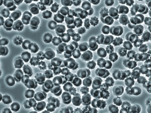
Up to now, blood chemistries have been the prima­ry mode of diagnosis or analysis for the presence of pathology. In the view presented here, the bright-field microscope, is used to easily and inexpensively reveal a disease state as reflected by changes in certain aspects of blood composition and clotting ability. DIC is char­acterized by the abnormal presence in the blood of fib­rin monomer. When allowed to clot, blood containing such an abnormal artifact will exhibit distortions of normal patterns. The presence in the blood of soluble fragments of the extracellular matrix and soluble fibronectin, as well as other factors, will also create abnormal blood clotting patterns as described below.

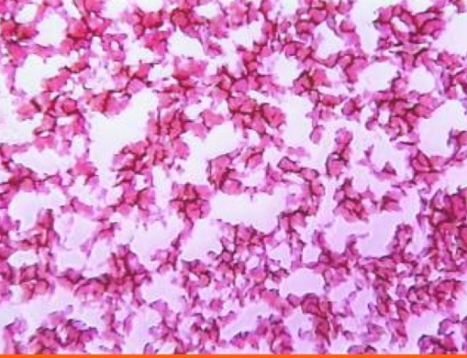
A small amount of blood from a fingertip is con­tacted with a microscope slide. A series of drops is allowed to dry and clot in a normal manner. Under the compound microscope, the pattern seen in healthy subjects is essentially the same—a dense mat of red areas interconnected by dark, irregular lines, completely filling the area of the drop. The blood of people under mycotoxic/oxidative stress exhibits a variety of characteristic patterns which deviate from nor­mal, but with one striking, common abnormality: “clear” or white areas, in which the fibrin net/red blood cell conglomerate is missing.

BowelCancerLive Blood Dried Blood_0166


[Micrograph 3; An abnormal clot with striking ‘clear’ or white areas or protein polymerization as seen in the hyper coagulated blood of a patient with lower bowel imbalances]

Why the fibrin net is missing may be understood from the following: Two peptides—A and B—in the central protein bead of the fibrinogen structure become bound in the cross-linking process. There are two ways this can be configured: (1) Thrombin is capable of activating peptides A and B, resulting in the formation of a polymer loosely held together only by hydrogen bonds; (2) With peptides A and B acti­vated normally, the resulting hard clot is insoluble, indicating that the peptides are linked by covalent bonds. The difference in bonds results from factor XIII, an enzyme which links the two fibrin strands with a glutamine-lysine peptide bond.

Additional research has shown that the release of sialic acid from fibrinogen inhibits the action of factor XIII, resulting in a soft, white clot. In addition, acetic aldehyde has been shown to inactivate factor XIII directly. The soft clotting, compounded by other polymeric aggregations (described below), results in clear areas in the dry specimens. In the opposite extreme, high serum levels of calcium, for the pur­pose of neutralizing MAT, activates factor XIII, lead­ing to excessive cross-linking of fibrin to form a clot harder than normal. This is reflected in the MOST pattern characteristic of definite hypercalcemia— that of a series of cracks in the clot radiating outward from the center, resembling the spokes of a wheel. High serum calcium is the body’s attempt to com­pensate for the acidity of mycotoxic stress by pulling this alkalizing mineral from bone into the blood. This demand creates endocrine stress in turn, because reabsorption of bone is mediated by parathormone (PTH). Therefore, this clotting pattern indicates cal­cium deficiency and thyroid/parathyroid imbalance.

calciumpattern


[Micrograph 4: A mineral deficiency or more specifically a calcium deficiency pattern associated with an imbalance of they thyroid and/or parathyroid}

Advanced research has shown that there are seven carbohydrate chains in fibrinogen (each terminated by sialic acid). A second action of factor XIII is to ferment a large amount of carbohydrate during clot­ting. Because carbohydrate is most often water solu­ble, the loss of this material undoubtedly adds to the insolubility of a clot, while pathological retention contributes to the softness of the abnormal clot.

Clinical experience demonstrates that the MOST is a reliable indicator of exotoxic and mycotoxic stress and, concurrently, of various disorganizing symptoma­tologies associated with fermentative and oxidative processes. As various cellular degradation occurs, the blood-borne phenomena which accompany such symptoms as diabetes, arthritis, heart attack, stroke, atherosclerosis and cancer show up in the MOST, often with sialic acid beads in the clear areas of poly­merized proteins. (Determination of the liberation of sialic acid from carbohydrate has been approved by the U.S. Food and Drug Administration as an accept­ed indicator for cancer, and is clinically available.)

sialicacid

[Micrograph 5: Sialic acid beads are seen inside the protein
polymerization of the hypocoagulated blood as black dots]

The extent and shape of the clear areas are reflec­tive of particular symptomatologies which have arisen from the way in which the disease condition manifests in a given individual. This observation is borne out by having the patient undergo appropriate alkalizing therapy. With success of treatment based on the patient’s freedom from symptoms, sense of well-being, and live blood exams discussed in the main text of Sick and Tired, Reclaim Your Inner Terrain, Appendix C,[7] repeated analysis with the MOST reveals a progressively improving clotting pattern.

[Micrographs 6 and 7: Medically diagnosed cancer patient with large polymerized protein pools (PPP) in the hypo-coagulated blood above. In the picture below PPP’s have significantly reduced in size and the blood is moving to a more hyper-coagulated state as a result of reducing acid loads with an alkaline lifestyle and diet (7, 70)]

Because of its very nature, the MOST is emi­nently suited to reveal and measure the presence in the blood of abnormal substances, clotting factors, and disorganization of cells due to an inverted way of living, eating, and thinking, which gives rise to MAT. The MOST indicates both the direct and indirect activity of MAT on blood clotting, endothelium, and the extracellular matrix (described next), as well as on biochemical pathways, including hormonal ones. The generation of excessive MOBS in response to EMPO and MAT, the inability that accompanies all degenerative symptoms to neutralize or eradicate EMPO and MAT, and the recognized hyper- and hypocoagulable states seen in various symptomatolo­gies, will beyond doubt be revealed in the MOST.

Aspergillusnigercrystal


[Micrograph 8 and 9: Medically Diagnosed HIV/AIDS micrograph showing above an Aspergullus niger mold crystal using dark field microscopy and below a hypocoagulated blood clot with systemic protein polymerizations measuring in excess of 40 microns using bright field microscopy}

HIV


As mentioned, hormones are easily fermented, and this will show up as a hypocoagulated blood pattern in the MOST. It is my opinion, this hypocoagulated blood appears in the MOST as misty clouds of protein polymerizations throughout the clot, as seen in the accompanying picture.

poorfibrin

[Micrograph 10: Poor fibrin interconnection in the clot associated with endocrine or hormonal imbalance]

The MOST from Solubilized Extracellular Matrix

There is now a clearer picture of the biochemical rationale for correlating abnormal blood clotting patterns with the presence of degenerative symptoms.  A link between symptoms and the distorted clotted blood patterns has been delineated in the MOST.
Another reason for the abnormal clotting patterns accompanying pathological states, in addition to insufficient bonding of fibrinogen peptides as seen in the MOST, is presence in the blood of water-soluble fragments of the extracellular matrix.

Extracellular Matrix Degradation by MAT

The extracellular matrix (EM) is a three-dimen­sional gel, binding cells together and composed of five or more major constituents: collagen (protein), hyaluronic acid (polysaccharide), proteoglycans (pro- tein/polysaccharide), fibronectin and laminin. Also included are glycosaminoglycans and elastin.[67] In every degenerative disease studied by this author, evidence has been found for MAT activity destruc­tive of EM.

One of the proteolytic enzymes activated in response to EMPO and MAT is alpha-1 antitrypsin (capable of neutralizing MAT), normally not active in the presence of the enzyme trypsin. The active por­tion of this anti-exotoxin and antimycotoxin contains the amino acid methionine, which includes a C-S-C linkage. When chelated by the hydroxyl radical (one of the MOBS oxidants), methionine’s central sulfur atom acquires one or two oxygen atoms (forming the sulfone or sulfoxide respectively). The fermentation of methionine is a secondary effect of immune response to an alarming situation, intended to neutral­ize MAT and prevent degradation of the EM. Once alpha-1 antitrypsin is exhausted, MAT will have more access to the EM. If the EM is damaged beyond repair, then the enzyme trypsin is released to disorganize and recycle the cells involved.[68]

A similar scenario holds for the enzymes collage- nase and elastase. Thus, the absence of alpha-1 antitrypsin in the presence of EMPO and MAT activates three enzymes which degrade the extracellular matrix. Degradation of the EM by enzymes and MAT puts into the blood the water-soluble fragments (proteins and glycoproteins) of normally insoluble EM components (see Table 11). The presence of these fragments modifies the normal clotting pattern (described below), as seen in the M/OST, and is therefore an indication of EM degradation, which is always found with degenerative symptoms. (Also present is fibrin monomer, which has been found in the blood of patients suffering from collagen dis­ease.[69] See Table 11.)

Fibronectin is a molecule in EM having several binding sites for various long-chain molecules— heparin (a sulfonated polysaccharide) and collagen, for example. As such, it functions as a cellular glue, bind­ing cells together as well as various components of the EM. A soluble form of fibronectin is normally found free in the blood, and enters into the formation of a blood clot through the action of factor XIII. This form of fibronectin binds to fibrin. Elevated, bound-serum fibronectin results from EM fragmentation by MAT, and accompanies degenerative symptoms such as arthritis and emphysema (collagen diseases).

Water-soluble fragments of the EM bound by fibronectin form a three-dimensional network or gel in the pathologically clotted blood (fibrin and com­ponents of the blood clotting cascade). Since fibronectin binds to both fibrin and collagen, the two polymeric networks are superimposed and intermin­gled, resulting in a modification of the normal clot­ting pattern. Exactly how the pattern is modified depends upon the nature of the collagen abnormally present, the nature and extent of hyaluronate pre­sent, and the degree to which EM fibronectin has been released by MAT.

Conclusion

Thus, it is easily seen that there are many forms which the pattern of clotted blood may take, depending on the individual and the internal terrain that produced the modifying substances. The MOST reveals not only the presence of exotoxic and mycotoxic stress, but indicates as well the nature of the symptom(s) resulting from the stress (see Table 12). Since MAT underlie the entire complex of events which degrade the extracellular matrix, I must conclude that the absence of these exotoxins, endotoxins and mycotoxins would provide substantial improvements in tissue integrity and the overall physiology and functionality of the organism or animal and human.

­

­

References

[1]  Jones, T.W., “Observations on some points in the anatomy, physiology and pathology of the blood.”  British Foreign Medical Review, 1842. 14 : 585.

[2] Trousseau, A., Phlegmasis alba delens. “Clinque Medicale de L’Hotel Dieu de Paris.”, 1865, 3:94

[3]  Virchow, R., “Hypercoagulability: A review of its development, clinical application, and recent progress.”  Gesammelte Abhandlungen our Wussenschaftlichen Medizin, 1856, 26:477.

[4]  Rapaport, S.I., “Blood Coagulation and its Alterations in Hemorrhagic, and Thrombotic Disorders.”  The Western Journal of Medicine, 1993; 158: 153.

[5]  Hamilton, P.J. et al., “Disseminatied Intravascular Coagulation: A Review.”  Journal of Clinical Pathology, 1978, 31: 609

[6] The Harper Collins Illustrated Medical Dictionary, 1994, p.13.

[7] Young, RO, “Sick and Tired, Reclaim Your Inner Terraine,” Woodland Publishing, 1999.

[8] BeChamp, A., “The Blood and Its Third Anatomical Element,”  Hikari Omni Publishing, 1999.

[9]  Schwerdtle, C, Arnoul, F, Enerlein, G, “Introduction to Darkfield Diagnostics”, Semmelweis-Verlag (2006).

[10]  Hawk, BO, Thoma, GE, Inkley, JJ, The Evaluation of the Bolen Test as a Screening Test for Malignancy*, cancerres.aacrjournals.org on December 5, 2015. © 1951 American Association for Cancer Research.

[11]  Uchida, K., “Role of Reactive Aldehyde in Cardiovascular Diseases”,  Labortory of Food and Biodynamics, Nagoya University Graduate School of Bioagricultural Sciences, Nagoya, Japan , Free Radical Biology and MedicineVolume 28, Issue 12, 15 June 2000, Pages 1685–1696

 [12] Chang JCvan der Hoeven LHHaddox CH, “Glutathione reductase in the red blood cells”,  Ann Clin Lab Sci. 1978 Jan-Feb;8(1):23-9.

[13] Kutzing, MK, Firestein, BL, “Altered Uric Acid Levels and Disease States”, Department of Cell Biology and Neuroscience (M.K.K., B.L.F.), Graduate Program in Biomedical Engineering (M.K.K.), Rutgers University, Piscataway, New Jersey. Address correspondence to: Dr. Bonnie L. Firestein, Department of Cell Biology and Neuroscience, Rutgers University, Piscataway, NJ 08854-8082. E-mail: firestein@biology.rutgers.edu

[14] Claudino, M,. Ceolin,,DS, Alberti, S.,  Cestari, TM,  Spadella, CT, Fischer Rubira-Bullen, IR, Gustavo Pompermaier Garlet, Gerson Francisco de Assis, ” Alloxan-Induced Diabetes Triggers the Development of Periodontal Disease in Rats”,  Published: December 19, 2007. DOI: 10.1371/journal.pone.0001320

[15] Young RO (2015), “Alkalizing Nutritional Therapy in the Prevention and Reversal of any Cancerous Condition. Int J Complement Alt Med 2(1): 00046. DOI: 10.15406/ijcam.2015.02.00046

[16] Heloise Pöckel FernandesCarlos Lenz Cesar, and  Maria de Lourdes Barjas-Castro, “Electrical properties of the red blood cell membrane and immunohematological investigation”, Rev Bras Hematol Hemoter. 2011; 33(4): 297–301. doi:  10.5581/1516-8484.20110080 PMCID: PMC3415751

[17] Harris, JO, “The Relationship Between the Surface Charge and the Absorption of Acid Dyes by Bacterial Cells”, Department of Bacteriology, Kansas Agricultural Experiment Station, Manhattan,Kansas, Received for publication March 3, 195.

[18] Young, RO, “Metabolic and Dietary Acids are the Fuel That Lights the Fuse that Ignites Inflammation that Leads to Cancer”. https://www.linkedin.com/pulse/metabolic-dietary-acids-fuse-ignites-inflammation-causes-young. 2015.

[19] Snaders, R, “Did Bacteria Spark Evolution of Multicellular Life?” Berkeley News, Research, Science and Environment,  October 24, 2012.

[20] Wenner, M, “Humans Carry More Bacterial Cells than Human Ones”. Scientific American, November 30th, 2007.

[21} Animals and humans respond to MAT as a poison.

[22]  Morrison, D.C. et al. The effects of bacterial endotox­ins on host mediation systems. American Journal of Pathology, 1978; 93: 526.

[23]  Ibid.

[24]  Ibid.

[25]  Van Deventer, S.J.H. et al. Intestinal Endotoxemia. Gastroenterology, 1988; 94(3): 825-831.

[26]  Morrison, D.C. et al., op. cit.

[27]  Ibid.

[28]  Hu, T. et al. Synthesis of tissue factor messenger RNA and procoagulant activity in breast cancer cells in response to serum stimulation. Thrombosis Research, 1993; 72: 155.

[29]  Rapaport, op. cit. (Ref. 4).

[30]  Ibid.

[31]  Mackman et al. Lipopolysaccharides—mediated tran­scriptional activation of the human tissue factor gene in THP-1 monocytic cells requires both activator protein 1 and nuclear factor kappa B binding sites. Journal of Experimental Medicine, 1991; 174: 1517.

[32]  Yamada, O. et al. Deleterious effects of endotoxins on cultured endothelial cells: An in vitro model of vascular injury. Inflammation, 1981; 5: 115.

[33]  Colucci, M. et al. Cultured human endothelial cells: An in vitro model of vascular injury. Journal of Clinical Investigation, 1983; 71: 1893.

[34]  Cho, T.H. et al. Effects of Escherichia coli toxin on structure and permeability of myocardial capillaries.

[35]  Acta Pathologica Japonica, 1991; 41: 12.

[36]  Rapaport, op. cit. (Ref. 4).

[37]  Ibid.

[38]  Margolis, J. The interrelationship of coagulation of plasma and release of peptides. Annals of the New York Academy of Sciences, 1963; 104: 133.

[39]  23-25. Ibid.

[40]  Morrison, D.C. et al., op. cit.

[41]  Rapaport, op. cit. (Ref. 4).

[42]  Alberts, B. et al, eds. Molecular Biology of the Cell. New York: Garland Publishing, Inc., 1989 (2nd ed.), p. 818.

[43]  Rapaport, op. cit. (Ref. 4).

[44] Bertz, A., et al. Modulation by cytokines of leukocyte endothelial cell interactions. Implications for thrombo­sis. Biorheology, 1990; 27: 455.

[45]  Rapaport, op. cit. (Ref. 4).

[46]  Nachman, R.L. et al. Hypercoagulable states. Annab of Internal Medicine, 1993; 119: 819.

[47]  Ibid.

[48]  Tallman, M.S., et al. New insights into the pathogene­sis of coagulation dysfunction in acute promyelocytic leukemia. Leukemia and Lymphoma, 1993; IT. 27.

[49]  Silberberg, J.M., et al. Identification of tissue factor in two human pancreatic cancer cell lines. Cancer Research, 1989; 49: 5443.

[50]  Grimstad, I.A. et al. Thromboplastin release, but not content, correlates with spontaneous metastasis of can­cer cells. International Journal of Cancer, 1988; 41: 427.

[51]  Gunji, Y. et al. Role of fibrin coagulation in protection of murine tumor cells from destruction by cytotoxic cells. Cancer Research, 1988; 48: 5216.

[52]  Sugiyama, S. et al. The role of leukotoxin (9, 10- epoxy-12-octadecenoate) in the genesis of coagulation abnormalities. Life Sciences, 1988; 43: 221.

[53]  Ibid.

[54]  White, A. et al, eds. Principles of Biochemistry. McGraw-Hill Book Co., New York, 1964, p. 648.

[55]  Mueller, H.E. et al. Increase of microbial neu­raminidase activity by the hydrogen peroxide concen­tration. Experientia, 1972; 23: 397.

[56]  Young, Robert O. Fermentology and oxidology. The study of fungus-produced mycotoxic species and the activation of the immune system and release of microzymian oxidative buffering species (MOBS). Self- published: InnerLight Biological Research Foundation, Alpine, Utah, 1994.

[57]Chandler, WL. et al. Evaluation of a new dynamic vis­cometer for measuring the viscosity of whole blood and plasma. Clinical Chemistry, 1986; 32: 505.

[58]  Saleem, A. et al. Viscoelastic measurement of clot for­mation: A new test of platelet function. Annals of Clinical and Laboratory Science, 1983; 13: 115.

[59]  Spillert, C.R. et al. Altered coagulability: An aid toselective breast biopsy. Journal of the National Medical Association, 1993; 85: 273.

[60]  Bowie, E.J. et al. The clinical pathology of intravascular coagulation. Bibliotheca Haematologica, 1983; 49: 217.

[61]  Muller-Berghaus, G. et al. The role of granulocytes in the activation of intravascular coagulation and the pre­cipitation of soluble fibrin by endotoxin. Blood, 1975; 45: 631.

[62]  Bick, R.L. Disseminated intravascular coagulation. Hematology/Oncology Clinics of North America, 1993; 6: 1259.

[63]  Bredbacka, S. et al. Laboratory methods for detecting disseminated intravascular coagulation (DIC): New aspects. Acta Anaesthesiologica Scandinavica, 1993; 37: 125.

[64]  Sigma Diagnostics, St. Louis, MO 63178; tel: 314- 771-5765.

[65]  Nachman, R.L. et al. Detection of intravascular coag­ulation by a serial-dilution protamine sulfate test. Annals of Internal Medicine, 1971; 75: 895.

[66]  Breen, F.A. et al. Ethanol gelation: A rapid screening test for intravascular coagulation. Annals of Internal Medicine, 1970; 69: 1197.

[67] Hay, E.D., ed. Cell Biology of Extracellular Matrix. New York: Plenum Press, 1981, p. 653.

[68]  Carp, H. et al. In vitro suppression of serum elastase- inhibitory capacity by ROTS generated by phagocytos- ing polymorphonuclear leukocytes. Journal of Clinical Investigation, 1979; 63: 793.

[69]  Wilson, C.L. The alternatively spliced V region con­tributes to the differential incorporation of plasma and cellular fibronectins into fibrin clots. Journal of Cell Biology, 1992; 119: 923.

[70] Young, RO, Young, SR, “The pH Miracle Revised and Updated”, Hachette Publishing, 2010.

Tables

Table 1

Expression of Sialic Acid/Galactose [MAT] from Cell and Protein Degeneration (From All Serum Proteins, RBC/WBC and Other Cell Surfaces)

  1.  Carbohydrate, Proteins, and Fats From Diet, Body Cells or Reserves
  2. As cells breakdown or ferment they give birth to bacteria, yeast, fungus and mold [EMPO] and their associated metabolic acidic waste [MAT]
  3. Exotoxins, Endotoxins, and Mycotoxins [MAT]
  4. Acetyl Aldehyde, Ethyl Alcohol, Uric Acid, Alloxan, Lactic Acid are examples of MAT
  5. MAT  Ferments Other Body Cells and their Extracellular Membranes and Proteins
  6. MAT Modifies Glycoprotein
  7. Binds to liver Galactosidase
  8. Creating an Increase in Cell and Protein Fermentation and Degeneration and Increased Amounts of Exotoxins, Endotoxins and Mycotoxins [MAT]

Table1a

Table 2

Expression of Sialic Acid [MAT] From the Fermentation of Degeneration of Insulin Producing Pancreatic Beta-Cells in Type I, Type II and Type III Diabetes

  1. Pancreatic Insulin producing Beta-Cells with no or minimal Surface Sialic Acid [MAT]A Physical and/or Emotional Disturbance Occurs from Lifestyle and/or Diet
  2. Normal regulation of Insulin Production
  3. A Physical and/or Emotional Disturbance Occurs from Lifestyle and/or Dietary choicesdd
  4. Leads to cellular fermentation and degeneration and the birth of EMPO
  5. This lead to increased abnormal amounts of MAT that the immune system, the alkaline buffering system and the elimination organs has to deal with
  6. Fermenting and degenerating Insulin Producing Beta Cells
  7. Giving Rise to Surface Cell Sialic Acid [MAT}
  8. Increased Amounts of Sialic Acid Activates the Immune Response [MOBS] and Sialidase [AB]
  9. Leads to Lowered or No Insulin Production
  10. Symptoms of Type I, Type II or Type III Expressed
  11. The insulin producing beta cells of the Islets of Langerhans express silica acid on their surface as a break down metabolite.  I have suggested that when insulin producing beta cells are physically disturbed by MAT they begin to disorganize and express sialic acid on the surface of the cell.  This indicates the death of the cell and insulin production will stop.

Table2a

Table 3

HIGH BLOOD PRESSURE, ATHEROSCLEROSIS, HEART ATTACKS, STROKES, and CONGESTIVE HEART FAILURE

  1. A Physical and/or Emotional Disturbance Occurs from Lifestyle and/or Dietary choices
  2. Leads to cellular fermentation and degeneration and the birth of EMPO
  3. This lead to increased abnormal amounts of MAT that activates the immune system to chelate the MAT.
  4. Increased amounts of MAT will cause endothelial breakdown and the expression of Sialic acid.
  5. Increased Amounts of Sialic Acid and damage to the endothelial will cause a reduction in the negative surface-charge leading to the release of Glycoproteins.
  6. The release of Glycoproteins will cause the activation of Factor XII and the blood clotting cascade.
  7. This cause the creation and formation of fibrin monomers and the increase of Platelet Deposition out of the red blood cells for clotting purposes
  8. The immune system will activate and MOBS will be released as well as sodium bicarbonate, calcium, lipids and other alkaline buffers to reduce metabolic acidity.
  9. The build-up of fibrin monomers in the clotting cascade will lead to fibrin nets and clots causing an increase in blood pressure and the risk of blockages potentially causing a Stroke or Heart Attack.

Table3a

Table 4

DISSEMINATED INTRAVASCULAR COAGULATION RESULTING
FROM INTRACELLULAR DISORGANIZATION OR FERMENTATION WHICH GIVES RISE TO MAT
 AND EMPO

  1. A Physical and/or Emotional Disturbance Occurs from Lifestyle and/or Dietary choices
  2. Leads to cellular fermentation and degeneration and the birth of EMPO
  3. This lead to increased abnormal amounts of MAT that activates the Tumor Necrosis Factor (TNF).
  4. Increased amounts of TNF activates the Tissue Factor Gene (TF)
  5. Increased Amounts of TF causes the release of Thromboplastin.
  6. The release of Thromboplastin activates the release of clotting Factors VII (VIIa) and trace amounts of Factor Xa into the blood.
  7. This activates the release of Factors IX and X to IXa and the increase of Factor Xa.
  8. The activation of the blood clotting cascade leads to Disseminated Intravascular coagulation and the clotting or thickening of the blood inside the blood vessels.
  9. The DIC or hyper-coagulation will mask the fermentation of healthy cells to unhealthy cells or cancerous cells.
  10. As the unhealthy cells or cancerous cells increase the body will go into preservation mode and begin forming fibrin nets to encapsulated these unhealthy cells to protect healthy body cells.
  11. As body and blood cells breakdown from MAT this causes an increase of MAT and EMPO leading to systemic latent tissue acidosis and a potential metastatic cancerous condition.

Table4a

 Table 5

DISSEMINATED INTRAVASCULAR COAGULATION RESULTING
IN CELLULAR DISORGANIZATION OR FERMENTATION/OXIDATON AND THE INCREASE OF MAT AND EMPO

  1. A Physical and/or Emotional Disturbance Occurs from Lifestyle and/or Dietary choices.
  2. Leads to cellular fermentation and degeneration and the birth of EMPO
  3. This lead to increased abnormal amounts of MAT that activates the Tumor Necrosis Factor (TNF).
  4. Increased amounts of TNF activates the Tissue Factor Gene (TF)
  5. Increased Amounts of TF causes the release of Thromboplastin.
  6. The release of Thromboplastin activates the release of clotting Factors VII and Factor Xa in the blood.
  7. This activates the release of Factors IX and X to IXa and the increase of Factor Xa.
  8. The activated blood clotting cascade leads to Disseminated Intravascular coagulation and the clotting or thickening of the blood inside the blood vessels.
  9. The DIC or hyper-coagulation will mask the fermentation of healthy cells to unhealthy cells or cancerous cells.
  10. As the unhealthy cells or cancerous cells increase the body will go into preservation mode and begin forming fibrin nets to encapsulated the unhealthy cells.
  11. This leads to tumor formation of the unhealthy or cancerous cells.
  12. As the body and blood cells breakdown this causes an increase of MAT and EMPO leading to an increased risk of  systemic metastatic cancer.

Table5aTable 6

ENDOTHEIAl CELL CONVERSION FROM AN
ANTITHROMBOTIC STATE TO A PROCOAGULANT STATE
CELLULAR DISORGANIZING PATHWAY

  1. A Physical and/or Emotional Disturbance Occurs from Lifestyle and/or Dietary choices
  2. Leads to cellular fermentation and degeneration and the birth of EMPO
  3. This leads to increased abnormal amounts of MAT that damages the protective endothelial cover cells leading to a reduction of PGI2
  4. The absence of PGI2 causes the release of Interleukin-1 and/or Tumor Necrosis Factor (TNF).
  5. In addition the loss of protective endothelial cover cells leads to Tissue Factor Gene Activation and the release of Thrombin causing a pro-coagulate state leading to DIC
  6. Another pathway to DIC would be the loss of protective endothelial cover cells and the absence of PGI2 causes the suppression of Thromomodulin, Protein C leading to procogradulation and DIC.

Talble6

 Table 7

ENDOTHELIAL CELL CONVERSION
FROM AN ANTITHROMBOTIC STATE
(NORMAL PATHWAY)

Table7

Table 8

MECHANISM OF DISSEMINATED INTRAVASCULAR COAGULATION GENERATED BY MAT

Table8Table 9

ACTIVATION OF SIALIDASE AND MICROZYMIAN OXIDATIVE BUFFERING SPECIES (MOBS) BY EMPO AND MAT

Table9

Table 10

DISSEMINATED INTRAVASCULAR COAGULATION RESULTING FROM PHAGOCYTIC OXIDATIVE BURST

Table10

Table 11

MOST BLOOD TEST and DISSEMINATED INTRAVASCULAR COAGULATION WITH SOLUBILIZED EXTRACELLULAR MATRIX

Table11

Table 12

TYPICAL SOURCES OF FERMENTATION INSULT (MAT) IN BIOLOGICAL SYSTEMS INITIATING DIC

Table12

Table 13

POSITIVE CHARGE OF CANCEROUS CELLS AND TUMORS AND THE FORMATION OF FIBRIN NETS AND TREES IN RESPONSE TO MAT

Table13

Scientific Breakthrough! The Most Powerful Selective Anti-Oxidant, Alkalizer, Energizer and Cell Hydrator

The Incredible Power of Molecular Reduced Magnesium ... Bringing Cutting Edge Nutritional Science To You Now!
Dr. Robert Young

Naturopathic Practitioner – The pH Miracle Ti Sana Detox Medical Spa

The Incredible Power of Molecular Reduced Magnesium … Bringing Cutting Edge Nutritional Science To You Now!

Nov 11, 2015

Scientific Breakthrough!

The Most Powerful Selective Anti-Oxidant, Alkalizer, Energizer and Cell Hydrator in the World!

With An Instant Impact on Alkalinity, Blood Quality and Energy!

My mission is to make optimal health as easy and as cost effective as possible. I am  constantly researching to find the most effective and natural ways to achieve and maintain optimal health, fitness and vitality for myself, my family, my friends and my clients. So I am extremely excited to share with you mu latest and greatest breakthrough research in nutritional science!

My 30 years of research has shown that nearly every health condition and chronic dis-ease have three factors often associated with them:

  • Excess metabolic and dietary acidity in the interstitial body fluids
  • That excess interstitial acidity triggers tissue inflammation
  • This tissue inflammation activates cell signaling  to release anti-oxidants or acid buffers to protect the tissues, glands and organs.

Medical Science has been looking at a way of tackling these three issues for quite some time now. 

I have recently discovered that molecular activated and reduced magnesium (MgOH-) that is potentiated with lithium ions tackles all of these problems! It acts as a super anti-oxidant or a super anti-acid, a super anti-inflammatory, a super neutralizer of excess metabolic, dietary, respiratory and environmental acids, acting alone as its own cell signaling molecule!

In our body’s, activated and reduced magnesium that carries an extra electron (MgOH-) acts as a very powerful and selective anti-oxidant helping to prevent cellular damage from acid caused inflammationprotecting DNA and combating out of control metabolic and dietary acids that are the primary cause of cellular degeneration.

According to my recent research published in the medical journal, “The International Journal of Complimentary and Alternation Medicine”, entitled, “Alkalizing Nutritional Therapy in the Prevention and Reversal of Any Cancerous Condition!”, I suggest:

As deficiencies are corrected in the intracellular and interstitial fluids with key alkalizing nutritional treatments, patients see the difference  in the improved interstitial pH and chemistry counts through follow-up tests using quantitative non-invasive 3-D FBBES.  They also feel the difference physiologically and functionally with increased energy and vitality.

This is how I know proper alkalizing nutritional support in any cancerous condition is important in the prevention and treatment of cancer, the metastasis of cancer, and the shrinking of a cancerous cyst or mass without chemotherapy and-or radiation. The best part about these alkalizing nutritional treatments is they are helpful in most, if not in all cancerous conditions.”  (To read the full article go to: https://www.linkedin.com/pulse/alkalizing-nutritional-therapy-prevention-reversal-any-young-6057087895716507648?trk=mp-reader-card)

There are now well over 1000 studies from peer reviewed medical journals discussing the countless health benefits from elemental magnesium.

Activated and reduced magnesium (MgOH-) has been shown to reduce inflammation and joint discomfortincrease stamina and energy.

Activated and reduced magnesium (MgOH-) has shown promise to be cardio protectiveneuro protective, offer intestinal protectionskin rejuvenation and many more conditions caused by metabolic and dietary acids that are not properly eliminated via the four channels of elimination – skin, lungs, bowels and kidneys.

Activated and reduced magnesium (MgOH-) also acts as a powerful cell signalling molecule to maintain our cellular communications system. The interference in this through excess metabolic and dietary acidity is the cause of many health and fitness problems in the body.

The Benefits of Activated and Reduced Magnesium

The health benefits of activated and reduced magnesium are new to the world.

This was because, until NOW, the delivery was only attainable through:

  • Electron-enriched ionized water (inefficiently delivered from water current electrical ionization)
  • Electrons release through hydroxyl Gas from a metallic cylinder under high pressure

The problem with these delivery systems is that electrons disappear very quickly and therefore are not very accessible to our cells to buffer or neutralize metabolic and/or dietary acids.

Basic chemistry has demonstrated that in the stomach, the generation of electrons carried by sodium bicarbonate is more complete and faster than any other means.

I took it upon myself to find some way of getting a highly accessible form of elections stabilized by magnesium that was  easy to use and highly bioavailable to the body.

I discovered the first reduced form of magnesium potentiated by lithium that would deliver a concentration of free-electron for buffering the dietary, respiratory, environmental and metabolic acids that cause ALL sickness and disease.

I have found that this is the most efficient and easiest way of getting the benefits of activated and reduced magnesium loaded with electron energy.

Benefits Include:

  • FAST RED BLOOD CELL TRANSFORMATION: Immediate improved difference which is viewable using phase contrast microscopy in just five minutes.
  • ALKALIZING EFFECTs: Pure Energy(TM) is a potent alkalizer serving to effectively neutralise all types of metabolic and dietary acids in our body including lactic acid.
  • NEGATIVE-CHARGED ORP EFFECT: Pure Energy(TM) has a high Negative-Charged Oxidative Reduction Potential.  My electron tests show an ORP of up to -1000mV!  In this case the negative-charge is good, not bad and represents a concentration of free electron energy! As we age, we oxidize or ferment, like an old car that rusts. Things that oxidize have a positive ORP or positive-charge. Therefore if we want to slow down the aging (rusting or fermenting) process, then it’s a good to ingest foods, liquids or supplements that carry a negative-charged ORP. This was only available before with an expensive water ionizers – but is now available in supplement form to all through my latest invention called Pure Energy(TM)
  • BIO-AVAILABLE:  Pure Energy(TM) is activated and reduced magnesium potentiated with lithium and is 100% bio-available, so it will act on ALL of the body’s fluids, tissues, glands and organs.
  • ANTI-INFLAMMATORY: Pure Energy(TM) acts as a powerful anti-inflammatory in the body because it neutralizes the metabolic, respiratory, dietary and environmental acids that cause inflammation.
  • ENHANCED CELLULAR HYDRATION: Pure Energy(TM) has been shown to increase cellular hydration by enhancing the ability to move alkalizing extracellular water into the cell.
  • ENERGY PRODUCTION: Pure Energy(TM) when added to distilled or purified water will activate and reduce and potentiated with lithium as it releases electron-energy which becomes available to the mitochondria in our cells.  Activated and reduced magnesium potentiated with lithium also increases electron stores in the liver and may improve functioning of all organs in the body by increasing stores of available electron energy.
  • ANTI-AGING: Aging is created by the body being broken down by metabolic and dietary acids. By having a continual supply of electrons released from activated and reduced magnesium potentiated with lithium, the body can use the increased electrons to neutralize the acids that cause aging and slow down the aging process.
  • SPORTS RECOVERY & LEGAL PERFORMANCE ENHANCER: By increasing alkalizing cellular hydration, reducing the acids that cause inflammation and most importantly reducing the lactic acid by up to 18% that causes inflammation that leads to pain, Pure Energy(TM) is a powerful (and fully permittedsports performance and recovery enhancer.
  • POWERFUL ANTIOXIDANT: Due to its extremely small size (0.24 Trillionth of a Meter), it can spread throughout the body in seconds and penetrate all tissue, cells, and cell components providing rapid protection from metabolic and dietary acids.
  • SELECTIVE ANTIOXIDANT: Activated and reduced magnesium has special SELECTIVE properties that allow the abundant release of electrons to deal with the “bad” acid but leave the “good” alkalizing buffers, such as sodium bicarbonate and hydrogen peroxide to do their tissue protective jobs.
  • HOLISTIC ANTIOXIDANT: Due to its molecular size, Pure Energy(TM) can provide protection to the inside and outside of body cellsexternal surface of the cell membrane (lipid-bi-layer), the extra-cellular matrixplasmainterstitial fluids and all external surfaces of cells, organs, and all tissue.
  • PURE ENERGY(TM) ENABLES THE PRODUCTION OF YOUR BODY’S OWN ANTIOXIDANTS: Acts as a natural Nrf2 transcription factor activator that allows the body to make its own antioxidant compounds (e.g., superoxide dismutase (SOD), catalase, and glutathione peroxidase).
  • CELL SIGNALLING: Cell signalling is a way the body can send a message to different parts of the body to supply it with required red blood cells. The released hydroxyl ions or OH- that releases an extra electron has recently been found to act as a cell signalling molecule.  The release of this electron from reduced and activated magnesium acts as a powerful antioxidant and anti-inflammatory.
  • NATURAL & SAFEPure Energy(TM) contains natural mineral ingredients and tested to be 100% safe, even at high doses. There are no known negative direct or side-effects.
  • Pure Energy(TM) can ONLY be obtained by calling this special phone number: 760-484-3797 or you can order on line at: http://store.phoreveryoung.com/products/pure-energy?variant=10089006916, or email us at: phmiraclelife@gmail.com
  • www.phoreveryoung.com and www.phoreveryoung.wordpress.com

How To Prepare the Pure Energy(TM) Magnesium for Activation and Reduction:

1) Take a 12 ounce surgical stainless steel bottle and fill the bottle to the top with distilled or purified water.  Make sure there is no air at the top of the bottle where electrons might escape.

2) Drop one Pure Energy(TM) magnesium tablet in the water and immediately seal the bottle with the screw on top in order to trap all the electrons inside.

3) Let the distilled or purified water sit for 2 hours while the magnesium activates and reduces.

4) When you are ready to drink the Pure Energy(TM) activated and reduced magnesium water you can take off the lid or you can flip up the straw to drink.  Drink the whole 12 ounces once opened.

Please note that the Pure Energy(R) activated reduced magnesium water is stable and drinkable for up to 1 year as long as the cap or the lid has not been opened.

Recommendations:

Drink one 12 ounce Pure Energy(TM) activated reduced magnesium water in the morning and one 12 ounce Pure Energy(TM) activated reduced magnesium water in the afternoon.

To order your Pure Energy(TM) go to: http://store.phoreveryoung.com/products/pure-energy?variant=10089006916

You will receive with your Pure Energy(TM) order, free shipping, Dr. Robert O. Young’s “Alkalizing Nutritional Therapy” book, a special container for your Pure Energy(TM) tablets and two stainless steel 12 ounce bottles, with each order of one month supply or a minimum of 60 tablets of Pure Energy(TM).

From Beer to Greens

12111976_895303250518987_8060493019611639627_n

Aquí las consecuencias de una Dieta Alcalina, de seguir a la naturaleza, de actuar de acuerdo a tu biología, de dedicarse tiempo, de levantarse más temprano, de limpiar el extractor, de aguantar el “bullying”, de manejar el stress, de soltar, de enfocarse, de amarse……

Cambio de insumos = cambio de resultados, la ciencia (naturaleza) es tan simple que solo con seguir sus pasos podemos sanar y vivir mejor!!

Aplausos y admiración para Noel, su esfuerzo y disciplina, por tomar el toro por los cuernos, por poner su salud en sus propias manos, y claro para sus super coaches VIP, Sofia ReyesOfelia Reyes y Sofía De la Rosa

Sus resultados son un motivo más para seguir trabajando de “salmones” nadando contracorriente, pero seguimos avanzando.

Read morehttp://phoreveryoung.wordpress.com
Follow us: @drrobertyoung on twitter and The pH Miracle Fan Club on Facebook

The Cure for Cancer? That’s an easy question to answer! The Cure for Cancer is Found in its Prevention NOT in its Treatment! – Dr. Robert O. Young

Do you know what rotten apples, grapefruit or bananas look like? If you do then you know what cancer cells look like. Cancer cells are nothing more that healthy cells that are spoiling because of a compromised environment! Look at the picture below and you will see colorized cancerous body cells rotting in their toxic acidic environment.

What compromises the internal environment of a human body that causes body cells to begin spoiling and rotting? The answer is simple! The body’s build-up of acidic metabolic and dietary waste that has not been properly eliminated through the four channels of elimination – urination, defecation, respiration and perspiration!

Cancer is not a noun but an adjective that describes what is happening to body cells in an acidic environment due to an acidic lifestyle and diet. www.phoreveryoung.com or http://www.phmiracle.com

To learn more about Dr. Robert O. Young go to: https://www.linkedin.com/in/drrobertoyoung

To read more of Dr. Young’s articles go to: www.phoreveryoung.wordpress.com and http://www.articlesofhealth.blogspot.com

To join Dr. Young on Twitter go to: @drrobertyoung

To watch more videos on YouTube go to: https://www.youtube.com/user/pHMiracleCenter

To Join Dr. Robert O. Young on Facebook go to:

1) The pH Miracle Fan Club: https://www.facebook.com/groups/50864627953/

2) Dr. Robert O. Young Fan Club: https://business.facebook.com/Dr.Robert.O.Young?business_id=10152751050143317

3) The pH Miracle Center: https://business.facebook.com/ThepHMiracle?business_id=10152751050143317

4) The pH Miracle Medical Association: https://www.facebook.com/pHMiracleMedicalAssociation?fref=ts
To purchase Dr. Young’s books or nutritional productts go to: www.phoreveryoung.com for wholesale or www.phmiracle.com for retailTo become a pH Miracle Coach or Microscopist go to: http://www.phoreveryoung.com or http://www.phmiracle.com or call: 760-751-8321 or 760-484-1075

To become a distributor for Dr. Robert O. Young’s nutritional and water products call: 760-751-8321 or 760-484-1075

To purchase a water purification system and ionizer go to: http://www.phoreveryoung.com or call: 760-751-8321 or 760-484-1075

Summary of Double Blind Research Study on the Negative Effects of Everyday Lifestyle Stressors on the Human Cell and the QLink Device to Buffer Stress1

Live and Dried Blod Analysis - Copyright ROY

Introduction

A double blind study was conducted by Robert Young2, PhD, DSc, microbiologist, to investigate and validate the potential of the Clarus QLink Pendant as a device for buffering everyday stress and maintaining the integrity of the human cell.

images-7

Materials and Methods

Human blood cells are examined for cellular organization and disorganization in which live and dried blood samples are tested before and after wearing QLink pendants.

In this study, two blood microscopy tests are used to examine the organization of matter in the human blood. These tests are the “Unchanged Blood Test” , also referred to as the live blood test and the “Mycotoxic Oxidative Stress Test” 3, also referred to as the dried blood test. (See page 2, Figures 1 & 2)

The Live Blood Test examines the unstained blood. Blood is drawn from the test subject and placed on a microscope slide for examination. From the live blood micrographs, the organization and disorganization of matter present in the blood plasma such as red blood cells, white blood cells, bacteria, yeast, mold and acid crystals can be seen and characterized.

Live Blod Analysis - Crystals .Copyright ROY

The Dried Blood Test examines the coagulation of blood. Blood is drawn from the fingertip and allowed to dry and clot in a normal manner. Dr Young states that the states of imbalance are reflected by changes in blood composition and clotting ability4. When blood is allowed to clot, the pattern seen in normal healthy subjects is essentially the same. This pattern is characterized by a dense mass of red areas interconnected by dark, irregular lines, completely filling the area of the drop. (figure 2) However, blood containing abnormal artifacts will distort the normal pattern. The blood of a subject under mycotoxic oxidative stress will exhibit a variety of characteristic patterns that deviate from the normal pattern. One common characteristic observed in these abnormal patterns is the presence of “clear” or white areas seen in the dried blood. These clear areas indicate the absence of the fibrin net or red blood cell conglomerate typically found in the normal healthy blood.5 These areas are the result of cellular disorganization from factors such as EMF exposure, poor diet, etc.

Live and Dried Blod Analysis - Copyright - ROY

References:

1 Young, Robert, Ph.D., D.Sc., Research Study for Everyday Lifestyle Stressors and the QLink Pendant, January 2001.

2 Founder of Robert O. Young Research Center, Salt Lake City, Utah

3 Young, Robert, Ph.D., D. Sc., Sick and Tired, Pathological Blood Coagulation, Woodland Publishing, January 2000.

4 The medical term disseminated intravascular coagulation is characterized by the abnormal presence of fibrin monomer in the blood.

5 These white areas are referred to as polymerized protein puddles and are the result of cellular disorganization from disturbing factors like low EMF. These polymerized protein puddles will appear in various areas of the red blood cell conglomerate based upon their molecular weight, indicating cellular disorganization in specific areas of the body.

Figure 1. Live Blood Test Micrograph Figure 2. Dried Blood Test Micrograph

To micrographs go to: http://www.qlinkproducts.com/pdf/young.pdf

A total of 16 adult volunteer subjects were tested. Personal history interviews were conducted for each individual case study prior to testing. The 16 subjects were divided into two groups, A & B. Subjects were given an inactive QLink (Group A) or real QLink (Group B) to wear continuously for 72 hours. Neither the test subjects nor the microbiologists conducting the blood tests knew which were wearing the dummy or the real QLink pendants.

Results and Conclusions

Live and dried blood samples are examined for cellular integrity and organization. Baseline micrographs were taken for each subject prior to wearing the real QLink or the dummy. After approximately 72 hours of wearing the QLink or dummy, each test subject was retested. The Baseline micrographs were compared against the micrographs taken 72 hours later. Significant differences or changes seen between the two conditions are highlighted in the micrographs taken. (Typical micrographs are shown in Appendix A).

To micrographs go to: http://www.qlinkproducts.com/pdf/young.pdf

images-8

Based upon microscopic examination of the blood, the blood micrographs (see Example Micrographs in Appendix A) taken and comments from each individual test subject, Dr Young6 made the following conclusions.

Group A (Dummy QLink):

To micrographs go to: http://www.qlinkproducts.com/pdf/young.pdf

“In all 8 subjects, there was little or no impact on the morphology of the red and white blood cells and the red blood cell conglomerate while wearing QLink A. In addition, all 8 reported little or no notable improvement in increased energy and/or health experiences. No perceived beneficial effects were reported while wearing QLink A.”

6 Individual case study comments and conclusions are detailed in the original report by Robert Young, Research Study for Everyday Lifestyle Stressors and the QLink Pendant, January 2001.

2  Group B (Real QLink):

To micrographs go to: http://www.qlinkproducts.com/pdf/young.pdf

“In all 8 subjects, there was a significant impact on the morphology of the red and white blood cells and the red blood cell conglomerate while wearing QLink B. After 72 hours the red blood cells appeared to be more round and symmetrical which is the normal healthy profile. The major white blood cells (neutrophils) were healthy, active and streaming, collecting and removing morbid matter. There appears to be a reduction in the presence of bacteria, yeast, acid crystals and colloid symplasts in the blood plasma. The red blood cell conglomerate appeared hyper-coagulated which represents the health normal profile rather than hypo-coagulated, an abnormal profile in which clear or white areas of protein mass are present in the conglomerate.”

A notable improvement in the morphology of the live and dried blood was seen in all 8 subjects in Group B as viewed in the micrographs. This improvement is hypothesized to be a direct result of the QLink pendant mediating disturbing lifestyle stressors. The QLink achieves this by maintaining the integrity of the energy fields that surround each individual blood cell. In addition, all 8 test subjects reported experiences of increased energy and/or health.

3  Appendix A

To micrographs go to: http://www.qlinkproducts.com/pdf/young.pdf

Example Blood Micrographs from Test Subject in Group A – Dummy QLink

Without QLink A – Baseline Live Blood Test

Without QLink A – Baseline Live Blood Test

With QLink A ~72 Hours Later Live Blood Test

With QLink A – ~72 Hours Later Live Blood Test

Dr Young reports little or no visible changes in the live or dried blood tests between the baseline micrographs and the micrographs taken after wearing QLink A.

4  Appendix A

To micrographs go to: http://www.qlinkproducts.com/pdf/young.pdf

Example Blood Micrographs from Test Subject in Group B – Real QLink

Without QLink B – Baseline With QLink B ~72 Hours Later Live Blood Test Live Blood Test

In the examples above, Dr Young reports significant visible changes between the Baseline and With QLink micrographs. The baseline micrographs show irregular shaped red blood cell organization with a “stacking” condition while the With QLink micrograph indicates more symmetrical, round shaped red blood cells. The cells also appear separate and free flowing, which is necessary in delivering oxygen and the removal of cellular waste.

Without QLink B – Baseline With QLink B ~ 72 Hours Later Dried Blood Test Dried Blood Test

To micrographs go to: http://www.qlinkproducts.com/pdf/young.pdf

In the above micrographs, Dr Young reports significant changes between the Baseline and With QLink Dried Blood Test. Evidence of several “clear” or white areas through the center of the red blood cell conglomerate is seen in the Baseline micrograph. Dr Young suggests that this indicates an abnormal blood clot or hypo-coagulation condition associated with lack of exercise, diet, adrenal and psychological stress. The With QLink micrograph indicates hyper-coagulation of the dried blood, which is a normal profile. Note that the large white protein mass in the center has filled in.

Say NO-MAM to Mammography!

Is X-Ray Mammography Accelerating The Epidemic of Breast Cancer?
Dr. Robert Young

 M.Sc., D.Sc.,Ph.D.,N.D.

Naturopathic Physician at the pH Miracle Ti Sana Medical Spa, Arlate, Italy

Is X-Ray Mammography Accelerating The Epidemic of Breast Cancer?

While a growing body of research now suggests that x-ray mammography is causing more harm than good in the millions of women who subject themselves to breast screenings, annually, without knowledge of their true health risks, the primary focus has been on the harms associated with over-diagnosis and over-treatment, and not the radiobiological dangers of the procedure itself.

In 2006, a paper published in the British Journal of Radiobiology, titled “Enhanced biological effectiveness of low energy X-rays and implications for the UK breast screening programme,” revealed the type of radiation used in x-ray-based breast screenings is much more carcinogenic than previously believed:

Recent radiobiological studies have provided compelling evidence that the low energy X-rays as used in mammography are approximately four times – but possibly as much as six times – more effective in causing mutational damage than higher energy X-rays. Since current radiation risk estimates are based on the effects of high energy gamma radiation, this implies that the risks of radiation-induced breast cancers for mammography X-rays are underestimated by the same factor.[1]

In other words, the radiation risk model used to determine whether the benefit of breast screenings in asymptomatic women outweighs their harm, underestimates the risk of mammography-induced breast and related cancers by between 4-600%.

The authors continued:

Risk estimates for radiation-induced cancer – principally derived from the atomic bomb survivor study (ABSS) – are based on the effects of high energy gamma-rays and thus the implication is that the risks of radiation-induced breast cancer arising from mammography may be higher than that assumed based on standard risks estimates.

This is not the only study to demonstrate mammography X-rays are more carcinogenic than atomic bomb spectrum radiation. There is also an extensive amount of data on the downside of x-ray mammography.

Sadly, even if one uses the outdated radiation risk model (which underestimates the harm done),* the weight of the scientific evidence (as determined by the work of The Cochrane Collaboration) actually shows that breast screenings are in all likelihood not doing any net good in those who undergo them.

In a 2009 Cochrane Database Systematic Review,** also known as the Gøtzsche and Nielsen’s Cochrane Review, titled “Screening for breast cancer with mammography,” the authors revealed the tenuous statistical justifications for mass breast screenings: http://www.greenmedinfo.com/article/x-ray-mammography-every-woman-whose-life-prolonged-10-womens-lives-will-be-shortened-ie

Screening led to 30% overdiagnosis and overtreatment, or an absolute risk increase of 0.5%. This means that for every 2000 women invited for screening throughout 10 years, one will have her life prolonged and 10 healthy women, who would not have been diagnosed if there had not been screening, will be treated unnecessarily. Furthermore, more than 200 women will experience important psychological distress for many months because of false positive findings. It is thus not clear whether screening does more good than harm.[2]

In this review, the basis for estimating unnecessary treatment was the 35% increased risk of surgery among women who underwent screenings. Many of the surgeries, in fact, were the result of women being diagnosed with ductal carcinoma in situ (DCIS), a “cancer” that would not exists as a clinically relevant entity were it not for the fact that it is detectable through x-ray mammography. DCIS, in the vast majority of cases, has no palpable lesion or symptoms, and some experts believe it should be completely reclassified as a non-cancerous condition.

A more recent study published in the British Medical Journal in 2011 titled, “Possible net harms of breast cancer screening: updated modeling of Forrest report,” not only confirmed the Gøtzsche and Nielsen’s Cochrane Review findings, but found the situation likely worse:

This analysis supports the claim that the introduction of breast cancer screening might have caused net harm for up to 10 years after the start of screening.[3]

So, let’s assume that these reviews are correct, and at the very least, the screenings are not doing any good, and at worst, causing more harm than good. The salient question, however, is how much more harm than good? If we consider that, according to data from Journal of the National Cancer Institute (2011), a mammogram uses 4 mSv of radiation vs. the .02 mSv of your average chest x-ray (which is 200 times more radiation), and then, we factor in the 4-600% higher genotoxicity/carcinogenicity associated with the specific “low-energy” wavelengths used in mammography, it is highly possible that beyond the epidemic of over-diagnosis and over-treatment, mammograms are planting seeds of radiation-induced cancer within the breasts of millions of women.*** 

With the advent of non-ionizing radiation based medical diagnostic technologies, such as thermography and ultrasound, it has become vitally important that patients educate themselves about the alternatives to x-ray mammography that already exist.  Until then, we must use our good sense – and research like this – to inform our decisions, and as far as the unintended adverse effects of radiation go, erring on the side of caution whenever possible.

Using 3-D Full Body Bio-Electro Scanning (FBBES), Full Body Thermography (FBT) and Full Body Ultrasound (FBU) to Determine the Best Possible Strategy for Preventing and/or Reversing a Cancerous Breast Condition [7]

In modern day oncology, surgeons biopsy the lymph nodes to determine how cancer is spreading or provide staging. Lymphocytes, a type of white blood cell that is found in these lymph nodes which are catch-basins for acidic waste and cancerous cells are responsible for breaking-down and removing cellular acidic waste and cancerous cells. Impaired lymphocytes and/or congested lymph nodes are at least one major factor in the many areas I test for functionality.

The lymphatic system, the lymph nodes and the lymphocytes themselves must be functional in preventing and reversing any cancerous condition.

Using electrodes attached to the head, hands and feet I am able to test the functionality of the lymphatic system, circulatory system, muscular system, skeletal system, endocrine system, neurological system, reproductive system, vascular system, digestive system,  and respiratory system.  interstitial chemistry, interstitial pH for metabolic acidosis and the electro-conductivity of the cells to determine the state of health of ALL organs, glands and tissues in the prevention and reversal of any cancerous condition.[7]

I also test clinically for nutritional deficiencies and metabolic alkalosis or acidosis by measuring the  interstitial chemistry, interstitial pH and the electro-conductivity.  Measuring the pH of the interstitial fluids is more revealing of a cancerous condition since the blood is always trying to maintain its delicate alkaline pH of 7.365 and will not vary much.  Based upon my theory that cancer is a compromised acidic environment of the interstitial fluids which may negatively affect the state of health of ALL body cells which make up the organs, glands and tissues.  It is significantly more important to measure interstitial and intracellular fluids than blood fluids in order to obtain a correct chemistry and pH when making nutritional recommendations in the prevention and treatment of a cancerous condition. [4, 5, 6,7,8,9]

The following are quantitative measurements in healthy patients, without cancer, comparing Blood fluids with Intracellular and Interstitial fluids of the body compartments as a benchmark which I use to determine deficiencies in alkalizing minerals, protein and whether or not the patient is in metabolic acidosis or a pre-cancerous or cancerous condition (Note: all cancer patients are in interstitial metabolic acidosis, low in interstitial sodium and high in interstitial calcium and potassium): [8,9]

1) Sodium: Na+ mEq/l

Venous blood: 130, Arterial blood: 137, Capillary blood: 135, Intracellular fluid: 10 and Interstitial fluid: 135

2) Potassium: K+ mEq/l

Venous blood: 3.2, Arterial blood: 3.5, Capillary blood: 4, Intracellular fluid: 140 and Interstitial fluid: 3.17

3) Calcium: Ca++ mEq/l

Venous blood: 2.5, Arterial blood: 2.2, Capillary blood: 2.3, Intracellular fluid: 0.0001 and Interstitial fluid: 1.55

4) Magnesium: Mg mEq/l

Venous blood: 0.64, Arterial blood: 0.62, Capillary blood: 0.60, Intracellular fluid: 58 and Interstitial fluid: 0.50

5) Chloride: Cl- mEq/l

Venous blood: 104, Arterial blood: 101, Capillary blood: 103, Intracellular fluid: 4 and Interstitial fluid: 106

6) Bicarbonate: HCO3 mEq/l

Venous blood: 22, Arterial blood: 24, Capillary blood: 23, Intracellular fluid: 10 and Interstitial fluid: 24

7) Phosphorus: P mE/l

Venous blood: 2.5, Arterial blood: 2.3, Capillary blood: 2, Intracellular fluid: 75 and Interstitial fluid: 0.70

8) Sulfate: SO4 mEq/l

Venous blood: 0.8, Arterial blood: 0.6, Capillary blood: 0.5, Intracellular fluid: 2 and Interstitial fluid: 0

9) Glycemia mg/dl

Venous blood: 1, Arterial blood: 1, Capillary blood: 1.01, Intracellular fluid: 0.20 and Interstitial fluid: 0.90

10) Cholesterol mg/dl

Venous blood: 0.66, Arterial blood: 0.630, Capillary blood: 0.676, Intracellular fluid: 0.2 and Interstitial fluid: 0.188

11) Partial Pressure of Oxygen or PO2 mmHg

Venous blood: 80, Arterial blood: 90, Capillary blood: 89, Intracellular fluid: 20 and Interstitial fluid: 87.2

12) Carbon Dioxide Or PCO2

Venous blood: 46, Arterial blood: 40, Capillary blood: 42, Intracellular fluid: 50 and Interstitial fluid: 46

13) pH or potential of hydrogen

Venous blood: 7.36, Arterial blood: 7.4, Capillary blood: 7.38, Intracellular fluid: 7.2 and Interstitial fluid: 7.36

14) Protein g/dl

Venous blood: 72, Arterial blood: 74, Capillary blood: 73.7, Intracellular fluid: 68 and Interstitial fluid: 20.6

As I correct the deficiencies in the intracellular and interstitial fluids targeted with key alkalizing nutritional treatments, patients see the difference through follow-up tests using quantitative non-invasive 3-D Full Body Bio-Electro scanning.  They also feel the difference physiologically and functionally.[4,5,6,7]

This is how I know proper alkalizing nutritional support in any cancerous condition is important in the prevention and treatment of cancer, the  metastasis of cancer and the shrinking of a cancerous cyst or mass without chemotherapy and/or radiation. The best part about these alkalizing nutritional treatments is they are helpful in most, if not in all cancerous conditions.[4, 5,6,7]

The following case study is with one of my patients who was diagnosed by biopsy with inflammatory ductal cell carcinoma who reversed her cancerous condition without chemotherapy, radiotherapy, and surgery.[7]

Using breast thermography and tumor location and size measured by breast ultrasound you can see the week by week thermography progress of a 14.2cm tumor in the left breast reduce to less than 2cm in 7 weeks of treatment using ANI protocol as outlined in this article and in Chapter 11 of the pH Miracle revised and updated book. (4,5,6,7)

The safest, painless, non-invasive, affordable full body screening tests are a combination of a Medical Diagnostic Ultrasound and Thermography, which may give the Physician about 95% accuracy in detecting breast cancer.[7]

Thermography is a physiological, non-invasive screening procedure that detects and records infrared heat emissions from the pre-cancerous or cancerous area, which can aid in the early detection of abnormal changes in body tissues, organs and glands. Thermography offers information that no other procedure can provide. The procedure is based on the principle that chemical and blood vessel activity in both pre-cancerous or cancerous tissue and the area surrounding a developing cancer is almost always higher in temperature than in the normal tissue.

Since pre-cancerous and cancerous masses are highly metabolic tissues, they need an abundant supply of nutrients to maintain their growth. The cells release substances that stimulate the formation of new blood vessels (neoangiogenesis). This process results in an increase in surface temperatures of the affected tissue, organ or gland.

The most promising aspect of medical diagnostic thermography is its ability to spot abnormalities years before the tumor is seen on any anatomical test. Since thermal imaging detects changes at the cellular level, this test can detect activity 8 to 10 years before any other test. This makes it unique in that it affords the physician the opportunity to view changes before the actual formation of the cancerous tumor.

Studies have shown that by the time a tumor has grown to sufficient size to be detectable by physical examination or mammography, it has in fact been growing for about seven years achieving more than 25 doublings of the malignant cell colony. At 90 days there are two cells, at one year there are 16 cells, and at five years there are 1,048,576 cells–an amount that is still undetectable by a mammogram. Thermography has the ability to provide the patient with future risk assessment. If discovered, certain thermographic risk markers can warn the patient that she/he needs to work closely with their physician with regular checkups to monitor her  health. [7]

Summary

Full-body ultrasound is an anatomical non-invasive, painless screening test without ionized radiation. Ultrasound, also known as sonography, uses sound waves to outline a part of the body. For this test, a small instrument called a transducer is placed on the skin (which is often first lubricated with ultrasound gel) and emits sound waves off body tissues. The echoes are converted by a computer into an image that is displayed on a computer screen.

Ultrasound imaging is “real-time,” meaning that it can show exactly what’s happening in the tissue, organ or gland at that moment, help to distinguish between cysts (fluid-filled sacs) and solid masses, detect increased vascularity around or within the mass, see the shape, exact size and location of the mass, cyst, calcification or dilated mammary ducts.

These safe medical diagnostic tests can be done on early bases for a regular check up, or more often if the problem was detected, to monitor a noninvasive alkalizing nutritional treatment progress.

Early detection, which includes self examination and safe, painless, non-invasive medical diagnostic Full Body Bio-electro Scan(FBBES) Full Body Thermography (FBT) and Full Body Ultrasound (FBU) screenings with no ionizing radiation coupled with a supportive alkalizing nutritional diet and ANI whether or not the patient is receiving chemotherapy and/or radiation, I have found clinically that this approach in a precancerous or cancerous condition will saves lives![4,5,6,7]

If you have questions concerning any specific acidic cancerous condition or to learn more about ANI and a alkalizing nutritional dietary and lifestyle protocol in the prevention and reversal of any precancerous or cancerous condition, please read The pH Miracle revised and update, The pH Miracle for Cancer and Reverse Cancer NOW. [4,5,6] http://www.phoreveryoung.com  You can also email: phmiraclelife@gmail.com

Additional Resources:

Is X-ray Mammography Findings Cancer or Benign Lesions? 

The Dark Side of Breast Cancer Awareness Month

Does Chemo & Radiation Actually Make Cancer More Malignant?

*This discrepancy in radiation risk models/estimates follows from two fundamental problems: 1) the older risk model was based on higher-energy radiation emissions, such as are given off from atomic bomb blasts 2) it was a crude model, developed before the discovery of DNA and a full understanding of radiotoxicity/genotoxicity.

** Keep in mind that the Cochrane Database Review is at the top of the “food chain” of truth, in the highly touted “evidence-based model” of conventional medicine. Cochrane Database Reviews are produced by The Cochrane Collaboration, which is internationally recognized as the benchmark for high quality, evidence-based information concerning the effectiveness (or lack thereof) of common health care interventions. The organization, comprised of over 28,000 dedicated people from over 100 countries, prides itself on being an “independent” source of information, and historically has not been afraid to point out the corrupting influence of industry, which increasingly co-opts  the biomedical research and publishing fields.

***The low-energy wavelengths cause double strand breaks within the DNA of susceptible cells, which the cell can not repair. Through time these mutations result in “neoplastic transformation”; radiation has the ability to induce a cancerous phenotype within formerly healthy cells that has cancer stem cell-like (CSC) properties.

References:

[1] Enhanced biological effectiveness of low energy X-rays and implications for the UK breast screening programme. Br J Radiol. 2006 Mar ;79(939):195-200. PMID: 16498030 

[2] Screening for breast cancer with mammography. Cochrane Database Syst Rev. 2009(4):CD001877. Epub 2009 Oct 7. PMID: 19821284

[3] Possible net harms of breast cancer screening: updated modelling of Forrest report. BMJ. 2011 ;343:d7627. Epub 2011 Dec 8. PMID: 22155336

[4] Robert O. Young, Ph.D., D.Sc., ND and Shelley Redford Young, LMT, “The pH Miracle revised and updated,”Hachett Publishing, Boston, USA, June, 2010.

[5] Robert O. Young, Ph.D., D.Sc., ND and Shelley Redford Young, LMT, “The pH Miracle for Cancer,” Hikari Omni Publishing, Valley Center, California, September, 2015.

[6] Robert O. Young, Ph.D., D.Sc., ND, Shelley Redford Young, LMT and Matt Traverso, “Reverse Cancer Now,” Hikari Omni Publishing, Valley Center, California, July, 2014.

[7] Galina Migalko, MD, ND, Universal Medical Imaging Group, Valley Village, California, http://www.universalmedicalimaging,com

[8] Reference Studies: Niels Fough-Anderson, Burton M, Attura, BElla T. Attura, Ole Siggard-Andersen, Clinical Chemistry, 41/10, 1522-1525, (1995)

[9] Gilariyi M., Bcriyi C., Fekete J, Ikreriyi K., Kovach AGB, “Ion Concentration in Subcutaneous Interstitial Fluid Measured versus Expected Values.” AMJ of Physiololgy 1988.

– See more at: www.phoreveryoung.com and http://www.phoreveryoung.wordpress.com

The Cure for Cancer? That’s an easy question to answer! The Cure for Cancer is Found in its Prevention NOT in its Treatment! – Dr. Robert O. Young

Do you know what rotten apples, grapefruit or bananas look like? If you do then you know what cancer cells look like. Cancer cells are nothing more that healthy cells that are spoiling because of a compromised environment! Look at the picture below and you will see colorized cancerous body cells rotting in their toxic acidic environment.

What compromises the internal environment of a human body that causes body cells to begin spoiling and rotting? The answer is simple! The body’s build-up of acidic metabolic and dietary waste that has not been properly eliminated through the four channels of elimination – urination, defecation, respiration and perspiration!

Cancer is not a noun but an adjective that describes what is happening to body cells in an acidic environment due to an acidic lifestyle and diet. www.phoreveryoung.com
To learn more about Dr. Robert O. Young go to: https://www.linkedin.com/in/drrobertoyoung
To read more of Dr. Young’s articles go to: www.phoreveryoung.wordpress.com
To join Dr. Young on Twitter go to: @drrobertyoung
To watch more videos on YouTube go to: https://www.youtube.com/user/pHMiracleCenter
Join Dr. Young on Facebook at: The PH Miracle Medical Association or The pH Miracle
To purchase Dr. Young’s books or nutritional productts go to: www.phoreveryoung.com or www.phmiracle.com

Monsanto and GMO Crops Totally Banned in Russia

No-GMO-Flag-Russia-1

GMO crops totally banned in Russia… powerful nation blocks Monsanto’s agricultural imperialism and mass poisoning of the population

(NaturalNews) At the same time that Monsanto’s corruption is infiltrating every corner of U.S. academia, government regulators and corporate-controlled media, Russia has just announced a total ban on the cultivation of GMO crops.

“A senior Russian government member told reporters the cabinet decided that any food production in the country will completely exclude any genetically-modified organisms or parts thereof,” reports RT.com.

RT reports:

According to official statistics the share of GMO in the Russian food industry has declined from 12 percent to just 0.01 percent over the past 10 years, and currently there are just 57 registered food products containing GMO in the country. The law ordering obligatory state registration of GMO products that might contact with the environment will come into force in mid-2017.

This puts Russia in a powerful position of producing nearly 100% non-GMO foods for both domestic consumption and export. Most consumers around the world, when given a choice, prefer to eat non-GMO foods. In the United States, the criminally-run food industry front group — the Grocery Manufacturers of America — is desperately trying to block all GMO food labeling in order to keep consumers in the dark about what they’re eating. Nearly the entire mainstream media, likewise, has also been bought off by the biotech industry and refuses to cover the truth about GMOs. (Which is why sites like GMO.newsare becoming so popular among independent thinkers.

Hungary also rejecting America’s Monsanto imperialism to produce GMO-free food for Europe

Hungary is also working hard to produce GMO-free food products for its own people. As reported on GMO.news:

<>Hungary’s Ministry of Agriculture believes that keeping Hungarian agriculture GMO-free is a matter of “extremely high strategic importance.” In fact, Hungary’s Constitution states that:

Hungary shall promote the effective application of the right referred to in Paragraph (1) by an agriculture free of genetically modified organisms, by ensuring access to healthy foodand drinking water, by organising safety at work and healthcare provision, by supporting sports and regular physical exercise, as well as by ensuring the protection of the environment.

Monsanto has infiltrated every institution in America: academic, media, government…

Monsanto, widely known as the world’s most evil corporation, has turned the United States into a massive GMO experiment by recruiting a literal mafia of academics by offering them monetary bribes disguised as “grants.” The University of Florida’s disgraced agricultural scientist Dr. Kevin Folta was recently exposed for financial collusion with Monsanto — a fact that even the New York Times could not ignore.

See the full document dump of Kevin Folta’s once-secret emails with Monsanto at this link.

Monsanto’s genetically modified seeds not only pose a risk of runaway genetic pollution and the collapse of food crops; the heavy use of cancer-causing glyphosate goes hand in hand with GM crops. Glyphosate is the deadly chemical that the Seralini study showed causing the growth of massive tumors in lab rats:

Here are some of the shocking findings from the Seralini study which was, of course, viciously attacked by the Monsanto Mafia and its cabal of paid-off, corrupt scientists:

• Up to 50% of males and 70% of females suffered premature death.

• Rats that drank trace amounts of Roundup (at levels legally allowed in the water supply) had a 200% to 300% increase in large tumors.

• Rats fed GM corn and traces of Roundup suffered severe organ damage including liver damage and kidney damage.

• The study fed these rats NK603, the Monsanto variety of GM corn that’s grown across North America and widely fed to animals and humans. This is the same corn that’s in your corn-based breakfast cereal, corn tortillas and corn snack chips.

Learn more about the scientific evidence showing the dangers of GMOs and glyphosate at GMOevidence.com. Or visit GMwatch.org or GMO.news to stay informed.

California has recently announced its intention to add glyphosate to its list of “known carcinogens,” and the World Health Organization recently declared glyphosate a “probable carcinogen.”

While the United States government openly colludes with Monsanto and the biotech industry to poison the American people with toxic GMOs and glyphosate, Russiaapparently realizes that poisoning your own population is bad government.

Go figure.

Sources:
http://www.rt.com/politics/315844-government…
http://gmo.news/2015-09-18-hungary-plans-to-…
http://www.naturalnews.com/051214_Kevin_Folt…
http://www.naturalnews.com/051098_Monsanto_i…
http://gmo.news/2015-09-15-academic-hall-of-…
http://www.naturalnews.com/037249_GMO_study_…
http://www.gmoseralini.org/en/
http://www.gmoevidence.com/
http://gmwatch.org

Does The Radiation From A Cell-Phone or a Cel-Phone Router Destroy Life?

After this you will think twice before putting your cell phone near your head!

cell phone

Five girls in the 9th grade in a school in Denmark have performed a very interesting experiment, which drew huge attention of people worldwide. The images that are placed on the Internet caused a stormy reaction of many researchers, biologists and experts on radiation from England, the Netherlands and Sweden.

The experiment consisted in the fact that they watched the development of seed of identical plants in two rooms with identical position to the sun, an equal amount of water when they were watered and the same temperature. In one room was placed router for wireless internet that emits the same type of radiation as an ordinary mobile phone, and in another room there was none.

After 12 days, the germinated seeds next to the router, hadn’t grown, and some of them were mutated or completely dead. The plants resulting from the seeds that were in a room without radiation progressed normally and were completely healthy. The young researcher actually wanted to draw attention to how mobile phones, which most of us at keep beside the bed close to the head at night, help with sleep and concentration, but such a thing was impossible to measure in school, so they opted to show on the plants.

Ever since they saw the result of the experiment, they no longer sleep with their phone next to the bed. “It’s scary that radiation has such a negative impact on living creatures, and everyone must pay attention to this. During the night, turn your phone off or put it in another room also, turn your computer off before going to sleep, “– says Leah Nielsen, one of the young scientists.

f80aa-umig_-_ph_miracle_cellphoneuse

What to say after all of this? The experiment proved how much radiation is harmful, and you may want to consider carefully whether or not you’ll continue to keep your phone close to you during the night (or day).

source: worldtruth.tv

– See more at: www.phoreveryoung.com

The Cure for Cancer? That’s an easy question to answer! The Cure for Cancer is Found in its Prevention NOT in its Treatment! – Dr. Robert O. Young

Do you know what rotten apples, grapefruit or bananas look like? If you do then you know what cancer cells look like. Cancer cells are nothing more that healthy cells that are spoiling because of a compromised environment! Look at the picture below and you will see colorized cancerous body cells rotting in their toxic acidic environment.

What compromises the internal environment of a human body that causes body cells to begin spoiling and rotting? The answer is simple! The body’s build-up of acidic metabolic and dietary waste that has not been properly eliminated through the four channels of elimination – urination, defecation, respiration and perspiration!

Cancer is not a noun but an adjective that describes what is happening to body cells in an acidic environment due to an acidic lifestyle and diet. www.phoreveryoung.com
To learn more about Dr. Robert O. Young go to: https://www.linkedin.com/in/drrobertoyoung
To read more of Dr. Young’s articles go to: www.phoreveryoung.wordpress.com
To join Dr. Young on Twitter go to: @drrobertyoung
To watch more videos on YouTube go to: https://www.youtube.com/user/pHMiracleCenter
Join Dr. Young on Facebook at: The PH Miracle Medical Association or The pH Miracle
To purchase Dr. Young’s books or nutritional productts go to: www.phoreveryoung.com or www.phmiracle.com