Category Archives: Vaccines

Has the Existence of Polio, Measles, HIV, CMV, EBV, Hep C, Ebola, the Flu, and Now Zika Viruses Been Demonstrated and Scientifically Proven?

Dismantling The Viral Theory

[Electron micrograph of the so-called Polio virus that has never been demonstrated scientifically to cause the symptoms of paralysis.  Illustration has been colorized for effect]

The first isolation of a virus was achieved in 1892 by Russian bacteria hunter Dimitri Iwanowski, who gathered fluid from diseased tobacco plants. He passed this liquid through a filter fine enough to retain bacteria; yet to Iwanowski’s surprise, the bacteria-free filtrate easily made healthy plants sick. In 1898 a Dutch botanist, Martinus Willem Beijerinck, repeating the experiment, also recognized that there was an invisible cause and named the infectious agent “tobacco mosaic virus.” In the same year as Beijerinck’s report, two German scientists purified a liquid containing filterable viruses that caused foot-and-mouth disease in cattle (viruses were at one time called “filterable viruses,” but eventually the term “filterable” came to apply only to viruses, and was dropped). Walter Reed followed in 1901 with a filtrate responsible for yellow fever, and soon dozens of other disease-causing viruses were found.

0-69

In 1935 another American, Wendell M. Stanley, went back to the beginning and created pure crystals of tobacco mosaic virus from a filtered liquid solution. He affirmed that these crystals could easily infect plants, and concluded that a virus was not a living organism, since it could be crystallized like salt and yet remained infectious. Subsequently, bacteriologists all over the world began filtering for viruses, and a new area of biology was born-virology.

Historically, medical science has vacillated on the question of whether a virus is alive. Originally it was described as nonliving, but is currently said to be an extremely complex molecule or an extremely simple microorganism, and is usually referred to as a parasite having a cycle of life. (The term “killed” is applied to certain viral vaccines, thus implying an official conviction that viruses live.) Commonly composed of either DNA or RNA cores with protein coverings, and having no inherent reproductive ability, viruses depend upon the host for replication. They must utilize the nucleic acids of living cells they infect to reproduce their proteins (i.e., trick the host into producing them), which are then assembled into new viruses like cars on an assembly line. Theoretically, this is their only means of surviving and infecting new cells or hosts.

The Replicating Virus Theory

Then it was discovered that, when bacteria slowly begin to die, bacteria create tiny, apparently lifeless forms of survival, the so-called spores. It was then suspected that these spores were toxic and that they were the so-called pathogenic poisons. This was then refuted, since the spores are rapidly developing into bacteria when their vital resources are being restored. When scientists in the laboratory observed that the weak, highly inbred bacteria perished very quickly while turning into much smaller structures than the spores, it was first believed that the bacteria were being killed by the alleged pathogenic poisons, called viruses, and that the viruses were thereby replicating.

[The micrograph above was done using Dark Field Microscopy showing red blood cells and the evolution of bacterial pHages and bacterial spores (the white spots0 from red blood cell biological transformation]

The Replicating Virus Theory

Then it was discovered that, when bacteria slowly begin to die, bacteria create tiny, apparently lifeless forms of survival, the so-called spores. It was then suspected that these spores were toxic and that they were the so-called pathogenic poisons. This was then refuted, since the spores are rapidly developing into bacteria when their vital resources are being restored. When scientists in the laboratory observed that the weak, highly inbred bacteria perished very quickly while turning into much smaller structures than the spores, it was first believed that the bacteria were being killed by the alleged pathogenic poisons, called viruses, and that the viruses were thereby replicating.

The Invention of Bacterial Viruses

Due to the belief that these – at the time of their discovery still invisible- structures were killing the bacteria, they were called phages/bacteriophages, “eaters of bacteria”. Only later it was determined that merely highly inbred and therefore almost non-viable bacteria can be made to turn into phages, or bacteria which are being destroyed so fast that they do not have time to form spores.

The introduction of the electron microscopy led to the discovery of the structures resulting from the biological transformation or pleomorphism of bacteria when these were suddenly dying or when the metabolism of the highly inbred germs was overwhelmed by processes triggered by the adding of “phages”. It was also discovered that there are hundreds of types of different-looking “phages”. The discovery of phages, the so-called bacterial “viruses”, reinforced the wrong assumption and the belief that there were human and animal viruses that looked the same and had the same structure. This is not and cannot be the case, for several different reasons.

After introducing chemical examination techniques in biology, it was discovered that there are thousands of types of phages and that phages of one type always have the same structure. They consist of a particular molecule, made of nucleic acid, which is covered in a shell of proteins of a given number and composition. It was only later discovered that merely the bacteria which had been highly inbred in the test tube could turn into phages themselves, by contact with phages, but this never applied to natural bacteria or bacteria which had just been isolated from their natural environment. In this process, it was discovered that these “bacterial viruses” actually serve to provide other bacteria with important molecules and proteins, and that the bacteria themselves emerged from such structures.

Before it could be established that the “bacterial viruses” cannot kill natural bacteria, but they are instead helping them to live and that bacteria themselves emerge from such structures, these “phages” were already used as models for the alleged human and animal viruses. It was assumed that the human and animal viruses looked like the “phages”, were allegedly killing cells and thereby causing diseases, while at the same time producing new disease poisons and in this way transmitting the diseases. To date, many new or apparently new diseases have been attributed to viruses if their origin is unknown or not acknowledged. This reflex found an apparent confirmation in the discovery of the “bacterial viruses”.

It is important to note that the theories of fight and infection were accepted and highly praised by a majority of the specialists only if and when the countries or regions where they lived were also suffering from war and adversity. In times of peace, other concepts dominated the world of science.[272]

It is very important to note that the theory of infection – starting from Germany – has only been globalized through the third Reich, when the Jewish researchers, most of which had opposed and refuted the politically exploited theories of infection, were removed from their positions.[273]

The Detection of Phages and Biological Transformation

The existence of phages can be proved rapidly

Screen Shot 2019-02-04 at 1.16.39 PM

[Bacterial pHage being born out of a blood and/or body cell.  A biological process known as pleomorphism]

First step: their presence is confirmed through an effect, namely the transformation of bacteria into phages, and also through an electron micrograph of those phages. The control experiments show that phages do not appear if bacteria do not change or if bacteria randomly start decomposing due to extrinsic sudden annihilation, without forming phages.

0-68

[The micrographs (micrographs #1 through #6] above show the cellular transformation of red blood cells, using pHase contrast microscopy, into rod bacteria, cell-wall deficient bacteria, Y-form yeast and then bacteria pHages]

Second step: the liquid containing the phages is concentrated and applied on another liquid, which has a high concentration at the bottom of the test tube and a low concentration at the top of the test tube. The test tube with the phages is then powerfully spun (centrifuged) and all the particles gather according to their mass and weight to the place of their own density. The density is the ratio of weight (mass) per unit of volume, expressed as Kg/l or g/mg, respectively. That is why this concentration and purification step for particles with the same density is called density gradient centrifugation.

The layer where many particles of the same density gather becomes “cloudy”, which is called a “band.” This step is being documented, then the particles concentrated, purified and sedimented in a “band” are removed with a syringe needle. The extracted concentrated amount of particles is called an isolate. A fast and simple electron micrograph will confirm the presence of phages in the isolate, which at the same time is an indication for the purity of the isolate, if the micrograph shows no other particles but the phages. The appearance and the diameter of the phages will also be established with the help of this micrograph.

The control experiment performed for this step consists in treating and centrifuging the liquid from bacteria which did not form any phages, where no phages appear at the end of the procedure.

After the step of successfully isolating the phages, the decisive biochemical characterization of the phages follows. The biochemical characterization of their composition is essential for identifying the specific type of phage, since different types of phages often appear to be similar. The isolate obtained through the density gradient centrifugation is now divided in two parts. One part is used to determine the size, type and composition of the nucleic acid; in a separate procedure, the other part is used to determine the amount, size and morphology of the proteins of the phages. Since the 1970s, these tests have been simple standard techniques that are learned by every biology student in their first semesters.

These tests represent the biochemical characterization of the phages. In almost every case, these results have been and are being published in only one publication, since a phage has a very simple structure which is very easy to analyze. The control experiments for these tests use liquid from bacteria which do not form phages and thus cannot present any biochemical proof. The existence of approximately two thousand different types of phages have been scientifically demonstrated this way

The So-Called Pathogenic Viruses

The “bacteriophages,” correctly defined as incomplete mini spores and building blocks of the bacteria, have been scientifically isolated, while the so-called pathogenic viruses have never been observed in humans or animals or in their body fluids and have never been isolated and subsequently biochemically analyzed. To date, none of the researchers involved in virology research seems to have realized this very important point.

The use of electron microscopy and the biochemistry were very slowly returning to normal after 1945 and no one had realized that not one pathogenic virus had ever been isolated in humans or animals; thus, as of 1949 researchers started applying the same idea used for the (bacterio) phages, in order to replicate the human and animal “viruses.” John Franklin Enders, born in 1897 in the family of a rich financier, was active in various fraternities after having finished his studies, then he worked as a real estate agent and studied foreign languages for four years before turning to bacterial virology, which fascinated him. He then simply transferred the ideas and concepts that he learned in this area of research to the supposed pathogenic viruses in humans.

UnScientific Experiments and Interpretations Gave Birth to Virology

With his unscientific experiments and interpretations that he had never confirmed through negative controls, Enders brought the entire “viral” infectious medicine to a dead end. It is important to note at this point that Enders, like many infectious diseases specialists, worked for the U.S. military, which had always been and remains to date a huge victim of the fear of contagions. It was mainly the U.S. military which spread its erroneous belief that besides chemical weapons there were also biological weapons in the form of bacteria and viruses.

In 1949, Enders announced that he had managed to cultivate and grow the alleged polio virus in vitro on various tissues. The American expert opinion believed everything immediately. What Enders did was to add fluids from patients with poliomyelitis to tissue cultures which he claimed to have had sterilized, then he alleged that the cells were dying because of the virus, that the virus was replicating in this way and that a vaccine could be harvested from the respective culture. At that time, summer polio epidemics (polio = flaccid paralysis) were very frequent during summer and they were believed to be caused by the polio virus. A vaccine was to help eradicate the alleged virus. After the polio vaccine was introduced, the symptoms were then re-diagnosed among other things as multiple sclerosis, flaccid acute paralysis, aseptic meningitis etc. and later polio was claimed to have been eradicated. During his experiments, Enders et al. sterilized the tissue cultures in order to exclude the possibility of bacteria killing the cells. What he didn’t take into consideration was that the sterilization and the treatment of the cell culture when preparing it for the alleged infection was exactly what was destroying and killing the cells. Instead, he interpreted the cytopathic effects as the existence and the action of a so-called polio virus, without ever having isolated a single virus and describing its biochemistry. The necessary negative control experiments, which would have shown that the sterilization and the treatment of the cells prior to the “infection” in the test tube was killing the cells, have never been performed. However, for this “performance” Enders received the Nobel prize in 1954.

The Invention of the Polio Virus and ‘YES” the Measles Virus Too!

download-37

[Measles virus or a bacterial pHage?]

1954 is also the year in which Enders applied and introduced the same technique in order to allegedly replicate the measles virus. As he had been awarded the Nobel prize for the alleged polio virus the same year, all researchers believed his technique to be scientifically valid. Thus, to date, the entire concept of polio and measles has been based upon this unscientific technique and fraud.

Thus, the polio and measles vaccines do not contain viruses, but particles of dead monkey kidney tissue or human cancerous body cells. To date, no negative control experiments have been done with respect to the so-called polio and measles viruses either, which would have shown that it was the laboratory procedures that lead to the cytopathic effects on the cells.

Additionally, all claims and experiments made by Enders et al. and subsequent researchers lead to the only objective conclusion, that in fact they were observing and analyzing the cellular particles or fragments and the activity thereof in the test tube, misinterpreting these as particles and characteristics of the alleged polio and/or measles viruses.

ALL Viruses from HIV, EBV, CMV, Hepatitis C, West Nile Virus, Ebola, Measles, Zika, etc., are ALL Phantom Viruses

Viral Existence Has NEVER Been Scientifically Demonstrated and Never Proven!

The following explanations applies to all the so-called (human or animal) “pathogenic viruses”. The six papers provided by Dr. Bardens in the course of the “measles trial” as proof for the existence of the measles virus described in a didactically ideal way the various steps of the chain of misinterpretations up to the belief in the existence of a measles virus.

The first paper was published in 1954 by Enders et al.: “Propagation in tissue cultures of cytopathogenic agents from patients with measles” (Proc Soc Exp Biol Med. 1954 Jun; 86 (2): 277–286).

This publication can be found on the internet, like all the other publications presented at the measles trial. In that experiment, Enders et al. cut down dramatically on the nutrient solution and added cell-destroying antibiotics to the cell culture before introducing the allegedly infected fluid. The subsequent dying of the cells was then misinterpreted as presence and also isolation of the measles virus. No control experiments were performed to exclude the possibility that it was the deprivation of nutrients as well as the antibiotics which led to the cytopathic effects.

Enders’ and his colleagues’ blindness can be explained by the fact that he truly wanted to help people, while the ‘virus hysteria’ was intensifying after the war and during the cold war. It can also be explained by the fact that Enders and many of his colleagues had no idea about medicine or biochemistry and they were competing with the Soviet Union for the development of the first measles vaccine. Such a pressure for success can also explain why Enders and his colleagues ignored their own reservations and cautions expressed in 1954, when they had observed and noted that many cells also died after being treated normally (i.e. without being “infected”), which they thought to have been caused by unknown viruses and other factors.  All these facts and cautions were subsequently disregarded.

The second paper presented by the claimant in the ‘measles trial’ was published in 1959[274] and, for the reasons presented above, the authors concluded that the technique introduced by Enders was not appropriate for the isolation of ANY virus. This rebuttal is not only NOT being discussed by ALL the other researchers, but it is being ignored completely!

The ‘Viral Dogma’ of Pathogenic Viruses is Still Being Promoted Today!

In a third paper[275], the authors photographed typical cellular particles inside the cells and misinterpreted these as measles virus. They did not isolate any virus. For unexplained reasons, they failed to determine and describe the biochemical structure of what they were presenting as a virus in a separate experiment. In the short description of the methods used, one can read that the authors did not apply the standard isolation technique for viruses, i.e. the density gradient centrifugation. They simply centrifuged fragments of dead cells at the bottom of a test tube and then, without describing their biochemical structure, they misinterpreted the cellular debris as viruses.

From the way the experiments were performed, one can only conclude that cellular particles were misinterpreted as viruses. We find the same situation in the fourth[276] and the sixth[277] publication put forward by the claimant as proof of the existence of a measles virus. The fifth publication[278] is a review describing the consensus process as to which nucleic acid molecules from the dead cells would represent the so-called genome of the polio or measles virus. The result is that dozens of research teams work with short pieces of cell-specific molecules, after which -following a given model – they put all the pieces together on paper. However, this jigsaw puzzle made of so many pieces was never scientifically proven to exist as a whole and was never isolated from a virus, for a polio, measles, HIV or Hepatitis C, Ebola or Zika viruses have never been seen, neither in humans nor in a test tube. Referring to this publication, the court-appointed expert stated that it described the gold standard, i.e. the entire virus genome. It is obvious that the expert did not read this paper, whose authors stated that the exact molecular composition and functions of the measles virus genome will have to be the object of further research, which is why they had to rely on other virus models in order to achieve a consensus on the structure and functions of ANY virus genome. The easiest thing for anyone to notice is that in all of these publications, as well as in all other publications on the “measles virus” and other pathogenic viruses, including HIV, EBV, CMV, Ebola and Zika, no control experiments have ever been performed. No researchers used the density gradient centrifugation technique; instead, they only centrifuged cellular debris at the bottom of a test tube. This technique, used to collect all the particles from a fluid, is called pelletizing. From a logical and scientific perspective, it can be said that in all publications on the so-called “pathogenic viruses”, the researchers demonstrated in fact only particles and characteristics of cells. I would also like to point out that the so-called giant viruses[279] , i.e. an enwrapped nucleic acid can be found everywhere in the sea and in basic organisms. Like all bacterial phages, not only are they harmless, but they have beneficial functions. They can be also isolated by using the density gradient centrifugation, which proves their existence (see the graphic above).

I also recommend Prof. Lüdtke’s relevant review (1999).[280] He noted that at the early beginnings of virology, the majority of virologists always concluded that the structures they had mistaken for viruses turned out to be components of the cells and thus, they were only the result of the experiment and not the cause of the changes observed.

After the discovery and characterization of the phages and after introducing the dogma that the nucleic acid was the genome of all cells and viruses, the consensus was born, according to which such viruses must exist in humans and animals as well. In 1992, the dogma stating that the nucleic acid is the genotype of all cells was retracted in the scientific community. The ‘viral dogma’ of pathogenic viruses, however, is still being promoted today to the harm of billions of people. – for what?

The Bottom Line Concerning Phantom Viruses and the Polio and Measles Virus

[An Electron micrograph of the so-called Polio virus that has never been demonstrated scientifically to cause the symptoms of paralysis.  Illustration has been colorized for effect]

My bottom line still holds the truth that the terrain or internal environment is everything and the germ or so-called virus is NOTHING! The germ or so-called virus can only be a symptom of cellular breakdown due to an imbalance of the delicate alkaline pH balance of the body fluids and NOT the cause of that breakdown. That is why years ago I offered any scientist in the World a finders fee of 5 million US dollars if they could prove the existence of the HIV virus using Koch’s postulates. It has now been over 20 years and I am still waiting even though currently I no longer have the funds to pay the prize due to political assassination! It is unfortunate that a former 5 million US dollar prize offered 20 years ago was not enough money to change the current medical viral dogma that is currently paying out trillions of dollars to guess who?[281]

0-70

Click here to read more: http://medcraveonline.com/IJVV/IJVV-02-00032.php

To order your copy of Second Thoughts About Viruses, Vaccines and the HIV/AIDS Hypothesis go to: https://www.amazon.com/…/ref=dbs_a_def_rwt_hsch_vapi_taft_p…

Second Thoughts About Cover copy

Lecture in Dubai – The 2nd Annual Conference on Bacterial, Viral and Infectious Diseases

http://www.drrobertyoung.com/events.html

Screen Shot 2019-02-01 at 10.33.27 AM

Join Robert O Young PhD and Galina Migalko MD in Dubai on June 18th and 19th, 2019 for the Annual Conference on Bacterial, Viral and Infectious Diseases. They will be Key Note Speakers and doing a workshop on the New Biology.

ca08b-fbbf2b_73f5bd2e12004de8bab7331527f598b6mv2

For more information and to register go to: https://bacterialdiseases.infectiousconferences.com/organiz…

download-8

The following is the abstract for Dr. Young’s lecture:

The Dismantling of the Viral Theory

Robert O Young CPT, MSc, DSc, PhD, Naturopathic Practitioner

Abstract

There is now over 100 years of documented history and research on the Polio virus and whether or not its treatment by inoculation has been successful in eradicating Polio. I am suggesting in this article and in my lecture that there are significant findings based on historical and past and current research, including my own that the viral theory of Polio and possibly other modern-day diseases, such as Post-Polio Syndrome, Polio Vaccine-Induced Paralysis, Legionnaires, CNS disease, Cancer, HIV/AIDS and now Zika may be caused by acidic chemical poisoning from DDT (dichloro-diphenyl-trichloroethane) and other related DDT pesticides, acidic vaccinations, and other factors including lifestyle and dietary factors rather than from a lone infectious virus. I will present ten historical graphs outlining the history of Polio, the production of DDT, BHC, Lead, Arsenic, Polio vaccinations and the author’s theory that chemical poisoning, vaccination, and lifestyle and dietary choices are a more likely causes for the symptoms of Polio, neurological diseases, Cancer, HIV/AIDS and now Zika.

https://www.linkedin.com/…/lecture-dubai-annual-conference…/
https://bacterialdiseases.infectiousconferences.com/organiz…

References:

[1] Morton S. Biskind, MD. “Public Health Aspects of the New Insecticides”. American Journal of Digestive Diseases, New York, 1953, v 20, p331.

[2] Handbook of Pesticide Toxicology, edited by Wayland J. Hayes, Jr. and Edward R. Laws, Academic Press Inc., Harcourt Brace Jovanovich, Publishers, San Diego, 1991, p769

[3] Toxicological Profile: for DDT, DDE, and DDE. Agency for Toxic Substances and Disease Registry, September 2002.

[4] U. Beck, E. Löser “Chlorinated Benzenes and other Nucleus-Chlorinated Aromatic Hydrocarbons” Ullmann’s Encyclopedia of Industrial Chemistry, 2012, Wiley-VCH, Weinheim.

[5] Chlorobenzene”. Immediately Dangerous to Life and Health. National Institute for Occupational Safety and Health (NIOSH)

[6] U.S. Vital Statistics, U.S. Government Printing Office, Washington, D.C.

[7] Historical Statistics of the U.S., The U.S. Government Printing Office, Washington, D.C.

[8] Van Nostrand’s Encyclopedia of Science and Engineering (1995), vol. 5, p1725. The phrase “Pesticides As A Panacea: 1942-1962” is a subtitle found in Encyclopedia Britannica, Macropaedia (1986).

[9] Thomas, Robert E. (1955), Salt & Water, Power & People: A Short History of Hooker Electrochemical Co. Niagara Falls, NY: Hooker Chemical Co.

[10] Booth, Gerald (2000), “Ullmann’s Encyclopedia of Industrial Chemistry – Nitro Compounds, Aromatic”. doi:10.1002/14356007.a17_411. ISBN 3527306730

[11] Weber, Manfred; Weber, Markus; Kleine-Boymann, Michael (2004). “Ullmann’s Encyclopedia of Industrial Chemistry – Phenol”. doi:10.1002/14356007.a19_299.pub2. ISBN 3527306730.

[12] Haller, H. L., Bartlett, P. D., Drake, N. L., and others: The Chemical Composition of Technical DDT, American Chemical Society, Journal, volume 67, pages 1591- 1602, 1945.

[13] Jo-Yu Chin, Christopher Godwin, Chunrong Jia, Thomas Robins, Toby Lewis, Edith Parker, Paul Max, and Stuart Batterman, “Concentrations and Risks of p-Dichlorobenzene in Indoor and Outdoor Air,” Indoor Air, 2013 Feb; 23(1): 40–49, Published online 2012 Jul 18. doi: 10.1111/j.1600-0668.2012.00796.x.

[14] Duesberg, PH, “Inventing the AIDS Virus,” Regnery, (1996). ISBN 0-89526-399-8. [15] Icon Group International (Author), Chlorobenzene: Webster’s Timeline History, 1851 – 2007 May 17, 2010

[16] Ibid [17] Ibid

[18] Risse, GB (1988). Fee E, Fox DM, eds. Epidemics and History: Ecological Perspectives. in AIDS: The Burden of History. University of California Press, Berkeley. ISBN 0-520-06396-1.

[19] A Disease of Cleanliness: Polio in New York City, 1900-1990, in David Rosner, ed., Hives of Sickness: Public Health and Epidemics in New York City Rutgers University Press, 1995, pp. 115-130.

[20] McDonough, F., The Origins of the First and Second World Wars (Cambridge Perspectives in History), Cambridge University Press, August 28, 1997.

[21] Goel, A, Aggarwal, P, “Pesticide Poisoning,” Natl Med J India. 2007 Jul-Aug; 20(4):182-91.

[22] Ibid.

[23] Biskind, MS (1953) “Public Health Aspects of the New Insecticides,” American Journal of Digestive Diseases 20: 331-341.

[24] TIME Magazine, U.S. Edition, March 14, 1994 Vol. 143 No. 11. [25] Baily, J. W.: J. Am. Vet. M. A. 113: 251, Sept. 1948.

[26] Biden-Steele, K. and Stuckey, R. E.: “Poisoning by DDT Emulsion: Report of a Fatal Case”, Lancet, 2: 235-236, Aug. 17, 1946.

[27] Biskind, M. S.: “DDT Poisoning and X Disease in Cattle”, J. Am. Vet. M. A. 114: 20, Jan. 1949.

[28] Biskind, M. S.: “DDT Poisoning a Serious Public Health Hazard”, Am. J. Dig. Dis. 16: 73, Feb. 1949.

[29] Biskind, M. S.: “DDT Poisoning and the Elusive ‘Virus X’: A New Cause for Gastro- Enteritis”, Am. J. Dig. Dis. 16: 79, March 1949.

[30] Boyd, C. L.: “A Report on “XX Disease in Texas”, J. Am. Vet. M. A. 113: 463, Nov. 1948.

[31] Cameron, C. R., and Burgess, F.: “The Toxicity of DDT”, Brit. M. J. 1: 865-871, June 23, 1945.

[32] Carte; R. H., Hubanks, P. E., et al: “Effect of Cooking on the DDT Content of Beef”, Science, 107: 347, April 2, 1948.

[33] Case, R. A. M.: Toxic Effects of DDT in Man”, Brit. M. J., 2: 842-845, Dec. 15, 1945.

[34] Council on Pharmacy and Chemistry, A. M. A.: “Health Hazards of Pesticides”, J. A. M. A. 137: 1603, Aug. 28, 1948.

[35] Crescitelli, F., and Gillman, A.: “Electrical Manifestations of Cerebellum and Cerebral Cortex Following DDT Administration to Cats and Monkeys”, Am. J. Physiol., 147: 127- 137, Sept. 1946.

[36] Deederer, C.: “DDT Toxicity”, M.Rec. 161: 216-220, April 1948

[37] Domenici, T. J.: “Hepatitis without Jaundice and without Hepatomegaly”, N. Eng. J. Med. 240: 88, Jan. 20, 1949

[38] Dunn, J. E., Dunn, J. C., and Smith, R. S.: “Skin Sensitising Properties of DDT for 31

Guinea Pig”, Pub. Health Rep. 61: 1614-1620, 1949.

[39] Editorial: Pesticides: “Chemical Contaminants of Foods”, J.A.M.A. 137: 1604, Aug. 28, 1948.

[40] Fitzhugh, O. G., and Nelson, A. A.: “The Chronic Oral Toxicity of DDT”, J. Pharm.acol. and Exper. Therap. 89: 18-30, Jan. 1947.

[41] Gamier, G.: “Treatment of Scabies with DDT”, .Presse Med. 56: 458, June 23, 1948. [42] Garett, ii. M., “Toxicity of DDT for Man”, Alabama St. M. A. J., 17: 74, Aug. 1947.

[43] Globus, J. H.: “DDT Poisoning; Histopathologic Observations on the Central Nervous System in So-Treated Monkeys, Dogs, Cats and Rats”, J. Neuropath. 7: 418-431, Oct. 1948.

[44] Haymaker, W., Ginzler, A. M., and Ferguson, J. L.: “Toxic Effects of Prolonged Ingestion of DDT on Dogs, with Special Reference to Lesions in Brain”, Am. J. M. Sc. 212: 423, Oct. 1946.

[45] Hill, K. R., and Daniiani, C. R.: “Death Following Exposure to DDT, Report of a Case”, New Eng. J. Med., 235: 897-899, Dec. 19, 1946.

[46] Hill, K. 3. and Robinson, G.: “A Fatal Case of DDT Poisoning in a Child, with an Account of Two Accidental Deaths in Dogs”. Brit. M. J. 2: 845-847, Dee. 15, 1945.

[47] Ingle, L.: “Toxicity of Chlordane to White Rats”, J. Econ. Entomol. 40: 264-268, 1947.

[48] Jandorf, B. J;. Sanett, H. P., and Bodansky, Oscar: “Effect of Oral Administration of DDT on Metabolism of Glucose and Pyruvie Acid in Rat Tissues”, J. Pharmaeol. and Exper. Therap. 88: 333-337, Dec. 1946.

[49] Jenkins, D. W.: “A Review of the Insecticide Hexachloro-cyclohexane (‘666’)”, Office of Technical Services, U. S. Dept of Commerce, Washington, D • C., No. PB 4034, Med. Div. Rept. No. 56, Sept. 26, 1945.

[50] Kempe, H. E.: “Progress Report on Benzene Hexachloride for the Destruction of Sheep Scab Mites”, Vet. Med., Feb. 1948, pp. 76-79.

[51] Kirk, H.: Vet. Red. 58: 43, 1946.

[52] Kirk, H.: “DDT in Canine Practice”, Vet. Med. Feb. 1947, PP. 76-78.

[53] Lawhon, G. J., Jr.: “X Disease in South Carolina”, N. Am. Vet. 29: 643, Oct. 1948.

[54] Leider, M.: “Allergenic Eczematous Contact-Type Dermatitis Caused by DDT”, J. Invest. Dermatol. 8: 125-126., March 1947.

[55] Lillie, R. D., Smith, M. I., and Stohlman, E. F.: Pathologic Action of DDT and Certain of its Analogs and Derivatives”, Arch. Path. 43: 127-142, Feb. 1947.

[56] Mackerras, I. M., and West, R. F. K.: “DDT Poisoning in Man”, M. J. Australia, 1: 400-401, March 23, 1946.

[57] Mobbs, J. F.:” Toxicity of Hexaehloroeyclohexane in Scabies, J.A.M.A. 138: 1253, Dec. 25, 1948. Personal Communication.

[58] Morrill, C. C.: “Hyperkeratosi.s or X Disease”, N. Am. Vet. 29: 642, Oct. 1948.

[59] Neal, P. A., Sweeney, T. B., Spicer, S. S., and von Oettingen, W. F.: “The Excretion of DDT in Man, Together with Clinical Observations”, Pub. Health Rep., 61: 403, March 22, 1946.

[60] Neal, P. A., von Oettingen, W. F., Smith, W. W., et al: Toxicology and Potential Dangers of Aerosols, Mists and Dusting Powders Containing DDT”, Pub. Health Rep. Suppl. 177, 1944.

[61] Neal, P. A., von Oettingeu, W. F., Dunn, R. C., and Sharpless, N. E.: “Toxicology and Potential Dangers of Aerosols and Residues from Aerosols Containing 3 Percent of DDT. Second Report, ibid., Suppl. 183, 1945.

[62] Nelson, A. A., Draize, 3. H., Woodard, G., et al: “Histopathological Changes Following Administration of DDT to Several Species of Animals”, U. S. Pub. Health Rep. 59: 1009, Aug. 4, 1944.

[63] Neve, Helen: “Toxic Effects of DDT on a Cat”, Vet. Rec. 58: 43, 1946. Vet. Med., Feb. 1947, p. 78.

[64] Niedelman, M. L.: “Contact Dermatitis Due to DDT”, Occup. Med. 1: 391-395, April 1946.

[65] Radeleff, R. D.: “DDT Spray Outmodes Dipping Vat”, Vet. Med. Oct. 1947, pp. 372- 373.

[66] Radeleff, R. D.: “Chlordane Poisoning: Symptomatology and Pathology, Vet. Med. Aug. 1948, pp. 342-347.

[67] Robinson, J. H.: “Harvest Analysis of DDT Residues”, Food Packer, 29: 50-53, 1948.

[68] Riker, W. F., Jr., Huebner, Virginia, R., Raska, S. B., and Cattell, McKeen: “Studies on DDT, Effects on Oxidative Metabolism”, J. Pharmacol. and, Exper. Therap., 88: 327- 332, Dec. 1946.

[69] Sarrett, H. P., and Jandorf, B. J.: “Effects of Chronic DDT Intoxication in Rats on Lipids and Other Constituents of Liver”, ibid., 91: 340-344, Dec. 1947.

[70] Smith, M. I.: “Accidental Ingestion of DDT, with a Note on its Metabolism in Man”, J.A.M.A., 131: 519-520, Juno 8, 1946.

[71] Smith, M. I., and Stohlnian, E. F.: “Pharmacologic Action of 2, 2 his (p-Chlorophenyl) 1,1,1-Trichloroethane and its Estimation in the Tissues and Body Fluid”, Pub. Health Rep., 59: 984, July 28, 1944.

[72] SmIth, M. I., and Stohlman, E. F.: “Further Studies on the Pharmacologic Action of DDT”, ibid., 60: 289, March 16, 1945.

[73] Smith, N. 3.: “Death Following Accidental Ingestion of DDT”, J.A.M.A., 136: 469- 471, Feb. 14, 1948.

[74] Smith, R. F., Fullmes, O. H., and Messenger, P. S.: “DDT Residues on Alfalfa Hay and Seed Chaff”, J. Econ. Entomol. 41: 755-8, 1948.

[75] Strycker, G. V., and Godfroy, B.: “Dermatitis Resulting from Exposure to DDT”, J. Missouri St. M. A., 43: 384-386, June 1948.

[76] Taylor, E. L.: “Danger of Ununction with DDT”, Lancet, 2: 320, Sept. 8, 1945.

[77] Telford, H. S., and Guthrie, J. E.: “Transmission of the Toxicity of DDT Through the Milk of White Rats and Goats”, Science, 102: 647, Dec. 21, 1945.

[78] Thoungh, TI. C.: “Poisonous Effects of DDT on Humans”, Indian M. Ga:. 81: 432, Oct. 1946.

[79] U. S. Dept. Agriculture, “Bureau of Entomology and Plant Quarantine: Now Insecticides in Grasshopper Control”, Bull. E-722, May 1947. Bull. EC.1, March 1948.

[80] U. S. Dept. Agriculture, Bureau of Entomology and Plant Quarantine: “New Insecticides for Controlling External Parasites of Livestock”, Bull. E. 762, Dec. 1948.

[81] Westerfteld, C.: “The Use of DDT in Medicine-A Review”, Vet. Med., Oct. 1946, pp. 355-360.

[82] Wigglesworth, V. D.: “A Case of DDT Poisoning in Man”, Brit M. J. 1: 517, April 14, 1945.

[83] Wilson, J. B.: Are Pesticides Making Your Food Unsafer? Hygiea, Jan. 1949. p. 44.

[84] Woodard, G., Ofner, Ruth B., and Montgomery, C. M.: “Accumulation of DDT in the Body Fat and its Appearance in the Milk of Dogs”, Science, 102: 177-178, Aug. 17, 1945.

[85] Wright, C. S., Doan, C. A., and Haynie, H. C.: “Agranulocytosis Occurring after Exposure to DDT Pyrethrum Aerosol Bomb”, Am. J. Med., 1: 562-567, Nov. 1946.

[86] The Pesticide Residues Amendment of 1954, Pub. L. No. 83-518, ch. 559, 68 Stat. 511 [codified at 21 USC § 346a (1981)]; and the Food Additives Amendments of 1958, Pub. L. No. 85-529, Ch. 4.72 Stat. 1785 [codified at 21 USC § 348 (1981)], respectively.

[87] 20 Fed. Reg. 750 (1955) [codified until repealed at 21 CFR § 120. 1(f) (1956). [88] DDT Regulatory History: A Brief Survey (to 1975). United States Environmental

Protection Agency (EPA).

[89] Ibid.

[90] TIME Magazine, U.S. Edition, March 14, 1994 Vol. 143 No. 11.

(91] Handbook of Pesticide Toxicology, edited by Wayland J. Hayes, Jr. and Edward R. Laws, Academic Press Inc., Harcourt Brace Jovanovich, Publishers, San Diego, 1991.

[92] Peter Duesberg and Brian J. Ellison, Inventing the AIDS Virus, Regnery Pub.,1996. [93] Ibid.

[94] Biskind, MS (1953) “Public Health Aspects of the New Insecticides,” American Journal of Digestive Diseases 20: 331-341.

[95] Peter Duesberg and Brian J. Ellison, Inventing the AIDS Virus, Regnery Pub.,1996. [96] DDT Regulatory History: A Brief Survey (to 1975). United States Environmental

Protection Agency (EPA).

[97] Poliomyelitis: Fact sheet N°114″. World Health Organization. Sep 2016. Retrieved 14 Sep 2016.

[98] Ibid.

[99] DDT Regulatory History: A Brief Survey (to 1975). United States Environmental

Protection Agency (EPA).

[100] Ibid.

[101] Handbook of Pesticide Toxicology, edited by Wayland J. Hayes, Jr. and Edward R.

Laws, Academic Press Inc., Harcourt Brace Jovanovich, Publishers, San Diego, 1991.

[102] Rea WJ, Johnson AR, Fenyves E, Butler J. Related Articles: The environmental aspects of the post-polio syndrome. Birth Defects Orig Artic Ser. 1987;23(4):173-81. No abstract available. Pub Med ID: 3620615; UI: 87299998.

[103] Ibid.

[104] Casarett and Doull’s Toxicology (1996).

[105) Rea WJ, Johnson AR, Fenyves E, Butler J. Related Articles: The environmental aspects of the post-polio syndrome. Birth Defects Orig Artic Ser. 1987;23(4):173-81. No abstract available. Pub Med ID: 3620615; UI: 87299998.

[106] PubMed ID: 7611631, UI: 95336052 (London, May, 1995)

[107] Pub Med ID: 7611630, UI: 95336051 (Bethesda, MA, May, 1995)

[108] Pub Med ID: 8818905, UI: 96415998 (Lyon, France, Aug., 1996)

[109] Alfredo Morabia (1 January 2004). A History of Epidemiologic Methods and Concepts. Springer. pp. 133–4. ISBN 978-3-7643-6818-0. Retrieved 22 June 2013.

[110] Ibid.

[111] Morton S. Biskind, MD. “Public Health Aspects of the New Insecticides”. American

Journal of Digestive Diseases, New York, 1953, v 20, p331. [112] Ibid.

[113] Young RO (2016) Second Thoughts Concerning Viruses, Vaccines and the HIV/AIDS Hypothesis – Part 2. Int J Vaccines Vaccin 2(3): 00034. DOI: 10.15406/ijvv.2016.02.00034

[114] Dirt and Disease: Polio before FDR Rutgers University Press, 1992. [115] Ibid.

[116] Menkes, John H., Child Neurology, pg. 420, (1995).

[117] A Paralyzing Fear: The Story of Polio in America. Produced by Paul Wagner, Nina Gilden Seavey. Directed, written by Nina Gilden Seavey. Narration written by Stephen Chodorov. With: Narrator: Olympia Dukakis. Camera (Colorlab color), Allen Moore, Reuben Aaronson; editor, Catherine Shields; music, Paul Christianson; associate producers, Tom Wentworth, Malvina Anderson Martin. Reviewed on videocassette, N.Y., March 3, 1998. Running time: 90 min.

[118] FILM REVIEW; Once a Fear Beyond Fear Itself, by STEPHEN HOLDEN, Published: March 4, 1998, New York Times.

[119] Ibid.

[120] Duesberg, Peter and Ellison, Brian J., Inventing the AIDS Virus, Regnery Pub.,1996.

[121] Ibid.

[122] Ibid.

[123] Ibid.

[124] Ibid.

[125] Rose DR (2004). “Fact Sheet—Polio Vaccine Field Trial of 1954.” March of Dimes Archives. (2004).

[126] Ibid.

[127] American Journal of Digestive Diseases, 1953 20:330 [128] Ibid.

[129] Ibid.

[130] Jenkins, D. W.: “A Review of the Insecticide Hexachloro-cyclohexane (‘666’)”, Office of Technical Services, U. S. Department of Commerce, Washington, D.C., No. PB 4034, Med. Div. Rept. No. 56, Sept. 26, 1945.

[131] Biskind, M., “DDT Poisoning and the Elusive ‘Virus X’.” A New Cause for Gastroenteritis.” Am. J. Dig., Vol. 16, Num 3, pg. 79-84, (1949).

[132] Biskind, MS, Bieber, I, “DDT Poisoning A New Syndrome With Neuropsychiatric Manifestations,” American Journal of Psychotherapy, p261, (1949).

[133] Presented before the Select Committee to Investigate the Use of Chemicals in Food Products, United States House of Representatives, U.S. December 12, 1950 Westport, Conn.

[134] “Salk and Sabin: poliomyelitis immunisation”. J Neurol Neurosurg Psychiatry. 75 (11): 1552. doi:10.1136/jnnp.2003.028530. PMC 1738787. PMID 15489385.

[135] H. Rept. No. 2356, 82d Cong., 2d sess. 1 (1952), reprinted in A Legislative History of the Federal Food, Drug and Cosmetic Act and Its Amendments 499 (hereinafter Legislative History)

[136] Scobey, RR, “Is The Public Health Law Responsible For The Poliomyelitis Mystery?” Syracuse, N.Y., Archive of Pediatrics (May, 1951).

[137] White, Mark; Sharon M. McDonnell; Denise H.Werker; Victor M. Cardenas; Stephen B. Thacker (2001). “Partnerships in International Applied Epidemiology Training and Service,”. American Journal of Epidemiology 154 (11): 993–999. doi:10.1093/aje/154.11.993.

[138] Van Nostrand’s Encyclopedia of Science and Engineering, Van Nostrand Reinhold 1995, v 5, p1775

[139] “Salk and Sabin: poliomyelitis immunisation”. J Neurol Neurosurg Psychiatry. 75 (11): 1552. doi:10.1136/jnnp.2003.028530. PMC 1738787. PMID 15489385.

[140] Ralph R. Scobey, MD. “The Poison Cause of Poliomyelitis and Obstructions to Its Investigation.” Archive of Pediatrics, April 1952.

[141] The National Adipose Tissue Survey, reported in Handbook of Pesticide Toxicology, edited by Wayland J. Hayes, Jr. and Edward R. Laws, Academic Press Inc., Harcourt Brace Jovanovich, Publishers, San Diego, 1991, pg. 303.

[142] The National Adipose Tissue Survey, reported in Handbook of Pesticide Toxicology, edited by Wayland J. Hayes, Jr. and Edward R. Laws, Academic Press Inc., Harcourt Brace Jovanovich, Publishers, San Diego, 1991, pg. 303.

[143] Van Nostrand’s Encyclopedia of Science and Engineering (1995), vol. 5, pg.1725. [144] Offit, Paul A. (2007). The Cutter Incident: How America’s First Polio Vaccine Led to

the Growing Vaccine Crisis. Yale University Press. p. 38. ISBN 0-300-12605-0. [145] Albert Sabin to Henry Kumm, Sabin Papers, UC, Pittsburgh Press, 1954. [146] American Journal of Digestive Diseases, 1953 20:330.

[147] Trevelyan, B., Smallman-Raynor, M. and Cliff, A.D., The Spatial Dynamics of Poliomyelitis in the United States: From Epidemic Emergence to Vaccine-Induced Retreat, 1910–1971, Ann Assoc Am Geogr. 2005 Jun; 95(2): 269–293.

[148] Baicus, A., History of Polio Vaccination, World J Virol. 2012 Aug 12; 1(4): 108–114. Published online 2012 Aug 12. doi: 10.5501/wjv.v1.i4.108.

[149] Ibid.

[150] Women’s History Month: “Oveta Culp Hobby” by Senator Kay Bailey Hutchison

Humanities Texas, March 2012.

[151] Harry M. Marks, “The 1954 Salk Poliomyelitis Vaccine Field Trial,” Institute of the History of Medicine, Johns Hopkins University, Baltimore, MD: 2008.

152[ National Museum of American History, “Whatever Happened to Polio?” Time line, http://americanhistory.si.edu/polio/timeline/index.htm (accessed March 28,, 2012).

[153] Abid.

[154] Norrby E., Prusiner S.B., Polio and Nobel Prizes: looking vack 50 years. Ann Neurol.

2007 May;61(5):385-95.

[155] Eloise Batic, You Are There 1955: Ending Polio exhibit text (2012).

[156] Boston Herald newspaper, April 18, 1955, “Drug Companies Expecting Big Profit on

Salk Vaccine”,

[157] Washington Bureau of the Detroit Free Press reports, June 3, 1955.

[158] Michigan University. Poliomyelitis Evaluation Center (1955), An evaluation mof the 1954 poliomyelitis vaccine trials; summary report. Ann Arbor: n.p. , pp. 17-18 as quoted in Marks, Harry M. “The 1954 Salk Poliomyelitis Vaccine Field Trial.” Institute of the History of Medicine, Johns Hopkins University. Baltimore: 2008, p. 20.

[160] McBean E. The Poisoned Needle. Mokelumne Hill, California: Health Research,1957:1

[161] McBean E. The Poisoned Needle. Mokelumne Hill, California: Health Research, 1957:119.

[162] McBean E. The Poisoned Needle. Mokelumne Hill, California: Health Research,1957:1

[163] Offit, Paul A. The Cutter Incident: How America’s First Polio Vaccine Led to the Growing Vaccine Crisis, Yale University Press, 2005, pp. 100, 116–19, 133. ISBN 0-300- 10864-8

[164] Ibid.

[165] Smith, JS, “Patenting the Sun: Polio and the Salk Vaccine,” 1st Edition, William

Morrow & Co; 1st edition (April 1990).

[166] Offit PA (2005), “The Cutter incident, 50 years later” (PDF). N. Engl. J. Med. 352 (14): 1411–1412. doi:10.1056/NEJMp048180. PMID 15814877

[167] McBean E., The Poisoned Needle. Mokelumne Hill, California: Health Research,1957:1.

[168] Harris RJ et al Contaminant viruses in two live vaccines produced in chick cells. J Hyg (London) 1966 Mar:64(1) : 1-7

[169] McBean E. The Poisoned Needle. Mokelumne Hill, California: Health Research,1957:1

[170] Ibid.

[171] Ibid.

[172] Ibid.

[173] Ii. Results. American journal of public health and the nation’s health. 1955;45:15–48. [PMC free article] [PubMed]

[174] Harper’s Magazine. “’Who is responsible, and why, for the chaotic confusion over the polio inoculations?’ A noted medical journalist disentangles the essential facts.” August, 1955.

[175] Ibid.

[176] Ibid.

[177] American Cancer Society, Volume 8, Issue 1, Pages 1–218, (1955).

[178] Paul JR. A history of poliomyelitis. New Haven, CT: Yale University Press; 1971.

[179] Ibid.

[180] Ibid.

[181] Ibid.

[182] Rogers N. Dirt and disease: Polio before fdr. New Brunswick, NJ: Rutgers University Press; 1992.

[183] Ibid.

[184] Smith, Derek R; Leggat Peter A (2005). “Pioneering figures in medicine: Albert Bruce Sabin–inventor of the oral polio vaccine”. The Kurume medical journal. 52 (3): 111–6. doi:10.2739/kurumemedj.52.111. PMID 16422178

[185] Rose, David, March of Dimes Archives, August 26, 2010. http://www.marchofdimes.org/mission/a-history-of-the-march-of-dimes.aspx

[186] American Journal of Public Health and the Nations Health: May 1956, Vol. 46, No. 5: 547–562. Citation | PDF (2177 KB) | PDF Plus (744 KB)

[187]

[188] Sweet BH, Hilleman MR. The Vacuolating Virus: SV-40. As cited in The polio vaccine and simian virus 40 by Moriarty, T.J. http://www.chronicillnet.org/online/bensweet.html

[189] O’Hern M. Profiles: Pioneer Women Scientists. Bethesda, MD: National Institutes of Health.

[190] Curtis T, Manson P. Scientist’s Polio Fear Unheeded: How U.S. Researcher’s Warning Was Silenced. The Houston Post 1992:A1 and A12.

[191] Sweet BH, Hilleman MR. The Vacuolating Virus: SV-40. As cited in The polio vaccine and simian virus 40 by Moriarty, T.J. http://www.chronicillnet.org/online/bensweet.html

[192] Moriarty T.J. The polio vaccine and simian virus 40. Online News Index.

http://www.chronicillnet.org/online/bensweet.html

[193] Shah K, Nathanson N. Human exposure to SV40. American Journal of Epidemiology, 1976;103:1-12.

[194] Curtis T. The origin of AIDS: A startling new theory attempts to answer the question, “Was it an act of God or an act of man”, Rolling Stone, March 19,1992:57.

[195] Bookchin D, Schumaker J. Tainted Polio Vaccine Still Carries Its Threat 40 Years Later. The Boston Globe, January 26, 1997.

[196] Innis MD. Oncogenesis and poliomyelitis vaccine. Nature, 1968;219:972–3. [197] Soriano F, et al. Simian virus 40 in a human cancer. Nature, 1974; 249:421–4.

[198] Weiss AF, et al. Simian virus 40-related antigens in three human meningiomas with defined chromosome loss. Proceedings of the National Academy of Science, 1975;72(2):609–13.

[199] Scherneck S, et al. Isolation of a SV-40-like papovavirus from a human glioblastoma. International Journal of Cancer, 1979;24:523–31.

[200] Stoian M, et al. Possible relation between viruses and oromaxillofacial tumors. II. Research on the presence of SV40 antigen and specific antibodies in patients with oromaxillofacial tumors. Virologie, 1987;38:35–40.

[201] Stoian M, et al. Possible relation between viruses and oromaxillofacial tumors. II. Detection of SV40 antigen and of anti-SV40 antibodies in patients with parotid gland tumors. Virologie, 1987;38:41–6.

[202] Bravo MP, et al. Association between the occurrence of antibodies to simian vacuolating virus 40 and bladder cancer in male smokers. Neoplasma, 1988;35:285–8.

[203] O’Connell K, et al. Endothelial cells transformed by SV40 T-antigen causeKaposi’s sarcoma-like tumors in nude mice. American Journal of Pathology, 1991;139(4):743–9.

[204] Weiner LP, et al. Isolation of virus related to SV40 from patients with progressive multifocal leukoencephalopathy. New England Journal of Medicine, 1972;286:385–90.

[205] Tabuchi K. Screening of human brain tumors for SV-40-related T-antigen. International Journal of Cancer 1978;21:12–7.

[206] Meinke W, et al. Simian virus 40-related DNA sequences in a human brain tumor. Neurology 1979;29:1590–4.

[207] Krieg P, et al. Episomal simian virus 40 genomes in human brain tumors. Proceedings of the National Academy of Science 1981; 78:6446-50.

[208] Krieg P, et al. Cloning of SV40 genomes from human brain tumors. Virology 1984;138:336–40.

[209] Geissler E. SV40 in human intracranial tumors: passenger virus or oncogenic >hit- and-run= agent? Z Klin Med, 1986;41:493–5.

[210] Geissler E. SV40 and human brain tumors. Progress in Medical Virology, 1990;37:211–22.

[211] Bergsagel DJ, et al. DNA sequences similar to those of simian virus 40 in ependymomas and choroid plexus tumors of childhood. New England Journal of Medicine, 1992;326:988–93.

[212] Martini, M., et al. Human brain tumors and simian virus 40. Journal of the National Cancer Institute, 1995;87(17):1331.

[213] Lednicky JA, et al. Natural Simian Virus 40 Strains are Present in Human Choroid Plexus and Ependymoma Tumors. Virology, 1995;212(2):710–7.

[214] Tognon M, et al. Large T Antigen Coding Sequence of Two DNA Tumor Viruses, BK and SV-40, and Nonrandom Chromosome Changes in Two Gioblastoma Cell Lines. Cancer Genetics and Cytogenics, 1996;90(1): 17–23.

[215] Vilchez RA, et al. Association between simian virus 40 and non-hodgkin lymphoma. Lancet, (March 9, 2002), 359: 817–23.

[216] Carbone, M., et al. SV-40 Like Sequences in Human Bone Tumors. Oncogene, 1996;13(3):527–35.

[217] Pass, HI, Carbone, M., et al. Evidence For and Implications of SV-40 Like Sequences in Human Mesotheliomas. Important Advances in Oncology, 1996:89-108.

[218] Rock, Andrea. The Lethal Dangers of the Billion Dollar Vaccine Business, Money, December 1996:161.

[219] Carlsen, W. Rogue virus in the vaccine: Early polio vaccine harbored virus now feared to cause cancer in humans. San Francisco Chronicle, July 15,2001:7. Research by Susan Fisher, epidemiologist, Loyola UniversityMedical Center.

[220] National Institutes of Health. Zones of Contamination: Globe Staff Graphic.

[221] Bookchin D, Schumacher J. Tainted polio vaccine still carries its threat 40 years later. The Boston Globe, January 26, 1997.

[222] SV-40 Contamination of Polio Vaccine. Well Within Online, (February 3,2001, updated). http://www.nccn.net/~wwithin/polio.htm

[223] Rosa FW, et al. Absence of antibody response to simian virus 40 afterinoculation with killed-poliovirus vaccine of mothers offspring with neurological tumors. New England Journal of Medicine, 1988;318:1469.

[224] Rosa FW, et al. Response to: Neurological tumors in offspring after inoculation of mothers with killed poliovirus vaccine. New England Journal of Medicine, 1988, 319:1226.

[225] Martini F, et al. SV-40 Early Region and Large T Antigen in Human Brain Tumors, Peripheral Blood Cells, and Sperm Fluids from Healthy Individuals. Cancer Research, 1996;56(20):4820–5.

[226] Fisher, Barbara. Vaccine safety consumer group cites conflict of interest in government report on cancer and contaminated polio vaccine link. National Vaccine Information Center (NVIC); Press Release, January 27, 1998.

[227] National Cancer Institute (June 2001).

[228] The Landsteiner and Popper study, first published in Germany, was reported in Robert W Lovett, MD. The Occurrence of Infantile Paralysis in Massachusetts in 1908. Boston Medical and Surgical Journal, pg. 112, July 22, 1909.

[229] Young, RO (2016) Second Thoughts about Viruses, Vaccines, and the HIV/AIDS Hypothesis – Part 1. Int J Vaccines Vaccin 2(3): 00032. DOI: 10.15406/ijvv.2016.02.00032

[230] Young, RO (2016) Second Thoughts Concerning Viruses, Vaccines and the HIV/AIDS Hypothesis – Part 2. Int J Vaccines Vaccin 2(3): 00034. DOI: 10.15406/ijvv.2016.02.00034

[231] Young RO (2016) Second Thoughts Concerning Viruses, Vaccines and the HIV/AIDS Hypothesis – Part 3 HIV/AIDS and the Monomorphic Disease Model. Int J Vaccines Vaccin 2(3): 00035. DOI: 10.15406/ijvv.2016.02.00035

[232] Young RO (2016) Who Had Their Finger on the Magic of Life – Antoine Bechamp or Louis Pasteur?. Int J Vaccines Vaccin 2(5): 00047. DOI: 10.15406/ijvv.2016.02.00047

[233] Peter Duesberg and Brian J. Ellison, Inventing the AIDS Virus, Regnery Pub., 1996. [234] Gerald L. Geison, The Private Science Of Louis Pasteur, Princeton University Press, 1995.

[235] The Landsteiner and Popper study, first published in Germany, was reported in Robert W Lovett, MD. The Occurrence of Infantile Paralysis in Massachusetts in 1908. Boston Medical and Surgical Journal, pg. 112, July 22, 1909.

[236] Shaw D. Unintended casualties in war on polio. Philadelphia Inquirer June 6, 1993:A1.

[237] Moriarty T.J. The polio vaccine and simian virus 40. Online News Index. http://www.chronicillnet.org/online/bensweet.html

[238] Koprowksi H. Tin anniversary of the development of live virus vaccine. Journal of the American Medical Association 1960;174:972–6.

[239] Hayflick L, Koprowski H, et al. Preparation of poliovirus vaccines in a human fetal diploid cell strain. American J Hyg 1962;75:240–58.

[240] Hayflick L, Koprowski H, et al. Preparation of poliovirus vaccines in a human fetal diploid cell strain. American J Hyg 1962;75:240–58.

[241] Koprowski H. In a letter sent to the Congressional Health and Safety Subcommittee, April 14, 1961.

[242] Rock, Andrea. The Lethal Dangers of the Billion Dollar Vaccine Business, Money, December 1996:161.

[243] Scheibner V. Vaccination: 100 Years of Orthodox Research Shows that Vaccines represent a Medical Assault on the Immune System. Blackheath, NSW, Australia: Scheibner Publications, 1993153.

[244] Curtis T. Expert says test vaccine: backs check of polio stocks for AIDS virus. The Houston Post, March 22, 1992:A-21.

[245] Carlsen, W. Rogue virus in the vaccine: Early polio vaccine harbored virus now feared to cause cancer in humans. San Francisco Chronicle, July 15,2001:7. Research by Susan Fisher, epidemiologist, Loyola UniversityMedical Center.

[246] Neustaedter R. The Vaccine Guide. Berkeley, California: North Atlantic Books, 1996:107–8

[247] Curtis T. Expert says test vaccine: backs check of polio stocks for AIDS virus. The Houston Post, March 22, 1992:A-21.

[248] Essex M, et al. The origin of the AIDS virus. Scientific American, 1988; 259:64–71. [249] Karpas A. Origin and Spread of AIDS. Nature, 1990; 348:578.

[250] Kyle WS. Simian retroviruses, poliovaccine, and origin of AIDS. Lancet, 1992; 339:600–1.

[251] Elswood BF, Stricker RB. Polio vaccines and the origin of AIDS. Medical Hypothesis, 1994:42:347–54.

[252] Myers G, et al. The emergence of simian/human immunodeficiency viruses. AIDS Res Human Retro 1992:8:373–86.

[253] Curtis T. The origin of AIDS: A startling new theory attempts to answer the question “Was it an act of God or an act of man”, Rolling Stone, March 19,1992:57.

[254] O’Hern M. Profiles: Pioneer Women Scientists. Bethesda, MD: National Institutes of Health.

[255] Curtis T. Expert says test vaccine: backs check of polio stocks for AIDS virus. The Houston Post, March 22, 1992:A-21.

[256] Curtis T. Expert says test vaccine: backs check of polio stocks for AIDS virus. The Houston Post, March 22, 1992:A-21.

[257] Essex M, et al. The origin of the AIDS virus. Scientific American, 1988; 259:64–71. [258] Karpas A. Origin and Spread of AIDS. Nature, 1990; 348:578.

[259] Kyle WS. Simian retroviruses, poliovaccine, and origin of AIDS. Lancet, 1992; 339:600–1.

[260] Elswood BF, Stricker RB. Polio vaccines and the origin of AIDS. Medical Hypothesis, 1994:42:347–54.

[261] Workshop on Simian Virus-40 (SV-40): A Possible Human Polyomavirus. National Vaccine Information Center, January 27-28, 1997. http://www.909shot.com/polio197.htm (Includes a summary of evidence presented at the Eighth Annual Houston Conference on AIDS.)

[262] Martin B. Polio vaccines and the origin of AIDS: The career of a threatening idea. Townsend Letter for Doctors, January 1994:97–100.

[263] Curtis T. Did a polio vaccine experiment unleash AIDS in Africa? The Washington Post, April 5, 1992:C3+.

[264] Myers G, et al. The emergence of simian/human immunodeficiency viruses. AIDS Res Human Retro 1992:8:373–86.

[265] World Health Organization. T-lymphotropic retroviruses of nonhuman primates. WHO informal meeting. Weekly Epidemiology Records, 1985; 30:269–70.

[266] Ibid.

[267] Elswood BF, Stricker RB. Polio vaccines and the origin of AIDS. Medical

Hypothesis, 1994:42:347–54.

[268] Ohta Y, et al. No evidence for the contamination of live oral poliomyelitis vaccines with simian immunodeficiency virus. AIDS, 1989; 3:183–5.

[269] Huet T, et al. Genetic organization of a chimpanzee lentivirus related to HIV-1. Nature, 1990; 345:356–9.

[270] Desrosiers RC. HIV-1 origins: A finger on the missing link. Nature, 1990;345:288– 9.

[271] Sabin AB. Properties and behavior of orally administered attenuated poliovirus vaccine. Journal of the American Medical Association, 1957; 164:1216–23.

[272] Siehe Ausführungen zu Virchows Leben und Wirkung in WissenschafftPlus Nr. 5/2015 und Nr. 6/2015. 2 Anticontagionism between 1821 and 1867.

[273] Aufsatz von Erwin H. Ackerknecht in der Zeitschrift Bulletin of the History of Medicine, Volume XXII, The Johns Hopkins Press, 1948.

[274] Bech V, Magnus Pv. Studies on measles virus in monkey kidney tissue cultures. Acta Pathol Microbiol Scand. 1959; 42 (1): 75–85.

[275] Nakai M, Imagawa DT. Electron microscopy of measels virus replication. J. Virol. 1969 Feb; 3v (2): 187–97.

[276] Lund GA, Tyrell, DL, Bradley RD, Scraba DG. The molecular length of measles virus RNA and the structural organization of measles nucleocapsids. J. Gen. Virol. 1984 Sep;65 (Pt 9): 1535–42.

[277] Daikoku E, Morita C, Kohno T, Sano K. Analysis of Morphology and Infectivity of Measles Virus Particles. Bulletin of the Osaka Medical College. 2007; 53 (2): 107–14.

[278] Horikami SM, Moyer SA. Structure, Transcription, and Replication of Measles Virus. Curr Top Microbiol Immunol. 1995; 191: 35–50.

[279] Siehe WissenschafftPlus Nr. 1/2014.

[280] Zur Geschichte der frühen Virusforschung. Übersichtsarbeit von Prof. Karlheinz Lüdtke. Reprint 125 des MAX-PLANCK-INSTITUT FÜR WISSENSCHAFTSGESCHICHTE, 89 Seiten, 1999.

[281) The government of the United States of America holds patents on the following viruses: Ebola, Patent number #CA2741523A1, Swine Flu, Patent number 8124101, HIV, Patent number #5676977, the cure for cancer, Patent number #6630507.

Can the Chemistry Including the pH of the Blood and Interstitial Fluids of the Interstitium be Altered by YOUR Lifestyle and Dietary Choices?

A Micrograph of Live Blood Showing a Symplast of Yeast and Bacteria - A Symptom of Systemic Aciosis of the Interstitial Fluids
A Micrograph of Live Blood Showing a Symplast of Yeast and Bacteria – A Symptom of Systemic Acidosis of the Interstitial Fluids

Robert Young CPT. MSc, DSc, PhD, Naturopathic Practitioner

Research Scientist at the pH Miracle Center


The alkalinity of the blood above pH 7.365 (alkaline phosphate)—any rise—is a result or a compensatory reaction due to over-acidity in the interstitial fluids of the Interstitium as the blood attempts to maintain pH balance at 7.365. There is no exception for this rule of alkalinity. The body will ALWAYS overcompensate for the excess acidity in the interstitial fluids of the Interstitium by over-alkalizing the blood to maintain homeostasis. I call this the “teeter-totter” effect. Along comes the conventional medical attendant and perceives that there is too much alkalinity, when really there is not.[1]


This is an important concept to grasp, so let’s oversimplify a bit. The interstitial fluids of the Interstitium have become acidic. The blood “knows” that. So, it pours out extra alkalinity or alkaline phosphate into the blood and the blood pH spikes up to a higher than normal pH. It’s like when we get the bejeebers scared out of us by something innocent, we over-react. When suddenly alarmed, a person might scream, holler, faint, get mad, strike out, drop the vase, kick the dog, or even have a heart attack. The blood does the same thing. A knee-jerk reaction…well, actually, a blood-jerk reaction.

Alternatively, how many times have you heard of a car going off the shoulder of the road and the driver over-reacts, jerks the wheel back, and flies into the other lane of oncoming traffic. It happens all the time. Incidentally, if that does happen to you, you’re better off not to interfere. Stay on the shoulder. Let the wheel stay there for a moment. Slow the car down. But don’t overreact.

Mainstream medicine, not understanding the cause of the excessive alkalinity pouring into the body, may try to stop the rushing over-alkalization. But that’s the wrong move. We’re better off not to interfere.

Once more. When your little boy falls down, sees mama going out the door, or is scared of the boogey man, what happens? He not only cries, but how often do we see a child go into a big, fat over-reaction? Sometimes, they really get worked up. It’s a natural over-reaction to a typical situation.

Now it’s Dad’s turn to over-react. Along comes Dad and says to keep quiet, shut-up, don’t be such a little sissy, put a lid on it, grow up, stop that crying, OR ELSE…

Since I have digressed to make a point, I may as well digress all the way. Wrong move, Dad. If you do that often enough, the message you send to your child is don’t have feelings, don’t express your feelings, you are not acceptable, don’t act like a child even though you are a child, and don’t be who you are. So don’t over-react Dad. Better to let the child get it out, stay in the room, validate their feelings, and use a little Active Listening (www.gordontraining.com). Strong feelings can come and go…or come and stay. If you’re really klutzy, you could be orchestrating chronic emotional issues for a lifetime. Gee, thanks Dad.

Now, back to your blood. Interstitial fluids that surround every cell in the body are acidic. Here comes a flood of alkalinity—even so much that the pH rises and concerns the western medical establishment. But whatever it was that caused the pH to over-react must be understood. Acidic interstitial fluids of the Interstitium mean problems ahead, correct? Not only do we need alkalinity but lots of it. The acidic interstitial fluids that surround the cells will soon even out the rise in blood pH, and we will need additional alkalinity to wipe out the acidic interstitial fluid problem.

Liver cancer is not a disease of alkalinity but a disease of acidity. The body uses the calcium of the bones as well as other buffers (bicarbonate, hemoglobin, sodium, etc.) to chelate or neutralize acidity! That is why there are always microcalcifications in the liver before the liver cancer or tumor shows up. Why prior to the tumor? Because the body will always try to protect and preserve itself by buffering acids with the alkalinity of calcium. The bones are always affected in any cancer because the bones are an excellent source for calcium. So is liver cancer the disease? No.

Then is the loss of bone mass the disease or the calcium deposits in the liver the disease. NO?

Is the increase in the alkaline phosphates the disease? NO! NO!

These are all symptoms, not diseases!

Then the disease must be the over-acidity? Well yes, and well no.

Then what is the disease? The “yes” part I call acidosis or hyperacidity. That is an acceptable term for the condition. But it is really much more. The “no” part is that it’s more than acidity. It’s a psychological disorder. It’s a sociological malaise. It’s a cultural-anthropological phenomenon. And once people understand the truth and the scientific foundation of the New Biology, and once people understand the science of what I have been writing about for three the better part of three decades, it may than become to be understood as a “moral disease” as well.

And why is that, you ask? Is committing suicide a moral issue? Well, yes. Is drinking yourself to death a moral issue? Well, yes. Is allowing your children to become obese flying in the face of natural law? Well, yes, assuming you are aware of what’s happening and have other options.

If you say “yes” to these last few questions, then we are looking at a very, complex psychological, sociological, cultural, biological and moral phenomenon. Once you know and believe that over-acidity causes every disease and most dis-ease, then to ignore that fact is a form of suicide. When you eat poorly, you pull the trigger every day of your life, and eventually, the gun fires. The bullet might hit you square in the head like a massive heart attack, or it may kill you more slowly like a cancer, or it may simply put you in a fog for the next 15 years like Alzheimer’s or dementia.

This “disease-phenomenon” is an inverted way of living, eating, breathing and thinking!!! Yes, this is the cause of ALL disease—ALL that disturbs the central balance of organized matter that leads to excess acidity in the fluids of the body. It is ALL that leads to increases in alkaline phosphates. It is ALL micro-calcifications in the liver, ALL liver tumors, ALL liver cancer and ALL potential bone cancer!!!!

First, we must understand that ALL of the above sicknesses and diseases are NOT sicknesses or diseases but a symptom of systemic acidosis and catarrh that has built up in the blood and then interstitial fluids that has significantly effected the white blood cells’ ability (the janitorial and garbage collectors for the blood and tissues) to remove metabolic acids and morbid matter. When we are dealing with any symptom or any effect, we need to look to the cause. To understand the cause is not difficult nor is the understanding of the treatment.

The “New Biology” explains the cause and effect of all sickness and disease in addition to explaining how to improve the quality and quantity of life. For example, enervation (the deprivation of force or strength) and muscle weakness per se is not a disease. Weakness, or lost power, is not a disease; but, by causing a flagging of the elimination of tissue-waste which is toxic, the blood and then interstitial fluids become charged with acids.

I refer to this as systemic acidosis—poison in the blood and interstitial fluids that surround all body cells. This is disease and when the toxin accumulates beyond the toleration point, a crisis takes place. This means that the poison or acid is being eliminated—often through the skin, the third kidney. We can call this disease, but it is not. The only disease is systemic acidosis which localizes in the compartments of the Intersititum and effects the weakest parts of our body.

And what we call disease is symptoms produced by the forced vicarious elimination of acids through the mucous membranes. When the elimination takes place through the mucous membranes of the nose, it is called a cold—catarrh of the nose. And where these crises are repeated for years, the mucous membranes thicken and ulcerates, and the bones enlarge, closing the passages. At this stage, hay fever or asthma develops. When the throat and tonsils, or any of the respiratory passages, become the seat of the crises of acidity, we have croup, tonsillitis, pharyngitis, laryngitis, bronchitis, asthma, pneumonia, etc.

When the acids locate in the cranial cavity we have dementia, Parkinson’s, Alzheimer’s, muddle thinking, forgetfulness, and even depression. When the acids locate in the gastrointestinal tract we have IBS, gastrointestinal dysmotility, autonomic dysfunction, carotid stenosis and ischemic colitis. When the acids are expressed through the skin we have psoriasis and melanoma cancer. When the acids locate in the liver tissue we have micro-calcifications of these acids that lead to tumors and liver cancer.

What’s in the name?

All are symptoms of the expulsion of acids from the blood and the interstitial fluids that surround the tissues, organs and glands at the different points named. They are of the same character essentially and evolve from the one cause, namely, systemic acidosis, a crisis of toxemia. [2]

The description can be extended to every organ of the body, including the second largest organ, the skin (the Interstitium is the largest organ of the body). For any organ that is enervated below the average standard from stress of habit, from work, or worry, from injury, or any other cause, that organ may become the location of the crises of systemic acidosis. The symptoms presented differ with each organ affected. That fact gives color to the erroneous belief that every symptom-complex is a separate and distinct disease. But, thanks to the new light being shed by the “New Biology” upon nomenclature involved in the naming of a disease, every symptom-complex goes back to the one and only cause of all diseases, namely, systemic acidosis of the interstitial fluids of the Interstitium.[1]

To find the cause of all symptomologies including liver, pancreatic, bowels, prostate, lung, breast or bone cancer, start with colds and catarrh, and watch the pathology as it travels through the seven stages of acidity, from sensitivity, irritation (IBS), catarrh, inflammation, induration (lupus), ulceration and then to degeneration—cancer. How well could you try to find the cause of man by ignoring his conception, embryonic life, childhood, manhood, etc. Nature’s order is interfered with by enervation habits until systemic acidosis is established. Then a vaccination (seen in Gulf War Syndrome and Spanish Flu Epidemic) or an infection (actually an outfection) from any source will act as a firebrand. Sooner or later cause the most vulnerable organ (the bowels) will undergo organic change. The organ, however, has nothing to do with cause, and directing treatment toward the organ compounds the problem and is nonsense. Examples of this wrong thinking yields blood transfusions for pernicious anemia, gland hormonal treatment for gland impotency, the cutting out of stones, ulcers and tumors – truly procedures that in the near future will be seen as barbaric and unscientific.

There is no question that one of the most pernicious practices in vogue today is treating so-called disease with disease and immunizing or vaccinating with the products of disease. Current medical science calls this form of pathological thinking a “vaccination.” When the cause is not known, how is prevention or cure possible except by luck? Producing a mild form of smallpox using vaccine is the same as introducing a poison into a healthy person. It makes no sense. Certainly only pathological thinking can arrive at such conclusions. Vaccine or autogenous remedies (metabolic, dietary, environmental and respiratory acids) are made from the products of disease. The idea that disease can be made to cure itself is an end-product of pathological thinking! If prevention and cure mean producing disease, surely prevention and cure are not desirable. If prevention can be accomplished, then cures will not be needed! It is not disease, it is cause “in all its aspects” that we need to know before we can take steps to prevent or cure “disease.” Cause is constant, ever-present, and always the same. Only effects, and the object on which a cause acts, change. And the change is most inconstant.[3]

To illustrate: a catarrh of the stomach presents first irritation, then inflammation, then ulceration, and finally induration and cancer. Not all cases run true to form. Only a small percentage evolve to ulcer and fewer reach the cancer stage. More toxins exit via acute food poisoning or acute indigestion then by chronic diseases. Most Americans are challenged with the symptomatology of indigestion, which can include acid reflux, diarrhea and/or constipation. The proper way to study disease is to study health and every influence favorable or not favorable to its continuance. This is exactly what I have done and have published numerous peer-reviewed scientific articles on this subject. Our Western system of medicine has been preoccupied with the study of disease, not health. Disease is perverted health. Any influence that lowers energy becomes disease-producing.

Disease cannot be its own cause, neither can it be its own cure and certainly not is own prevention!

My personal discovery of the truth of ALL sickness and disease—that systemic acidosis of the interstitial fluids of the Interestitium is the cause of all so-called diseases—came about slowly, step by step, line upon line, precept upon precept, here a little and there a little. At first, I postulated that yeast and molds must be the general cause of disease. Then I decided that it was not yeast and mold but that the body becoming enervated. But wait a minute, enervation is not a disease; disease must be due to metabolic acids. I reasoned that localized or systemic acidosis in the interstitial fluids of the Interstitium is the true general cause of all disease and must be autogenerated. And if disease is due to autogenerated acids, what is the cause of that auto-generation?

How could I then measure the chemistry, including the pH of an organ that had never been studied and measured quantitatively before? The ability to test and quantify the chemistry, including the pH of the largest organ of the body, the interstitial fluids of the Interstitium was finally published by myself and Dr. Galina Migalko, a scientist and a medical doctor from the Ukraine and now living and working in the USA for over 20 years. Our peer-reviewed article on the chemistry and pH of the arterial blood, the venous blood, the interstitial fluids and the intracellular fluids and their relationship to ALL cancerous conditions was finally published by the International Journal of Complimentary and Alternative Medicine in November of 2015. Our article is entitled, “Alkalizing Nutritional Therapy in the Prevention and Reversal of Any Cancerous Condition,” which finally quantifies ALL of the chemistry, including the pH of all body fluids in a healthy and a sick or cancerous body. [1]

51vh5WeIL4L-1

So, what is the answer to the question that if ALL disease is due to autogenerated acids, then what is the cause of that auto-generation?

The answer is found in understanding the nature of matter and how it organizes and disorganizes. I realized that there must be a physical or emotional disturbance to organized matter before it can begin its disorganization. And when matter begins to disorganize, it gives rise to autogenerated acids that can be measured and quantified in the blood and interstitial fluids of the Interstitium. This is true for all matter that is disorganizing or breaking down!

To illustrate, take a physical injury to a joint which is often complicated with the prior symptom of rheumatism. The rheumatism previous to the injury was potentially in the blood and/or interstitial fluids. Just what change had taken place in the matter which, under stress of injury or shock of any kind, would caused a reaction with fever? I could not understand until the “Acid or pH Theory” of the interstitial fluids and blood suggested itself to my mind. After that, the cause of disease unfolded before me in an easy and natural manner. I called this new paradigm for ALL sickness and disease “The Cycle of Imbalance.” You can read about “The Cycle of Imbalance” in my book, “Sick and Tired, Reclaim You Inner Terrain.” You can order this book at: www.phoreveryoung.com.


In a few words, without acidosis of the interstitial fluids, there can be no sickness or disease and there can be NO CANCER! It is also true that without acidosis in the interstitial fluids there can be NO PAIN! Therefore, pain equals acid and acid equals pain. I knew that the waste products of cellular disorganization and metabolism were toxic and that the only reason why we were not poisoned by it was because it was removed from the organism as fast as it was produced.

Then we discovered that the acid was retained in the blood and then interstitial fluids of the Interstitium when there was a checking of elimination. Then, the cause of the checking had to be determined. In time, I thought out the cause of all sickness and disease. I knew that when we had normal energy, organic functioning was normal. Then came the discovery that enervation caused a checking of elimination.

Eureka! The cause of ALL sickness and disease is NOW found! Enervation checks elimination of the waste-products — ACIDS — of cellular disorganization and metabolism. Retention of metabolic ACIDS is the first and the only cause of sickness and disease! [4 through 52]

One of the first things to do to get rid of any so-called disease is to get rid of all the acid, for it is this state of the blood and tissues that makes disease possible. Infection, drugs and food poisoning may kill, but if they do not, they will be short-lived in a subject that is free from enervation and the acidic waste that causes sickness and disease. Conversely, the poisoning will linger in the system until the acid is overcome. Then and only then will elimination remove all traces of the outfection from the cells.

Syphilitic outfection is pronouncedly an acidic subject thrown into great virulency by poor nutrition, lifestyle and conventional treatment. The same is true with HIV/AIDS. The so-called infection is the least offender of the trio. Add fear (false evidence appearing real) and wrong eating and we have a formidable symptom complex that serves to justify all that professional syphilomaniacs say and write about the disease. Remove acidosis, drugging, fear, and vile eating, and there is little left. What is left can be easily thrown out of the body by Nature! Scientific research is being carried on vigorously in an attempt to find the cause of disease. The conception of disease is that the cause is individual. Here is where investigators meet their Waterloo. All of the so-called diseases are increasing symptom complexes due to repeated crises of acidosis of the interstitial fluids of the Interstitium. They have no independent existence! As soon as acidity is controlled, the symptoms disappear unless an organ has been forced by innumerable crises to degenerate. Even organic change, when the organ is not destroyed, will come back by correcting the lifesyle and getting rid of the true cause—the crisis of systemic acidosis discovered by myself and Dr. Galina Migalko, MD!

All symptoms of all so-called diseases have one origin. All diseases are ONE! Unity in all things is Nature’s plan. Polytheism is gone, and everything pertaining to it and coming out of it must go. So there is only one sickness, one disease, and NOW one treatment. The one sickness and disease is the over-acidification of the interstitial fluids of the Interstitium and then the blood due to an inverted way of living, eating, and thinking. The one treatment is to alkalize and energize with the pH Miracle Lifestyle and Dietary Plan. You can learn more about this program on our website or in our books, The pH Miracle, The pH Miracle for Diabetes, The pH Miracle for Weight Loss, The pH Miracle for Cancer, The Cancer Solution, Reverse Cancer NOW1, Back to the House of Health I and II and Sick and Tired which you can also purchase through Amazon.com, Barnes and Noble.

46673811_2197137003886541_4195248806905774080_n


The complete program is a 12 week program that includes the foods outlined in the foundational section of the book, “Back to the House of Health.” You start off the program with a 10 to 14 day liquid feast. You can eat as much and as often as you like as long as the food is green and purred. The soups found in the pH Miracle books such as the Broccoli Soup, Aspar/Zinc Soup, The Healing Soup and the Popeye Soup with lots of avocados are excellent to eat during the liquid feast. You also need to begin taking the nutritional supplements while drinking at least 4 to 6 liters of iJuice Super Greens a day. Start out gradually drinking 1 liter of Greens per day and then work up to 2, then 3, then 4, until you are drinking 6 liters a day.(www.ijuicenow.com)


When you take the nutritional supplements, take 5 drops 6 times a day of the liquid colloids under the tongue, (except the pH drops which are taken in purified water and NEVER taken under the tongue) away from meals, or taking 1 capsule 6 times a day of the capsule products with meals. I would suggest taking 4 capsules every 4 waking hours of the bowel cleansing formula. The bowel cleansing product helps to keep things moving through normal elimination.

After you complete the 10 to 14 day liquid feast, you can then begin introducing some solid food but it still needs to be as green as possible. I would suggest not only the vegetable soups, but steam fry vegetables and lots of green salads. Make sure you use only lemon or lime and good oils on your salads for the dressing. Another tip is to include liberal amounts of flax and olive oil in or with your soups and salads. I suggest a minimum of 5 to 6 tablespoons of good oils each day.


In conclusion, the medical world has been looking for a remedy to cure disease, notwithstanding the obvious fact that nature needs no remedy. She needs only an opportunity to exercise her own prerogative of self-healing.

Cures! There are NO cures! The subconscious builds health or disease according to OUR ORDER. If we send impulses of irritation, discontent, unhappiness, complaining, hate, envy, selfishness, greed, lust, and the biggest one of all pride, the subconscious builds us in the image of OUR ORDER!

The truth is that we need no doctor. We need to empower ourselves to effect a reconciliation between our subconscious creator and ourselves. What we need is to learn self-control, respect, poise, and relaxation! And when these impulses are sent over the sympathetic nerves to our subconscious creator, we will begin to receive images of a more ideal man or woman, until an approach to “Perfection is Attained.”

Sickness and disease, including the symptoms of heart disease, cancer, tumors, AIDS, diabetes, MS, lupus, HIV/AIDS, depression, hyperthyroidism, Wilson’s Syndrome, fybromyalgia, pain in every joint and muscle, chronic fatigue syndrome, muscle cramps, allergies (food), asthma, bronchitis, frequent colds, candida, hypoglycemia, allergic reaction to any chemical, chronic fatiguing, food cravings, indigestion, inflamed joints, insomnia, mood swings, gas, bloating, diverticulitis, irritable bowel, pneumonia, ulcers, stomach and bowel cramps and even memory loss is the culmination of years of abuse of nutrition and years of acids from faulty elimination by forcing the bowels to move. We don’t GET sick and tired we DO sick and tired! The most powerful way to eliminate acids in the interstitial fluids of the Interstitium and the blood is the pH Miracle Lifestyle and Dietary Plan.

0-8

You are the builder of tomorrow, and you need not pay a fortune-teller, a doctor, a lawyer, a preacher, or a banker to tell you what will happen to you tomorrow. Nothing will happen. The inevitable will come. You will inherit the fruits of today’s sowing. I hope you find these thoughts and suggestions helpful when dealing with any symptomatology, whether physical, emotional or spiritual.

To learn more about the work, research and findings of Robert O Young CPT, MSc, DSc, PhD, Naturopathic Practitioner go to: http://www.drrobertyoung.com

0-4


To learn more about testing the chemistry including the pH of the blood and the interstitial fluids go to: http://www.universalmedicalimaging.com

0-46

References

  1. Young RO,Migalko G (2015) Alkalizing Nutritional Therapy in the Prevention and Reversal of any Cancerous Condition. Int J Complement Alt Med 2(1): 00046. DOI: 10.15406/ijcam.2015.02.00046
  2. Metabolic and Dietary Acids are the Fuel that Lights the Fuse that Ignites Inflammation that Leads to Cancer!. Int J Complement Alt Med 3(6): 00094. DOI: 10.15406/ijcam.2016.03.00094
  3. Young RO (2016) Second Thoughts about Viruses, Vaccines, and the HIV/AIDS Hypothesis – Part 1. Int J Vaccines Vaccin 2(3): 00032. DOI: 10.15406/ijvv.2016.02.0003
  4. Ströhle A, Hahn A, Sebastian A. Estimation of the diet-dependent net acid load in 229 worldwide historically studied hunter-gatherer societies. American Journal of Clinical Nutrition2010;91(2):406–412.[PubMed]
  5. Sebastian A, Frassetto LA, Sellmeyer DE, Merriam RL, Morris RC., Jr. Estimation of the net acid load of the diet of ancestral preagricultural Homo sapiens and their hominid ancestors. American Journal of Clinical Nutrition2002;76(6):1308–1316. [PubMed]
  6. Frassetto L, Morris, Jr. R.C. RC, Jr., Sellmeyer DE, Todd K, Sebastian A. Diet, evolution and aging—the pathophysiologic effects of the post-agricultural inversion of the potassium-to-sodium and base-to-chloride ratios in the human diet. European Journal of Nutrition2001;40(5):200–213. [PubMed]
  7. Konner M, Boyd Eaton S. Paleolithic nutrition: twenty-five years later. Nutrition in Clinical Practice2010;25(6):594–602. [PubMed]
  8. Lindeman RD, Goldman R. Anatomic and physiologic age changes in the kidney. Experimental Gerontology1986;21(4-5):379–406. [PubMed]
  9. Reddy ST, Wang CY, Sakhaee K, Brinkley L, Pak CY. Effect of low-carbohydrate high-protein diets on acid-base balance, stone-forming propensity, and calcium metabolism. American Journal of Kidney Diseases2002;40(2):265–274. [PubMed]
  10. Malov YS, Kulikov AN. Bicarbonate deficiency and duodenal ulcer. Terapevticheskii Arkhiv1998;70(2):28–32. [PubMed]
  11. Ohman H, Vahlquist A. In vivo studies concerning a pH gradient in human stratum corneum and upper epidermis. Acta Dermato-Venereologica1994;74(5):375–379. [PubMed]
  12. Ferris DG, Francis SL, Dickman ED, Miler-Miles K, Waller JL, McClendon N. Variability of vaginal pH determination by patients and clinicians. Journal of the American Board of Family Medicine2006;19(4):368–373. [PubMed]
  13. Remer T, Manz F. Estimation of the renal net acid excretion by adults consuming diets containing variable amounts of protein. American Journal of Clinical Nutrition1994;59(6):1356–1361. [PubMed]
  14. Remer T. Influence of diet on acid-base balance. Seminars in Dialysis2000;13(4):221–226. [PubMed]
  15. Fenton TR, Eliasziw M, Tough SC, Lyon AW, Brown JP, Hanley DA. Low urine pH and acid excretion do not predict bone fractures or the loss of bone mineral density: a prospective cohort study. BMC Musculoskeletal Disorders2010;11, article 88 [PMC free article] [PubMed]
  16.  Boelsma E, van de Vijver LPL, Goldbohm RA, Klöpping-Ketelaars IAA, Hendriks HFJ, Roza L. Human skin condition and its associations with nutrient concentrations in serum and diet. American Journal of Clinical Nutrition2003;77(2):348–355. [PubMed]
  17. Ince BA, Anderson EJ, Neer RM. Lowering dietary protein to U.S. recommended dietary allowance levels reduces urinary calcium excretion and bone resorption in young women. Journal of Clinical Endocrinology and Metabolism2004;89(8):3801–3807. [PubMed]
  18. Boron WF. Regulation of intracellular pH. Advances in Physiology Education2004;28:160–179.[PubMed]
  19. Remer T, Manz F. Potential renal acid load of foods and its influence on urine pH. Journal of the American Dietetic Association1995;95(7):791–797. [PubMed]
  20. Fenton TR, Eliasziw M, Lyon AW, Tough SC, Hanley DA. Meta-analysis of the quantity of calcium excretion associated with the net acid excretion of the modern diet under the acid-ash diet hypothesis. American Journal of Clinical Nutrition2008;88(4):1159–1166. [PubMed]
  21. Sebastian A, Morris RC., Jr. Improved mineral balance and skeletal metabolism in postmenopausal women treated with potassium bicarbonate. New England Journal of Medicine1994;331(4):p. 279.[PubMed]
  22. Dawson-Hughes B, Harris SS, Palermo NJ, Castaneda-Sceppa C, Rasmussen HM, Dallal GE. Treatment with potassium bicarbonate lowers calcium excretion and bone resorption in older men and women. Journal of Clinical Endocrinology and Metabolism2009;94(1):96–102. [PMC free article][PubMed]
  23. Heaney RP, Dowell MS, Hale CA, Bendich A. Calcium absorption varies within the reference range for serum 25-hydroxyvitamin D. Journal of the American College of Nutrition2003;22(2):142–146.[PubMed]
  24. Schwalfenberg GK, Genuis SJ, Hiltz MN. Addressing vitamin D deficiency in Canada: a public health innovation whose time has come. Public Health2010;124(6):350–359. [PubMed]
  25. Lu KC, Lin SH, Yu FC, Chyr SH, Shieh SD. Influence of metabolic acidosis on serum 1,25(OH)2D3 levels in chronic renal failure. Mineral and Electrolyte Metabolism1995;21(6):398–402. [PubMed]
  26. Fenton TR, Lyon AW, Eliasziw M, Tough SC, Hanley DA. Phosphate decreases urine calcium and increases calcium balance: a meta-analysis of the osteoporosis acid-ash diet hypothesis. Nutrition Journal2009;8, article 41 [PMC free article] [PubMed]
  27. Hulley SB, Vogel JM, Donaldson CL, Bayers JH, Friedman RJ, Rosen SN. The effect of supplemental oral phosphate on the bone mineral changes during prolonged bed rest. Journal of Clinical Investigation1971;50(12):2506–2518. [PMC free article] [PubMed]
  28. Fenton TR, Lyon AW, Eliasziw M, Tough SC, Hanley DA. Meta-analysis of the effect of the acid-ash hypothesis of osteoporosis on calcium balance. Journal of Bone and Mineral Research2009;24(11):1835–1840. [PubMed]
  29. Supplee JD, Duncan GE, Bruemmer B, Goldberg J, Wen Y, Henderson JA. Soda intake and osteoporosis risk in postmenopausal American-Indian women. Public Health Nutrition2011:1–7.[PMC free article] [PubMed]
  30. Fenton TR, Tough SC, Lyon AW, Eliasziw M, Hanley DA. Causal assessment of dietary acid load and bone disease: a systematic review & meta-analysis applying Hill’s epidemiologic criteria for causality. Nutrition Journal2011;10(1, article 41) [PMC free article] [PubMed]
  31. Frassetto LA, Morris RC, Jr., Sebastian A. Dietary sodium chloride intake independently predicts the degree of hyperchloremic metabolic acidosis in healthy humans consuming a net acid-producing diet. American Journal of Physiology—Renal Physiology2007;293(2):F521–F525. [PubMed]
  32. Frings-Meuthen P, Buehlmeier J, Baecker N, et al. High sodium chloride intake exacerbates immobilization-induced bone resorption and protein losses. Journal of Applied Physiology2011;111(2):537–542. [PubMed]
  33. Cappuccio FP, Meilahn E, Zmuda JM, Cauley JA. High blood pressure and bone-mineral loss in elderly white women: a prospective study. Lancet1999;354(9183):971–975. [PubMed]
  34. Devine A, Criddle RA, Dick IM, Kerr DA, Prince RL. A longitudinal study of the effect of sodium and calcium intakes on regional bone density in postmenopausal women. American Journal of Clinical Nutrition1995;62(4):740–745. [PubMed]
  35. Morris RC, Jr., Schmidlin O, Frassetto LA, Sebastian A. Relationship and interaction between sodium and potassium. Journal of the American College of Nutrition2006;25(3):262S–270S. [PubMed]
  36. Barzel US, Massey LK. Excess dietary protein may can adversely affect bone. Journal of Nutrition1998;128(6):1051–1053. [PubMed]
  37. Heaney RP, Layman DK. Amount and type of protein influences bone health. American Journal of Clinical Nutrition2008;87(5):156S–157S. [PubMed]
  38. Dawson-Hughes B, Harris SS, Ceglia L. Alkaline diets favor lean tissue mass in older adults. American Journal of Clinical Nutrition2008;87(3):662–665. [PMC free article] [PubMed]
  39. Garibotto G, Russo R, Sofia A, et al. Muscle protein turnover in chronic renal failure patients with metabolic acidosis or normal acid-base balance. Mineral and Electrolyte Metabolism1996;22(1–3):58–61.[PubMed]
  40. Caso G, Garlick PJ. Control of muscle protein kinetics by acid-base balance. Current Opinion in Clinical Nutrition and Metabolic Care2005;8(1):73–76. [PubMed]
  41. Webster MJ, Webster MN, Crawford RE, Gladden LB. Effect of sodium bicarbonate ingestion on exhaustive resistance exercise performance. Medicine and Science in Sports and Exercise1993;25(8):960–965. [PubMed]
  42. McSherry E, Morris RC., Jr. Attainment and maintenance of normal stature with alkali therapy in infants and children with classic renal tubular acidosis. Journal of Clinical Investigation1978;61(2):509–527. [PMC free article] [PubMed]
  43. Frassetto L, Morris RC, Jr., Sebastian A. Potassium bicarbonate reduces urinary nitrogen excretion in postmenopausal women. Journal of Clinical Endocrinology and Metabolism1997;82(1):254–259.[PubMed]
  44. Wass JAH, Reddy R. Growth hormone and memory. Journal of Endocrinology2010;207(2):125–126.[PubMed]
  45. Frassetto L, Morris RC, Jr., Sebastian A. Long-term persistence of the urine calcium-lowering effect of potassium bicarbonate in postmenopausal women. Journal of Clinical Endocrinology and Metabolism2005;90(2):831–834. [PubMed]
  46. Vormann J, Worlitschek M, Goedecke T, Silver B. Supplementation with alkaline minerals reduces symptoms in patients with chronic low back pain. Journal of Trace Elements in Medicine and Biology2001;15(2-3):179–183. [PubMed]
  47. Zofková I, Kancheva RL. The relationship between magnesium and calciotropic hormones. Magnesium Research1995;8(1):77–84. [PubMed]
  48. Schwalfenberg G. Improvement of chronic back pain or failed back surgery with vitamin D repletion: a case series. Journal of the American Board of Family Medicine2009;22(1):69–74. [PubMed]
  49. Groos E, Walker L, Masters JR. Intravesical chemotherapy. Studies on the relationship between pH and cytotoxicity. Cancer1986;58(6):1199–1203. [PubMed]
  50. Smith SR, Martin PA, Edwards RHT. Tumour pH and response to chemotherapy: an in vivo 31P magnetic resonance spectroscopy study in non-Hodgkin’s lymphoma. British Journal of Radiology1991;64(766):923–928. [PubMed]
  51. Raghunand N, Gillies RJ. pH and chemotherapy. Novartis Foundation Symposium2001;240:199–211.[PubMed]
  52. Raghunand N, He X, Van Sluis R, et al. Enhancement of chemotherapy by manipulation of tumour pH. British Journal of Cancer1999;80(7):1005–1011. [PMC free article] [PubMed]


CDC confirms 62 cases of Post-Polio illness or Acute Flaccid Paralysis (AFP)!

The Centers for Disease Control and Prevention has confirmed 62 cases of a post polio neurological condition called acute flaccid myelitis, also known as AFM.  So far this year in the U.S. more than 90 percent of the cases involved children 18 or younger, with an average age of just 4 years old.

AFM is an illness that affects the nervous system, specifically the area of spinal cord called gray matter. It causes the muscles and reflexes in the body to become weak or even paralyzed. Cases of AFM are characterized by a sudden onset of arm or leg weakness and loss of muscle tone and reflexes.

Its symptoms are identical to those of poliomyelitis or polio which is associated with a so-called virus.  Years of research have shown that these cases of polio and post-polio are associated with chemical poisoning or organochlorines used as a pesticide in farming found in all of our food sources (except for organically grown food).

Table 1: This graph shows polio in the United States in a context rarely (if ever) portrayed since Dr. Morton Biskind, the environmental context. [1] “DDT” and “DDT-like chemicals” are selected for this graph as the least complex way to represent a broad overview of the evolution of the technology of, and potential for, mass acidic chemical poisoning. (US Vital Statistics, US Government Printing Office, Washington, D.C.) [2][6]

Additional symptoms can include facial drooping or weakness, difficulty swallowing and slurred speech.

This condition, which may be caused by acidic poisoning from DDT like chemicals found in all non-organic fruit, vegetables, poultry and meat can lead to paralysis and even death, but no deaths have been reported so far this year.

Some prominent organochlorines are chlorobenzene, PCBs (polychlorinated biphenyls) and DDT (dichloro-diphenyl-trichloroethane). [3] Chlorobenzene is a precursor, a foundational compound used in the production of many industrial organochlorines as a chemical pesticide. [4] In the U.S., high production of chlorobenzene began in 1915, soon after the beginning of World War I. [5]

The above graph is a compilation of new cases per year (not incidence, as portrayed elsewhere herein). The data for the last half of the 20th century was gathered from U.S. Vital Statistics. [6] The very earliest numbers, from 1887 to about 1904, and the post polio numbers, are interpolated from the general historical commentary regarding those periods. [7] (see bibliography on Homepage and NYC Health Commissioner Haden Emerson’s compilations). While the graph is not perfectly accurate, due to changing methods of diagnoses and record-keeping within the medical system, it does give a reliable overall picture of Polio cases in terms of known literature and records.

The source for the U.S. and Swiss discoveries of paralysis in calves is from Van Nostrand’s Encyclopedia of Science and Engineering (1995), vol. 5, p1725. The phrase “Pesticides as a Panacea: 1942-1962” is a subtitle found in Encyclopedia Britannica, Macropaedia (1986). [8] Refer to other graphs (Overview) for specific pesticide comparisons with Polio incidence.

In 1915 Hooker Electrochemical began massive, unprecedented production of chlorobenzene (8,200 metric tons per year) and Dow Chemical began large-scale production soon thereafter. [9]

Chlorobenzenes are the basis for picric acid explosive used in World War I. [10] They have also been used in the manufacture of wood treatments, war gas, herbicides, insecticides, bactericide, moth control, and polymer resins. [11] (Mono) chlorobenzene is the base compound for DDT production. [12] Currently in the U.S., 15 million pounds of p- dichlorobenzene production goes into room deodorants. [13] According to Peter Duesberg, CDC’s investigation into Legionnaires disease ignored toxic chemical causes and created a new false field of study regarding the Legionella bacterium. [[4]

The sudden surge of chlorobenzene production coincides in time and place (1915, Niagara Falls) to be considered as probable cause for the epidemic of central nervous system diseases that followed the next year in the New York City region. [15] This epidemic lasted only six months, June to November, with 82% of the cases occurring in just 8 weeks. [16] While Polio literature terms this a world-wide Polio epidemic, it was peculiarly a phenomena of the U.S. and was especially prominent in the New York City region. [17] This is strange behavior for a supposedly so-called predatory Poliovirus, in an era, a continent, wholly unprotected by so-called miracle vaccines!

The number of new cases for 1916 (40,485) were calculated by multiplying the U.S. incidence rate by the U.S. population. [18] The number seems too high because of Naomi Rogers’ statements that worldwide new cases in 1916 were 27,000, that two-thirds of world Polio new cases were in the U.S. and that New York City new cases were 9,000. [19] While this discrepancy exists, the data is still useful for showing relative case numbers and/or incidence for the early 20th century. [See Tables 1,3,4,5,8,9,10 and 11]

Both Polio epidemics occurred two years after the beginning of World War I and World War II, if we use the dates of the epidemics, 1916 and 1942. [20]

DDT and “DDT-like chemicals” are used to represent the major organochlorine pesticides and organochlorines of similar neurotoxic character. [21] Most of the industrial organochlorines can produce CNS disease symptoms similar to Polio. [22] [Refer to Tables 2, 3, 5, 6, 7, 8, 9,10 and 11] below to see the relationship between DDT and DDT-like chemical production and the incidence of Polio.

Other Poisonous DDT-Like Pesticide Composite

Just over three billion pounds of persistent pesticides are represented in the Table 2 above and 3 below. Virtually all peaks and valleys correlate with a direct one-to-one relationship with each pesticide as it enters and leaves the US market. Generally, pesticide production precedes polio incidence by 1 to 2 years. The variation may be to variations in reporting methods and the time it takes to move pesticides from factory to warehouse, through distribution channels, onto the food crops and to the dinner table. A composite of these graphs, of the persistent pesticides–lead, arsenic, and the dominant organochlorines (DDT and BHC) is presented in Table 10.

The four chemicals were not selected arbitrarily. These are representative of the major pesticides in use during the last major polio epidemic. They persist in the environment as neurotoxins that cause polio-like symptoms, polio-like physiology, and were dumped onto and into human food at dosage levels far above that approved by the FDA. They directly correlate with the incidence of various neurological diseases called “polio” before 1965. They were utilized, according to Dr. Biskind, in the “most intensive campaign of mass poisoning in known human history.” [23]

Critique of Pesticides and Polio Vaccination

It certainly appears, from the above graphs, that the vaccination programs arrived a few years too late to be credited for declining polio case numbers. The programs were close enough, however, for media to shoehorn them into their historical position. This quote from Time Magazine (March 28, 1994) is a typical example:

“The great postwar epidemic peaked in the U.S. in 1952, when more than 20,000 children were paralyzed by polio and it tapered off in the early ’60s, after the Salk vaccine and then the Sabin oral version were introduced.” [23]

This smooth, loaded phrase, framed with glossy photos and clever captions, goes down like several shots of Vodka and with the same physiological effects. However, if we contain our admiration, and review the actual data, we realize that the great Polio epidemic actually occurred from 1942 (or gradually, beginning decades earlier) to 1962, that is, it was not a “postwar epidemic”. (Refer to Table 1) The epidemic declined not “in the early ’60s”, but a full decade earlier, in the early 1950s. Polio cases per year did not “taper off… after the Salk vaccine” as Time would have us believe — new cases per year dove resolutely downward two years before the Salk vaccine field trials and four years before the vaccination programs were firmly underway. The decline of Polio actually occurred after heated discussions regarding the dangers of DDT that began with in-house government/industry reviews of DDT in 1951, following Dr. Morton Biskind and other’s criticism of pesticides which began in 1945. [23 to 85] These discussions were followed by a phase-out through industry compliance, a huge shift of sales to third-world countries, a phase-in of less-persistent pesticides, which was facilitated by legislation in 1954 and 1956, (86) a renewed public image regarding the proper use and dangers of pesticides, [87] the cancellation of DDT registration by 1968, [88] and eventually the official ban of many of the persistent organochlorine pesticides by 1972 (in U.S. and developed countries). [89]

Notice that while pesticide production directly correlates with new polio cases per year through every peak and valley, the Salk vaccine enters only after Polio’s decline. (Refer to Tables 1 and 4) Salk’s point of entry is not sufficient evidence to be routinely offered as proof for the victory of vaccines over the Poliovirus, as Time implies, [90] and as implied by Hayes and Laws, [91] and virtually all other presentations of polio history in whatever media or educational forum.

The molecular biologist, Peter Duesberg, in his attempt to give Modern Medicine some credence with regard to virus causality (before refuting HIV causality with AIDS), [92] apparently felt he could assume, in Inventing the AIDS Virus, that, …the sudden, frightening polio epidemic that exploded in the Western nations, brought home by troops returning from the Pacific theater in 1945. [93]

Yet a glance at the graphs in Tables 1 and 4 shows his statement to be inaccurate. Polio was entrenched in the U.S. long before returning troops, and the increased Polio cases per year correlate much more consistently with pesticide production than returning troops. A rise in new cases per year that peaked in 1945 can be clearly attributed to the government’s release of war surplus DDT to the public market in 1945, not vague data about “troops returning from the Pacific theater in 1945”. The troops were heavily treated with DDT years before the U.S. civilian population and as can be expected, in light of the acidic chemical poison-theory, the troops suffered unusually high Polio incidence rates when compared to the non-treated populations where they were stationed, and soldiers based in the U.S.. [94] The unusual drama and rash assumption that fills this excerpt of Peter Duesberg’s writings gives a sense that he has taken the whole package of ingrained Polio images for granted. [95]

Pesticide Phase-Out and Vaccinations Phase-In

DDT and BHC were phased out from the developed nations and at the same time vaccination programs were dramatically credited with saving these countries from the ravages of the Poliovirus. (96) However, the banned pesticides continued with higher than ever total distribution in the under-developed countries thanks to W.H.O.’s anti-mosquito campaigns, where to this day acute flaccid paralysis (AFP), Polio, and DDT/BHC still prevail. (97) DDT application, DDT phase-out programs, and Polio vaccination programs are all being directed in these countries concurrently by the World Health Organization with little or no success. (98)

Registration for DDT was canceled in 1968, and DDT was banned by the EPA in 1972 — after the major organochlorines (DDT, BHC) had been gradually phased out of the U.S. market by the chemical industry and replaced with the less environmentally persistent pesticides, the organophosphates. (99)

Post-Polio Pesticides

In 1983, via new legislation, DDT was allowed back into the U.S. marketplace, but only in pesticide blends. (100) Within only a few months of this re-entry, a new kind of polio epidemic suddenly occurred. (101) It was labeled “Post-Polio”, the re-emergence of Polio symptoms in former victims. (102) This has involved approximately 600,000 victims and is shown in Table I above. Like most of the data, this correlation is not even a whisper in the mainstream media.

Central nervous system diseases other than Polio continues in the U.S. and throughout the world: acute flaccid paralysis, chronic fatigue syndrome, encephalitis, meningitis, muscular sclerosis, and rarely in humans, rabies. (103)

The harsh realities of government policy are stated in Casarett and Doull’s Toxicology (1996): “Although government agencies and industry have been slow in their re-evaluation of a vast array of pesticides in use, reassessment often comes in the wake of or concomitant with some recently disclosed adverse environmental or health effect.” (104) This after-the-fact approach to pesticide poisoning is puzzling enough without questioning Casarett and Doull’s careful usage of the words: “often”, “some”, “recently”, and “disclosed”. The acidic chemical environmental correlations of “Post-Polio are overlooked.

Searching PubMed has not been successful. However, an online a paper entitled “The Environmental Aspects of The “Post-Polio” Syndrome”, was found. This article establishes a strong correlation between environmental acidic chemical factors and “Post-Polio”. (105)

No other similar articles are to be found, and no abstracts were available, although it can be ordered from PubMed. Poliovirus presence in “Post-Polio” according to immunity and vaccination theories, if anyone should be immune to Polio, it should be former Polio victims, however, numerous studies of “Post-Polio” victims have found evidence of active Poliovirus. (106) (107) (108)

Polio images are projected as if this data doesn’t exist. It does not appear that money is being directed into these kinds of research studies.

Farr’s Law

Farr’s Law requires, for an epidemic to be a valid example of contagion, that the epidemic increase its incidence rates exponentially. (109) Since Polio has been ubiquitous since the beginning of human history, its incidence rate should have peaked long ago and universal immunity conferred, if immunity was ever required, and if the Poliovirus was actually a predator or even existed! Polio’s non-compliance with Farr’s Law is explained by viro pathologists with a unique argument, the inverse of the argument usually given to support so-called germ theory. (110) The argument is that the Poliovirus, which has been intimate with mankind since the beginning of history, suddenly became estranged from humans because of modern hygiene, and thus humans lost their natural immunity to the virus. (111) So it is modern hygiene and the resulting lack of exposure to the virus that is said to have caused the Polio epidemics to rage as never before. (112) It is interesting that for only one brief moment, viro-pathologists are willing to become eco-nutritional types who appreciate the value of natural breast feeding and the importance of the internal microbiological ecology conferred positively upon humans as I have suggested in my pH Miracle books and other published articles. (113)

Three different promotions of their inverse or perverse argument follows:

1)   The prominent book on polio history by Naomi Rogers, where the inverse argument resides in the title, Dirt and Disease: Polio Before FDR. (114) The language style here is popular. (115)

2)   In Textbook of Child Neurology (1995), John H. Menkes promotes the inverse argument with scientific language style: “Poliomyelitis… is less likely to be symptomatic in areas with inadequate sanitation, because poor sanitation is conducive to exposure at an age when lingering

transferred maternal immunity can attenuate the clinical picture.” (116)

3)   In the propaganda film, A Paralyzing Fear: The Story of Polio in America. This was funded by the government and pharmaceutical firms and released in 1998. (117)

The New York Times (March 4, 1998) reviews the film. It reinforces the fundamental tenets of the Polio culture, beginning with a quotation from a section that portrays a “vintage film clip”: “My name is virus Poliomyelitis,” intones a cultivated, sinister male voice, as a camera pans over fair-weather clouds from which a hollow shadow emerges carrying the silhouette of a crutch. “I consider myself quite an artist, a sort of sculptor,” the voice continues. “I specialize in grotesques, twisting and deforming human bodies. That’s why I’m called The Crippler.”

Having dramatically demonized the Poliovirus, the medical cavalry rides to the rescue: …the epidemics grew steadily worse each year, with the number of new cases climbing from 5,000 in 1933 to 59,000 in 1952. (Refer to Tables 1 and 4)

Salvation came in 1954 with the Salk vaccine…And the inverse argument is now fit to print:

“The irony of the rise of polio in the 20th century, the movie reports, is that its prevalence was a result of improved sanitation. In grubbier times, babies and very young children developed antibodies to the disease, which had been around forever. A cleaner environment left increasing numbers of children with no natural immunity. (118)

So The New York Times review concisely presents the standard Polio images:

“the predatory virus, paralytic horror, epidemics, salvation via the Salk vaccine, and a unique exception from Farr’s Law.” (119)

I have my own personal concerns that anyone at NYT actually wrote this article, rather that it was probably supplied to the journalist as a suggested article, to be adjusted to the author’s style, thus essentially a customized press release.

The Epidemic Intelligence, Inventing The AIDS Virus (1996): The CDC’s disease-control mission was increasingly being regarded as obsolete, prompting serious discussions about abolishing the CDC altogether. (120) The situation changed in 1949 when the CDC brought on board Alexander Langmuir, an associate professor at the Johns Hopkins University School of Hygiene and Public Health. (121) Langmuir was the CDC’s first VIP, bringing with him both his expertise in epidemiology (the statistical study of epidemics) and his high-level connections — including his security clearance as one of the few scientists privy to the Defense Department’s biological warfare program……Langmuir and talked public officials and Congress into giving the CDC contingent powers to deal with potential emergencies… (122)

In July of 1951 he assembled the first class of the Epidemic Intelligence Service (EIS), composed of twenty-three young medical or public health graduates. After six weeks of intensive epidemiological training, these EIS officers were assigned for two years to hospitals or state and local health departments around the country. Upon completing their field experience, EIS alumni were free to pursue any career they desired, on the assumption that their loyalties would remain with the CDC and that they would permanently act as its eyes and ears. (123) The focus of this elite unit was on activism rather than research and was expressed in its symbol — a shoe sole worn through with a hole. According to British epidemiologist Gordon Stewart, a former CDC consultant, the EIS was nicknamed the “medical CIA.” (124)

To read and understand more about the Phantom virus called Polio please order Dr. Young’s book by clicking here: https://www.amazon.com/…/ref=dbs_a_def_rwt_hsch_vapi_taft_p…

 To order a hard copy of Dr. Young’s book go to: http://www.phoreveryoung.com

Do YOU Believe the Polio Viral Theory?

The first isolation of a virus was achieved in 1892 by Russian bacteria hunter Dimitri Iwanowski, who gathered fluid from diseased tobacco plants. He passed this liquid through a filter fine enough to retain bacteria; yet to Iwanowski’s surprise, the bacteria-free filtrate easily made healthy plants sick. In 1898 a Dutch botanist, Martinus Willem Beijerinck, repeating the experiment, also recognized that there was an invisible cause and named the infectious agent “tobacco mosaic virus.” In the same year as Beijerinck’s report, two German scientists purified a liquid containing filterable viruses that caused foot-and-mouth disease in cattle (viruses were at one time called “filterable viruses,” but eventually the term “filterable” came to apply only to viruses, and was dropped). Walter Reed followed in 1901 with a filtrate responsible for yellow fever, and soon dozens of other disease-causing viruses were found.

In 1935 another American, Wendell M. Stanley, went back to the beginning and created pure crystals of tobacco mosaic virus from a filtered liquid solution. He affirmed that these crystals could easily infect plants, and concluded that a virus was not a living organism, since it could be crystallized like salt and yet remained infectious. Subsequently, bacteriologists all over the world began filtering for viruses, and a new area of biology was born-virology.

Historically, medical science has vacillated on the question of whether a virus is alive. Originally it was described as nonliving, but is currently said to be an extremely complex molecule or an extremely simple microorganism, and is usually referred to as a parasite having a cycle of life. (The term “killed” is applied to certain viral vaccines, thus implying an official conviction that viruses live.) Commonly composed of either DNA or RNA cores with protein coverings, and having no inherent reproductive ability, viruses depend upon the host for replication. They must utilize the nucleic acids of living cells they infect to reproduce their proteins (i.e., trick the host into producing them), which are then assembled into new viruses like cars on an assembly line. Theoretically, this is their only means of surviving and infecting new cells or hosts.

The Replicating Virus Theory

Then it was discovered that, when bacteria slowly begin to die, bacteria create tiny, apparently lifeless forms of survival, the so-called spores. It was then suspected that these spores were toxic and that they were the so-called pathogenic poisons. This was then refuted, since the spores are rapidly developing into bacteria when their vital resources are being restored. When scientists in the laboratory observed that the weak, highly inbred bacteria perished very quickly while turning into much smaller structures than the spores, it was first believed that the bacteria were being killed by the alleged pathogenic poisons, called viruses, and that the viruses were thereby replicating.

The Invention of Bacterial Viruses

Due to the belief that these – at the time of their discovery still invisible- structures were killing the bacteria, they were called phages/bacteriophages, “eaters of bacteria”. Only later it was determined that merely highly inbred and therefore almost non-viable bacteria can be made to turn into phages, or bacteria which are being destroyed so fast that they do not have time to form spores.

The introduction of the electron microscopy led to the discovery of the structures resulting from the biological transformation or pleomorphism of bacteria when these were suddenly dying or when the metabolism of the highly inbred germs was overwhelmed by processes triggered by the adding of “phages”. It was also discovered that there are hundreds of types of different-looking “phages”. The discovery of phages, the so-called bacterial “viruses”, reinforced the wrong assumption and the belief that there were human and animal viruses that looked the same and had the same structure. This is not and cannot be the case, for several different reasons.

After introducing chemical examination techniques in biology, it was discovered that there are thousands of types of phages and that phages of one type always have the same structure. They consist of a particular molecule, made of nucleic acid, which is covered in a shell of proteins of a given number and composition. It was only later discovered that merely the bacteria which had been highly inbred in the test tube could turn into phages themselves, by contact with phages, but this never applied to natural bacteria or bacteria which had just been isolated from their natural environment. In this process, it was discovered that these “bacterial viruses” actually serve to provide other bacteria with important molecules and proteins, and that the bacteria themselves emerged from such structures.

Before it could be established that the “bacterial viruses” cannot kill natural bacteria, but they are instead helping them to live and that bacteria themselves emerge from such structures, these “phages” were already used as models for the alleged human and animal viruses. It was assumed that the human and animal viruses looked like the “phages”, were allegedly killing cells and thereby causing diseases, while at the same time producing new disease poisons and in this way transmitting the diseases. To date, many new or apparently new diseases have been attributed to viruses if their origin is unknown or not acknowledged. This reflex found an apparent confirmation in the discovery of the “bacterial viruses”.

It is important to note that the theories of fight and infection were accepted and highly praised by a majority of the specialists only if and when the countries or regions where they lived were also suffering from war and adversity. In times of peace, other concepts dominated the world of science.[272]

It is very important to note that the theory of infection – starting from Germany – has only been globalized through the third Reich, when the Jewish researchers, most of which had opposed and refuted the politically exploited theories of infection, were removed from their positions.[273]

The Detection of Phages and Biological Transformation

The existence of phages can be proved rapidly

First step: their presence is confirmed through an effect, namely the transformation of bacteria into phages, and also through an electron micrograph of those phages. The control experiments show that phages do not appear if bacteria do not change or if bacteria randomly start decomposing due to extrinsic sudden annihilation, without forming phages.

Second step: the liquid containing the phages is concentrated and applied on another liquid, which has a high concentration at the bottom of the test tube and a low concentration at the top of the test tube. The test tube with the phages is then powerfully spun (centrifuged) and all the particles gather according to their mass and weight to the place of their own density. The density is the ratio of weight (mass) per unit of volume, expressed as Kg/l or g/mg, respectively. That is why this concentration and purification step for particles with the same density is called density gradient centrifugation.

The layer where many particles of the same density gather becomes “cloudy”, which is called a “band.” This step is being documented, then the particles concentrated, purified and sedimented in a “band” are removed with a syringe needle. The extracted concentrated amount of particles is called an isolate. A fast and simple electron micrograph will confirm the presence of phages in the isolate, which at the same time is an indication for the purity of the isolate, if the micrograph shows no other particles but the phages. The appearance and the diameter of the phages will also be established with the help of this micrograph.

The control experiment performed for this step consists in treating and centrifuging the liquid from bacteria which did not form any phages, where no phages appear at the end of the procedure.

After the step of successfully isolating the phages, the decisive biochemical characterization of the phages follows. The biochemical characterization of their composition is essential for identifying the specific type of phage, since different types of phages often appear to be similar. The isolate obtained through the density gradient centrifugation is now divided in two parts. One part is used to determine the size, type and composition of the nucleic acid; in a separate procedure, the other part is used to determine the amount, size and morphology of the proteins of the phages. Since the 1970s, these tests have been simple standard techniques that are learned by every biology student in their first semesters.

These tests represent the biochemical characterization of the phages. In almost every case, these results have been and are being published in only one publication, since a phage has a very simple structure which is very easy to analyze. The control experiments for these tests use liquid from bacteria which do not form phages and thus cannot present any biochemical proof. The existence of approximately two thousand different types of phages have been scientifically demonstrated this way

The So-Called Pathogenic Viruses

The “bacteriophages,” correctly defined as incomplete mini spores and building blocks of the bacteria, have been scientifically isolated, while the so-called pathogenic viruses have never been observed in humans or animals or in their body fluids and have never been isolated and subsequently biochemically analyzed. To date, none of the researchers involved in virology research seems to have realized this very important point.

The use of electron microscopy and the biochemistry were very slowly returning to normal after 1945 and no one had realized that not one pathogenic virus had ever been isolated in humans or animals; thus, as of 1949 researchers started applying the same idea used for the (bacterio) phages, in order to replicate the human and animal “viruses.” John Franklin Enders, born in 1897 in the family of a rich financier, was active in various fraternities after having finished his studies, then he worked as a real estate agent and studied foreign languages for four years before turning to bacterial virology, which fascinated him. He then simply transferred the ideas and concepts that he learned in this area of research to the supposed pathogenic viruses in humans.

UnScientific Experiments and Interpretations Gave Birth to Virology

With his unscientific experiments and interpretations that he had never confirmed through negative controls, Enders brought the entire “viral” infectious medicine to a dead end. It is important to note at this point that Enders, like many infectious diseases specialists, worked for the U.S. military, which had always been and remains to date a huge victim of the fear of contagions. It was mainly the U.S. military which spread its erroneous belief that besides chemical weapons there were also biological weapons in the form of bacteria and viruses.

In 1949, Enders announced that he had managed to cultivate and grow the alleged polio virus in vitro on various tissues. The American expert opinion believed everything immediately. What Enders did was to add fluids from patients with poliomyelitis to tissue cultures which he claimed to have had sterilized, then he alleged that the cells were dying because of the virus, that the virus was replicating in this way and that a vaccine could be harvested from the respective culture. At that time, summer polio epidemics (polio = flaccid paralysis) were very frequent during summer and they were believed to be caused by the polio virus. A vaccine was to help eradicate the alleged virus. After the polio vaccine was introduced, the symptoms were then re-diagnosed among other things as multiple sclerosis, flaccid acute paralysis, aseptic meningitis etc. and later polio was claimed to have been eradicated. During his experiments, Enders et al. sterilized the tissue cultures in order to exclude the possibility of bacteria killing the cells. What he didn’t take into consideration was that the sterilization and the treatment of the cell culture when preparing it for the alleged infection was exactly what was destroying and killing the cells. Instead, he interpreted the cytopathic effects as the existence and the action of a so-called polio virus, without ever having isolated a single virus and describing its biochemistry. The necessary negative control experiments, which would have shown that the sterilization and the treatment of the cells prior to the “infection” in the test tube was killing the cells, have never been performed. However, for this “performance” Enders received the Nobel prize in 1954.

The Invention of the Polio Virus and ‘YES” the Measles Virus Too!

1954 is also the year in which Enders applied and introduced the same technique in order to allegedly replicate the measles virus. As he had been awarded the Nobel prize for the alleged polio virus the same year, all researchers believed his technique to be scientifically valid. Thus, to date, the entire concept of polio and measles has been based upon this unscientific technique and fraud.

Thus, the polio and measles vaccines do not contain viruses, but particles of dead monkey kidney tissue or human cancerous body cells. To date, no negative control experiments have been done with respect to the so-called polio and measles viruses either, which would have shown that it was the laboratory procedures that lead to the cytopathic effects on the cells.

Additionally, all claims and experiments made by Enders et al. and subsequent researchers lead to the only objective conclusion, that in fact they were observing and analyzing the cellular particles or fragments and the activity thereof in the test tube, misinterpreting these as particles and characteristics of the alleged polio and/or measles viruses.

ALL Viruses from HIV, EBV, CMV, Hepatitis C, West Nile Virus, Ebola, Zika Virus, etc. are ALL Phantom Viruses

Their Existence Has NEVER Been Scientifically Demonstrated!

The following explanations applies to all the so-called (human or animal) “pathogenic viruses”. The six papers provided by Dr. Bardens in the course of the “measles trial” as proof for the existence of the measles virus described in a didactically ideal way the various steps of the chain of misinterpretations up to the belief in the existence of a measles virus.

The first paper was published in 1954 by Enders et al.: “Propagation in tissue cultures of cytopathogenic agents from patients with measles” (Proc Soc Exp Biol Med. 1954 Jun; 86 (2): 277–286).

This publication can be found on the internet, like all the other publications presented at the measles trial. In that experiment, Enders et al. cut down dramatically on the nutrient solution and added cell-destroying antibiotics to the cell culture before introducing the allegedly infected fluid. The subsequent dying of the cells was then misinterpreted as presence and also isolation of the measles virus. No control experiments were performed to exclude the possibility that it was the deprivation of nutrients as well as the antibiotics which led to the cytopathic effects.

Enders’ and his colleagues’ blindness can be explained by the fact that he truly wanted to help people, while the ‘virus hysteria’ was intensifying after the war and during the cold war. It can also be explained by the fact that Enders and many of his colleagues had no idea about medicine or biochemistry and they were competing with the Soviet Union for the development of the first measles vaccine. Such a pressure for success can also explain why Enders and his colleagues ignored their own reservations and cautions expressed in 1954, when they had observed and noted that many cells also died after being treated normally (i.e. without being “infected”), which they thought to have been caused by unknown viruses and other factors.  All these facts and cautions were subsequently disregarded.

The second paper presented by the claimant in the ‘measles trial’ was published in 1959[274] and, for the reasons presented above, the authors concluded that the technique introduced by Enders was not appropriate for the isolation of ANY virus. This rebuttal is not only NOT being discussed by ALL the other researchers, but it is being ignored completely!

The ‘Viral Dogma’ of Pathogenic Viruses is Still Being Promoted Today!

In a third paper[275], the authors photographed typical cellular particles inside the cells and misinterpreted these as measles virus. They did not isolate any virus. For unexplained reasons, they failed to determine and describe the biochemical structure of what they were presenting as a virus in a separate experiment. In the short description of the methods used, one can read that the authors did not apply the standard isolation technique for viruses, i.e. the density gradient centrifugation. They simply centrifuged fragments of dead cells at the bottom of a test tube and then, without describing their biochemical structure, they misinterpreted the cellular debris as viruses.

From the way the experiments were performed, one can only conclude that cellular particles were misinterpreted as viruses. We find the same situation in the fourth[276] and the sixth[277] publication put forward by the claimant as proof of the existence of a measles virus. The fifth publication[278] is a review describing the consensus process as to which nucleic acid molecules from the dead cells would represent the so-called genome of the polio or measles virus. The result is that dozens of research teams work with short pieces of cell-specific molecules, after which -following a given model – they put all the pieces together on paper. However, this jigsaw puzzle made of so many pieces was never scientifically proven to exist as a whole and was never isolated from a virus, for a polio, measles, HIV or Hepatitis C, Ebola or Zika viruses have never been seen, neither in humans nor in a test tube. Referring to this publication, the court-appointed expert stated that it described the gold standard, i.e. the entire virus genome. It is obvious that the expert did not read this paper, whose authors stated that the exact molecular composition and functions of the measles virus genome will have to be the object of further research, which is why they had to rely on other virus models in order to achieve a consensus on the structure and functions of ANY virus genome. The easiest thing for anyone to notice is that in all of these publications, as well as in all other publications on the “measles virus” and other pathogenic viruses, including HIV, EBV, CMV, Ebola and Zika, no control experiments have ever been performed. No researchers used the density gradient centrifugation technique; instead, they only centrifuged cellular debris at the bottom of a test tube. This technique, used to collect all the particles from a fluid, is called pelletizing. From a logical and scientific perspective, it can be said that in all publications on the so-called “pathogenic viruses”, the researchers demonstrated in fact only particles and characteristics of cells. I would also like to point out that the so-called giant viruses[279] , i.e. an enwrapped nucleic acid can be found everywhere in the sea and in basic organisms. Like all bacterial phages, not only are they harmless, but they have beneficial functions. They can be also isolated by using the density gradient centrifugation, which proves their existence (see the graphic above).

I also recommend Prof. Lüdtke’s relevant review (1999).[280] He noted that at the early beginnings of virology, the majority of virologists always concluded that the structures they had mistaken for viruses turned out to be components of the cells and thus, they were only the result of the experiment and not the cause of the changes observed.

After the discovery and characterization of the phages and after introducing the dogma that the nucleic acid was the genome of all cells and viruses, the consensus was born, according to which such viruses must exist in humans and animals as well. In 1992, the dogma stating that the nucleic acid is the genotype of all cells was retracted in the scientific community. The ‘viral dogma’ of pathogenic viruses, however, is still being promoted today to the harm of billions of people. – for what?

The Bottom Line Concerning Phantom Viruses and the Polio Virus

My bottom line still holds the truth that the terrain or internal environment is everything and the germ or so-called virus is NOTHING! The germ or so-called virus can only be a symptom of cellular breakdown due to an imbalance of the delicate alkaline pH balance of the body fluids and NOT the cause of that breakdown. That is why years ago I offered any scientist in the World a finders fee of 5 million US dollars if they could prove the existence of the HIV virus using Koch’s postulates. It has now been over 20 years and I am still waiting even though currently I no longer have the funds to pay the prize due to political assassination! It is unfortunate that a former 5 million US dollar prize offered 20 years ago was not enough money to change the current medical viral dogma that is currently paying out trillions of dollars to guess who?

Click here to read more: http://medcraveonline.com/IJVV/IJVV-02-00032.php

To order your copy of Second Thoughts About Viruses, Vaccines and the HIV/AIDS Hypothesis go to: https://www.amazon.com/…/ref=dbs_a_def_rwt_hsch_vapi_taft_p…

Lecture in Dubai – The Annual Conference on Bacterial, Viral and Infectious Diseases

http://www.drrobertyoung.com/events.html

Join Robert O Young PhD and Galina Migalko MD in Dubai on December 5th and 6th, 2018 for the Annual Conference on Bacterial, Viral and Infectious Diseases. They will be Key Note Speakers and doing a workshop on the New Biology.

For more information and to register go to: https://bacterialdiseases.infectiousconferences.com/organiz…

The following is the abstract for Dr. Young’s lecture:

The Dismantling of the Viral Theory

Robert O Young CPT, MSc, DSc, PhD, Naturopathic Practitioner

Abstract

There is now over 100 years of documented history and research on the Polio virus and whether or not its treatment by inoculation has been successful in eradicating Polio. I am suggesting in this article and in my lecture that there are significant findings based on historical and past and current research, including my own that the viral theory of Polio and possibly other modern-day diseases, such as Post-Polio Syndrome, Polio Vaccine-Induced Paralysis, Legionnaires, CNS disease, Cancer, HIV/AIDS and now Zika may be caused by acidic chemical poisoning from DDT (dichloro-diphenyl-trichloroethane) and other related DDT pesticides, acidic vaccinations, and other factors including lifestyle and dietary factors rather than from a lone infectious virus. I will present ten historical graphs outlining the history of Polio, the production of DDT, BHC, Lead, Arsenic, Polio vaccinations and the author’s theory that chemical poisoning, vaccination, and lifestyle and dietary choices are a more likely causes for the symptoms of Polio, neurological diseases, Cancer, HIV/AIDS and now Zika.

https://www.linkedin.com/…/lecture-dubai-annual-conference…/
https://bacterialdiseases.infectiousconferences.com/organiz…

References:

[1] Morton S. Biskind, MD. “Public Health Aspects of the New Insecticides”. American Journal of Digestive Diseases, New York, 1953, v 20, p331.

[2] Handbook of Pesticide Toxicology, edited by Wayland J. Hayes, Jr. and Edward R. Laws, Academic Press Inc., Harcourt Brace Jovanovich, Publishers, San Diego, 1991, p769

[3] Toxicological Profile: for DDT, DDE, and DDE. Agency for Toxic Substances and Disease Registry, September 2002.

[4] U. Beck, E. Löser “Chlorinated Benzenes and other Nucleus-Chlorinated Aromatic Hydrocarbons” Ullmann’s Encyclopedia of Industrial Chemistry, 2012, Wiley-VCH, Weinheim.

[5] Chlorobenzene”. Immediately Dangerous to Life and Health. National Institute for Occupational Safety and Health (NIOSH)

[6] U.S. Vital Statistics, U.S. Government Printing Office, Washington, D.C.

[7] Historical Statistics of the U.S., The U.S. Government Printing Office, Washington, D.C.

[8] Van Nostrand’s Encyclopedia of Science and Engineering (1995), vol. 5, p1725. The phrase “Pesticides As A Panacea: 1942-1962” is a subtitle found in Encyclopedia Britannica, Macropaedia (1986).

[9] Thomas, Robert E. (1955), Salt & Water, Power & People: A Short History of Hooker Electrochemical Co. Niagara Falls, NY: Hooker Chemical Co.

[10] Booth, Gerald (2000), “Ullmann’s Encyclopedia of Industrial Chemistry – Nitro Compounds, Aromatic”. doi:10.1002/14356007.a17_411. ISBN 3527306730

[11] Weber, Manfred; Weber, Markus; Kleine-Boymann, Michael (2004). “Ullmann’s Encyclopedia of Industrial Chemistry – Phenol”. doi:10.1002/14356007.a19_299.pub2. ISBN 3527306730.

[12] Haller, H. L., Bartlett, P. D., Drake, N. L., and others: The Chemical Composition of Technical DDT, American Chemical Society, Journal, volume 67, pages 1591- 1602, 1945.

[13] Jo-Yu Chin, Christopher Godwin, Chunrong Jia, Thomas Robins, Toby Lewis, Edith Parker, Paul Max, and Stuart Batterman, “Concentrations and Risks of p-Dichlorobenzene in Indoor and Outdoor Air,” Indoor Air, 2013 Feb; 23(1): 40–49, Published online 2012 Jul 18. doi: 10.1111/j.1600-0668.2012.00796.x.

[14] Duesberg, PH, “Inventing the AIDS Virus,” Regnery, (1996). ISBN 0-89526-399-8. [15] Icon Group International (Author), Chlorobenzene: Webster’s Timeline History, 1851 – 2007 May 17, 2010

[16] Ibid [17] Ibid

[18] Risse, GB (1988). Fee E, Fox DM, eds. Epidemics and History: Ecological Perspectives. in AIDS: The Burden of History. University of California Press, Berkeley. ISBN 0-520-06396-1.

[19] A Disease of Cleanliness: Polio in New York City, 1900-1990, in David Rosner, ed., Hives of Sickness: Public Health and Epidemics in New York City Rutgers University Press, 1995, pp. 115-130.

[20] McDonough, F., The Origins of the First and Second World Wars (Cambridge Perspectives in History), Cambridge University Press, August 28, 1997.

[21] Goel, A, Aggarwal, P, “Pesticide Poisoning,” Natl Med J India. 2007 Jul-Aug; 20(4):182-91.

[22] Ibid.

[23] Biskind, MS (1953) “Public Health Aspects of the New Insecticides,” American Journal of Digestive Diseases 20: 331-341.

[24] TIME Magazine, U.S. Edition, March 14, 1994 Vol. 143 No. 11. [25] Baily, J. W.: J. Am. Vet. M. A. 113: 251, Sept. 1948.

[26] Biden-Steele, K. and Stuckey, R. E.: “Poisoning by DDT Emulsion: Report of a Fatal Case”, Lancet, 2: 235-236, Aug. 17, 1946.

[27] Biskind, M. S.: “DDT Poisoning and X Disease in Cattle”, J. Am. Vet. M. A. 114: 20, Jan. 1949.

[28] Biskind, M. S.: “DDT Poisoning a Serious Public Health Hazard”, Am. J. Dig. Dis. 16: 73, Feb. 1949.

[29] Biskind, M. S.: “DDT Poisoning and the Elusive ‘Virus X’: A New Cause for Gastro- Enteritis”, Am. J. Dig. Dis. 16: 79, March 1949.

[30] Boyd, C. L.: “A Report on “XX Disease in Texas”, J. Am. Vet. M. A. 113: 463, Nov. 1948.

[31] Cameron, C. R., and Burgess, F.: “The Toxicity of DDT”, Brit. M. J. 1: 865-871, June 23, 1945.

[32] Carte; R. H., Hubanks, P. E., et al: “Effect of Cooking on the DDT Content of Beef”, Science, 107: 347, April 2, 1948.

[33] Case, R. A. M.: Toxic Effects of DDT in Man”, Brit. M. J., 2: 842-845, Dec. 15, 1945.

[34] Council on Pharmacy and Chemistry, A. M. A.: “Health Hazards of Pesticides”, J. A. M. A. 137: 1603, Aug. 28, 1948.

[35] Crescitelli, F., and Gillman, A.: “Electrical Manifestations of Cerebellum and Cerebral Cortex Following DDT Administration to Cats and Monkeys”, Am. J. Physiol., 147: 127- 137, Sept. 1946.

[36] Deederer, C.: “DDT Toxicity”, M.Rec. 161: 216-220, April 1948

[37] Domenici, T. J.: “Hepatitis without Jaundice and without Hepatomegaly”, N. Eng. J. Med. 240: 88, Jan. 20, 1949

[38] Dunn, J. E., Dunn, J. C., and Smith, R. S.: “Skin Sensitising Properties of DDT for 31

Guinea Pig”, Pub. Health Rep. 61: 1614-1620, 1949.

[39] Editorial: Pesticides: “Chemical Contaminants of Foods”, J.A.M.A. 137: 1604, Aug. 28, 1948.

[40] Fitzhugh, O. G., and Nelson, A. A.: “The Chronic Oral Toxicity of DDT”, J. Pharm.acol. and Exper. Therap. 89: 18-30, Jan. 1947.

[41] Gamier, G.: “Treatment of Scabies with DDT”, .Presse Med. 56: 458, June 23, 1948. [42] Garett, ii. M., “Toxicity of DDT for Man”, Alabama St. M. A. J., 17: 74, Aug. 1947.

[43] Globus, J. H.: “DDT Poisoning; Histopathologic Observations on the Central Nervous System in So-Treated Monkeys, Dogs, Cats and Rats”, J. Neuropath. 7: 418-431, Oct. 1948.

[44] Haymaker, W., Ginzler, A. M., and Ferguson, J. L.: “Toxic Effects of Prolonged Ingestion of DDT on Dogs, with Special Reference to Lesions in Brain”, Am. J. M. Sc. 212: 423, Oct. 1946.

[45] Hill, K. R., and Daniiani, C. R.: “Death Following Exposure to DDT, Report of a Case”, New Eng. J. Med., 235: 897-899, Dec. 19, 1946.

[46] Hill, K. 3. and Robinson, G.: “A Fatal Case of DDT Poisoning in a Child, with an Account of Two Accidental Deaths in Dogs”. Brit. M. J. 2: 845-847, Dee. 15, 1945.

[47] Ingle, L.: “Toxicity of Chlordane to White Rats”, J. Econ. Entomol. 40: 264-268, 1947.

[48] Jandorf, B. J;. Sanett, H. P., and Bodansky, Oscar: “Effect of Oral Administration of DDT on Metabolism of Glucose and Pyruvie Acid in Rat Tissues”, J. Pharmaeol. and Exper. Therap. 88: 333-337, Dec. 1946.

[49] Jenkins, D. W.: “A Review of the Insecticide Hexachloro-cyclohexane (‘666’)”, Office of Technical Services, U. S. Dept of Commerce, Washington, D • C., No. PB 4034, Med. Div. Rept. No. 56, Sept. 26, 1945.

[50] Kempe, H. E.: “Progress Report on Benzene Hexachloride for the Destruction of Sheep Scab Mites”, Vet. Med., Feb. 1948, pp. 76-79.

[51] Kirk, H.: Vet. Red. 58: 43, 1946.

[52] Kirk, H.: “DDT in Canine Practice”, Vet. Med. Feb. 1947, PP. 76-78.

[53] Lawhon, G. J., Jr.: “X Disease in South Carolina”, N. Am. Vet. 29: 643, Oct. 1948.

[54] Leider, M.: “Allergenic Eczematous Contact-Type Dermatitis Caused by DDT”, J. Invest. Dermatol. 8: 125-126., March 1947.

[55] Lillie, R. D., Smith, M. I., and Stohlman, E. F.: Pathologic Action of DDT and Certain of its Analogs and Derivatives”, Arch. Path. 43: 127-142, Feb. 1947.

[56] Mackerras, I. M., and West, R. F. K.: “DDT Poisoning in Man”, M. J. Australia, 1: 400-401, March 23, 1946.

[57] Mobbs, J. F.:” Toxicity of Hexaehloroeyclohexane in Scabies, J.A.M.A. 138: 1253, Dec. 25, 1948. Personal Communication.

[58] Morrill, C. C.: “Hyperkeratosi.s or X Disease”, N. Am. Vet. 29: 642, Oct. 1948.

[59] Neal, P. A., Sweeney, T. B., Spicer, S. S., and von Oettingen, W. F.: “The Excretion of DDT in Man, Together with Clinical Observations”, Pub. Health Rep., 61: 403, March 22, 1946.

[60] Neal, P. A., von Oettingen, W. F., Smith, W. W., et al: Toxicology and Potential Dangers of Aerosols, Mists and Dusting Powders Containing DDT”, Pub. Health Rep. Suppl. 177, 1944.

[61] Neal, P. A., von Oettingeu, W. F., Dunn, R. C., and Sharpless, N. E.: “Toxicology and Potential Dangers of Aerosols and Residues from Aerosols Containing 3 Percent of DDT. Second Report, ibid., Suppl. 183, 1945.

[62] Nelson, A. A., Draize, 3. H., Woodard, G., et al: “Histopathological Changes Following Administration of DDT to Several Species of Animals”, U. S. Pub. Health Rep. 59: 1009, Aug. 4, 1944.

[63] Neve, Helen: “Toxic Effects of DDT on a Cat”, Vet. Rec. 58: 43, 1946. Vet. Med., Feb. 1947, p. 78.

[64] Niedelman, M. L.: “Contact Dermatitis Due to DDT”, Occup. Med. 1: 391-395, April 1946.

[65] Radeleff, R. D.: “DDT Spray Outmodes Dipping Vat”, Vet. Med. Oct. 1947, pp. 372- 373.

[66] Radeleff, R. D.: “Chlordane Poisoning: Symptomatology and Pathology, Vet. Med. Aug. 1948, pp. 342-347.

[67] Robinson, J. H.: “Harvest Analysis of DDT Residues”, Food Packer, 29: 50-53, 1948.

[68] Riker, W. F., Jr., Huebner, Virginia, R., Raska, S. B., and Cattell, McKeen: “Studies on DDT, Effects on Oxidative Metabolism”, J. Pharmacol. and, Exper. Therap., 88: 327- 332, Dec. 1946.

[69] Sarrett, H. P., and Jandorf, B. J.: “Effects of Chronic DDT Intoxication in Rats on Lipids and Other Constituents of Liver”, ibid., 91: 340-344, Dec. 1947.

[70] Smith, M. I.: “Accidental Ingestion of DDT, with a Note on its Metabolism in Man”, J.A.M.A., 131: 519-520, Juno 8, 1946.

[71] Smith, M. I., and Stohlnian, E. F.: “Pharmacologic Action of 2, 2 his (p-Chlorophenyl) 1,1,1-Trichloroethane and its Estimation in the Tissues and Body Fluid”, Pub. Health Rep., 59: 984, July 28, 1944.

[72] SmIth, M. I., and Stohlman, E. F.: “Further Studies on the Pharmacologic Action of DDT”, ibid., 60: 289, March 16, 1945.

[73] Smith, N. 3.: “Death Following Accidental Ingestion of DDT”, J.A.M.A., 136: 469- 471, Feb. 14, 1948.

[74] Smith, R. F., Fullmes, O. H., and Messenger, P. S.: “DDT Residues on Alfalfa Hay and Seed Chaff”, J. Econ. Entomol. 41: 755-8, 1948.

[75] Strycker, G. V., and Godfroy, B.: “Dermatitis Resulting from Exposure to DDT”, J. Missouri St. M. A., 43: 384-386, June 1948.

[76] Taylor, E. L.: “Danger of Ununction with DDT”, Lancet, 2: 320, Sept. 8, 1945.

[77] Telford, H. S., and Guthrie, J. E.: “Transmission of the Toxicity of DDT Through the Milk of White Rats and Goats”, Science, 102: 647, Dec. 21, 1945.

[78] Thoungh, TI. C.: “Poisonous Effects of DDT on Humans”, Indian M. Ga:. 81: 432, Oct. 1946.

[79] U. S. Dept. Agriculture, “Bureau of Entomology and Plant Quarantine: Now Insecticides in Grasshopper Control”, Bull. E-722, May 1947. Bull. EC.1, March 1948.

[80] U. S. Dept. Agriculture, Bureau of Entomology and Plant Quarantine: “New Insecticides for Controlling External Parasites of Livestock”, Bull. E. 762, Dec. 1948.

[81] Westerfteld, C.: “The Use of DDT in Medicine-A Review”, Vet. Med., Oct. 1946, pp. 355-360.

[82] Wigglesworth, V. D.: “A Case of DDT Poisoning in Man”, Brit M. J. 1: 517, April 14, 1945.

[83] Wilson, J. B.: Are Pesticides Making Your Food Unsafer? Hygiea, Jan. 1949. p. 44.

[84] Woodard, G., Ofner, Ruth B., and Montgomery, C. M.: “Accumulation of DDT in the Body Fat and its Appearance in the Milk of Dogs”, Science, 102: 177-178, Aug. 17, 1945.

[85] Wright, C. S., Doan, C. A., and Haynie, H. C.: “Agranulocytosis Occurring after Exposure to DDT Pyrethrum Aerosol Bomb”, Am. J. Med., 1: 562-567, Nov. 1946.

[86] The Pesticide Residues Amendment of 1954, Pub. L. No. 83-518, ch. 559, 68 Stat. 511 [codified at 21 USC § 346a (1981)]; and the Food Additives Amendments of 1958, Pub. L. No. 85-529, Ch. 4.72 Stat. 1785 [codified at 21 USC § 348 (1981)], respectively.

[87] 20 Fed. Reg. 750 (1955) [codified until repealed at 21 CFR § 120. 1(f) (1956). [88] DDT Regulatory History: A Brief Survey (to 1975). United States Environmental

Protection Agency (EPA).

[89] Ibid.

[90] TIME Magazine, U.S. Edition, March 14, 1994 Vol. 143 No. 11.

(91] Handbook of Pesticide Toxicology, edited by Wayland J. Hayes, Jr. and Edward R. Laws, Academic Press Inc., Harcourt Brace Jovanovich, Publishers, San Diego, 1991.

[92] Peter Duesberg and Brian J. Ellison, Inventing the AIDS Virus, Regnery Pub.,1996. [93] Ibid.

[94] Biskind, MS (1953) “Public Health Aspects of the New Insecticides,” American Journal of Digestive Diseases 20: 331-341.

[95] Peter Duesberg and Brian J. Ellison, Inventing the AIDS Virus, Regnery Pub.,1996. [96] DDT Regulatory History: A Brief Survey (to 1975). United States Environmental

Protection Agency (EPA).

[97] Poliomyelitis: Fact sheet N°114″. World Health Organization. Sep 2016. Retrieved 14 Sep 2016.

[98] Ibid.

[99] DDT Regulatory History: A Brief Survey (to 1975). United States Environmental

Protection Agency (EPA).

[100] Ibid.

[101] Handbook of Pesticide Toxicology, edited by Wayland J. Hayes, Jr. and Edward R.

Laws, Academic Press Inc., Harcourt Brace Jovanovich, Publishers, San Diego, 1991.

[102] Rea WJ, Johnson AR, Fenyves E, Butler J. Related Articles: The environmental aspects of the post-polio syndrome. Birth Defects Orig Artic Ser. 1987;23(4):173-81. No abstract available. Pub Med ID: 3620615; UI: 87299998.

[103] Ibid.

[104] Casarett and Doull’s Toxicology (1996).

[105) Rea WJ, Johnson AR, Fenyves E, Butler J. Related Articles: The environmental aspects of the post-polio syndrome. Birth Defects Orig Artic Ser. 1987;23(4):173-81. No abstract available. Pub Med ID: 3620615; UI: 87299998.

[106] PubMed ID: 7611631, UI: 95336052 (London, May, 1995)

[107] Pub Med ID: 7611630, UI: 95336051 (Bethesda, MA, May, 1995)

[108] Pub Med ID: 8818905, UI: 96415998 (Lyon, France, Aug., 1996)

[109] Alfredo Morabia (1 January 2004). A History of Epidemiologic Methods and Concepts. Springer. pp. 133–4. ISBN 978-3-7643-6818-0. Retrieved 22 June 2013.

[110] Ibid.

[111] Morton S. Biskind, MD. “Public Health Aspects of the New Insecticides”. American

Journal of Digestive Diseases, New York, 1953, v 20, p331. [112] Ibid.

[113] Young RO (2016) Second Thoughts Concerning Viruses, Vaccines and the HIV/AIDS Hypothesis – Part 2. Int J Vaccines Vaccin 2(3): 00034. DOI: 10.15406/ijvv.2016.02.00034

[114] Dirt and Disease: Polio before FDR Rutgers University Press, 1992. [115] Ibid.

[116] Menkes, John H., Child Neurology, pg. 420, (1995).

[117] A Paralyzing Fear: The Story of Polio in America. Produced by Paul Wagner, Nina Gilden Seavey. Directed, written by Nina Gilden Seavey. Narration written by Stephen Chodorov. With: Narrator: Olympia Dukakis. Camera (Colorlab color), Allen Moore, Reuben Aaronson; editor, Catherine Shields; music, Paul Christianson; associate producers, Tom Wentworth, Malvina Anderson Martin. Reviewed on videocassette, N.Y., March 3, 1998. Running time: 90 min.

[118] FILM REVIEW; Once a Fear Beyond Fear Itself, by STEPHEN HOLDEN, Published: March 4, 1998, New York Times.

[119] Ibid.

[120] Duesberg, Peter and Ellison, Brian J., Inventing the AIDS Virus, Regnery Pub.,1996.

[121] Ibid.

[122] Ibid.

[123] Ibid.

[124] Ibid.

[125] Rose DR (2004). “Fact Sheet—Polio Vaccine Field Trial of 1954.” March of Dimes Archives. (2004).

[126] Ibid.

[127] American Journal of Digestive Diseases, 1953 20:330 [128] Ibid.

[129] Ibid.

[130] Jenkins, D. W.: “A Review of the Insecticide Hexachloro-cyclohexane (‘666’)”, Office of Technical Services, U. S. Department of Commerce, Washington, D.C., No. PB 4034, Med. Div. Rept. No. 56, Sept. 26, 1945.

[131] Biskind, M., “DDT Poisoning and the Elusive ‘Virus X’.” A New Cause for Gastroenteritis.” Am. J. Dig., Vol. 16, Num 3, pg. 79-84, (1949).

[132] Biskind, MS, Bieber, I, “DDT Poisoning A New Syndrome With Neuropsychiatric Manifestations,” American Journal of Psychotherapy, p261, (1949).

[133] Presented before the Select Committee to Investigate the Use of Chemicals in Food Products, United States House of Representatives, U.S. December 12, 1950 Westport, Conn.

[134] “Salk and Sabin: poliomyelitis immunisation”. J Neurol Neurosurg Psychiatry. 75 (11): 1552. doi:10.1136/jnnp.2003.028530. PMC 1738787. PMID 15489385.

[135] H. Rept. No. 2356, 82d Cong., 2d sess. 1 (1952), reprinted in A Legislative History of the Federal Food, Drug and Cosmetic Act and Its Amendments 499 (hereinafter Legislative History)

[136] Scobey, RR, “Is The Public Health Law Responsible For The Poliomyelitis Mystery?” Syracuse, N.Y., Archive of Pediatrics (May, 1951).

[137] White, Mark; Sharon M. McDonnell; Denise H.Werker; Victor M. Cardenas; Stephen B. Thacker (2001). “Partnerships in International Applied Epidemiology Training and Service,”. American Journal of Epidemiology 154 (11): 993–999. doi:10.1093/aje/154.11.993.

[138] Van Nostrand’s Encyclopedia of Science and Engineering, Van Nostrand Reinhold 1995, v 5, p1775

[139] “Salk and Sabin: poliomyelitis immunisation”. J Neurol Neurosurg Psychiatry. 75 (11): 1552. doi:10.1136/jnnp.2003.028530. PMC 1738787. PMID 15489385.

[140] Ralph R. Scobey, MD. “The Poison Cause of Poliomyelitis and Obstructions to Its Investigation.” Archive of Pediatrics, April 1952.

[141] The National Adipose Tissue Survey, reported in Handbook of Pesticide Toxicology, edited by Wayland J. Hayes, Jr. and Edward R. Laws, Academic Press Inc., Harcourt Brace Jovanovich, Publishers, San Diego, 1991, pg. 303.

[142] The National Adipose Tissue Survey, reported in Handbook of Pesticide Toxicology, edited by Wayland J. Hayes, Jr. and Edward R. Laws, Academic Press Inc., Harcourt Brace Jovanovich, Publishers, San Diego, 1991, pg. 303.

[143] Van Nostrand’s Encyclopedia of Science and Engineering (1995), vol. 5, pg.1725. [144] Offit, Paul A. (2007). The Cutter Incident: How America’s First Polio Vaccine Led to

the Growing Vaccine Crisis. Yale University Press. p. 38. ISBN 0-300-12605-0. [145] Albert Sabin to Henry Kumm, Sabin Papers, UC, Pittsburgh Press, 1954. [146] American Journal of Digestive Diseases, 1953 20:330.

[147] Trevelyan, B., Smallman-Raynor, M. and Cliff, A.D., The Spatial Dynamics of Poliomyelitis in the United States: From Epidemic Emergence to Vaccine-Induced Retreat, 1910–1971, Ann Assoc Am Geogr. 2005 Jun; 95(2): 269–293.

[148] Baicus, A., History of Polio Vaccination, World J Virol. 2012 Aug 12; 1(4): 108–114. Published online 2012 Aug 12. doi: 10.5501/wjv.v1.i4.108.

[149] Ibid.

[150] Women’s History Month: “Oveta Culp Hobby” by Senator Kay Bailey Hutchison

Humanities Texas, March 2012.

[151] Harry M. Marks, “The 1954 Salk Poliomyelitis Vaccine Field Trial,” Institute of the History of Medicine, Johns Hopkins University, Baltimore, MD: 2008.

152[ National Museum of American History, “Whatever Happened to Polio?” Time line, http://americanhistory.si.edu/polio/timeline/index.htm (accessed March 28,, 2012).

[153] Abid.

[154] Norrby E., Prusiner S.B., Polio and Nobel Prizes: looking vack 50 years. Ann Neurol.

2007 May;61(5):385-95.

[155] Eloise Batic, You Are There 1955: Ending Polio exhibit text (2012).

[156] Boston Herald newspaper, April 18, 1955, “Drug Companies Expecting Big Profit on

Salk Vaccine”,

[157] Washington Bureau of the Detroit Free Press reports, June 3, 1955.

[158] Michigan University. Poliomyelitis Evaluation Center (1955), An evaluation mof the 1954 poliomyelitis vaccine trials; summary report. Ann Arbor: n.p. , pp. 17-18 as quoted in Marks, Harry M. “The 1954 Salk Poliomyelitis Vaccine Field Trial.” Institute of the History of Medicine, Johns Hopkins University. Baltimore: 2008, p. 20.

[160] McBean E. The Poisoned Needle. Mokelumne Hill, California: Health Research,1957:1

[161] McBean E. The Poisoned Needle. Mokelumne Hill, California: Health Research, 1957:119.

[162] McBean E. The Poisoned Needle. Mokelumne Hill, California: Health Research,1957:1

[163] Offit, Paul A. The Cutter Incident: How America’s First Polio Vaccine Led to the Growing Vaccine Crisis, Yale University Press, 2005, pp. 100, 116–19, 133. ISBN 0-300- 10864-8

[164] Ibid.

[165] Smith, JS, “Patenting the Sun: Polio and the Salk Vaccine,” 1st Edition, William

Morrow & Co; 1st edition (April 1990).

[166] Offit PA (2005), “The Cutter incident, 50 years later” (PDF). N. Engl. J. Med. 352 (14): 1411–1412. doi:10.1056/NEJMp048180. PMID 15814877

[167] McBean E., The Poisoned Needle. Mokelumne Hill, California: Health Research,1957:1.

[168] Harris RJ et al Contaminant viruses in two live vaccines produced in chick cells. J Hyg (London) 1966 Mar:64(1) : 1-7

[169] McBean E. The Poisoned Needle. Mokelumne Hill, California: Health Research,1957:1

[170] Ibid.

[171] Ibid.

[172] Ibid.

[173] Ii. Results. American journal of public health and the nation’s health. 1955;45:15–48. [PMC free article] [PubMed]

[174] Harper’s Magazine. “’Who is responsible, and why, for the chaotic confusion over the polio inoculations?’ A noted medical journalist disentangles the essential facts.” August, 1955.

[175] Ibid.

[176] Ibid.

[177] American Cancer Society, Volume 8, Issue 1, Pages 1–218, (1955).

[178] Paul JR. A history of poliomyelitis. New Haven, CT: Yale University Press; 1971.

[179] Ibid.

[180] Ibid.

[181] Ibid.

[182] Rogers N. Dirt and disease: Polio before fdr. New Brunswick, NJ: Rutgers University Press; 1992.

[183] Ibid.

[184] Smith, Derek R; Leggat Peter A (2005). “Pioneering figures in medicine: Albert Bruce Sabin–inventor of the oral polio vaccine”. The Kurume medical journal. 52 (3): 111–6. doi:10.2739/kurumemedj.52.111. PMID 16422178

[185] Rose, David, March of Dimes Archives, August 26, 2010. http://www.marchofdimes.org/mission/a-history-of-the-march-of-dimes.aspx

[186] American Journal of Public Health and the Nations Health: May 1956, Vol. 46, No. 5: 547–562. Citation | PDF (2177 KB) | PDF Plus (744 KB)

[187]

[188] Sweet BH, Hilleman MR. The Vacuolating Virus: SV-40. As cited in The polio vaccine and simian virus 40 by Moriarty, T.J. http://www.chronicillnet.org/online/bensweet.html

[189] O’Hern M. Profiles: Pioneer Women Scientists. Bethesda, MD: National Institutes of Health.

[190] Curtis T, Manson P. Scientist’s Polio Fear Unheeded: How U.S. Researcher’s Warning Was Silenced. The Houston Post 1992:A1 and A12.

[191] Sweet BH, Hilleman MR. The Vacuolating Virus: SV-40. As cited in The polio vaccine and simian virus 40 by Moriarty, T.J. http://www.chronicillnet.org/online/bensweet.html

[192] Moriarty T.J. The polio vaccine and simian virus 40. Online News Index.

http://www.chronicillnet.org/online/bensweet.html

[193] Shah K, Nathanson N. Human exposure to SV40. American Journal of Epidemiology, 1976;103:1-12.

[194] Curtis T. The origin of AIDS: A startling new theory attempts to answer the question, “Was it an act of God or an act of man”, Rolling Stone, March 19,1992:57.

[195] Bookchin D, Schumaker J. Tainted Polio Vaccine Still Carries Its Threat 40 Years Later. The Boston Globe, January 26, 1997.

[196] Innis MD. Oncogenesis and poliomyelitis vaccine. Nature, 1968;219:972–3. [197] Soriano F, et al. Simian virus 40 in a human cancer. Nature, 1974; 249:421–4.

[198] Weiss AF, et al. Simian virus 40-related antigens in three human meningiomas with defined chromosome loss. Proceedings of the National Academy of Science, 1975;72(2):609–13.

[199] Scherneck S, et al. Isolation of a SV-40-like papovavirus from a human glioblastoma. International Journal of Cancer, 1979;24:523–31.

[200] Stoian M, et al. Possible relation between viruses and oromaxillofacial tumors. II. Research on the presence of SV40 antigen and specific antibodies in patients with oromaxillofacial tumors. Virologie, 1987;38:35–40.

[201] Stoian M, et al. Possible relation between viruses and oromaxillofacial tumors. II. Detection of SV40 antigen and of anti-SV40 antibodies in patients with parotid gland tumors. Virologie, 1987;38:41–6.

[202] Bravo MP, et al. Association between the occurrence of antibodies to simian vacuolating virus 40 and bladder cancer in male smokers. Neoplasma, 1988;35:285–8.

[203] O’Connell K, et al. Endothelial cells transformed by SV40 T-antigen causeKaposi’s sarcoma-like tumors in nude mice. American Journal of Pathology, 1991;139(4):743–9.

[204] Weiner LP, et al. Isolation of virus related to SV40 from patients with progressive multifocal leukoencephalopathy. New England Journal of Medicine, 1972;286:385–90.

[205] Tabuchi K. Screening of human brain tumors for SV-40-related T-antigen. International Journal of Cancer 1978;21:12–7.

[206] Meinke W, et al. Simian virus 40-related DNA sequences in a human brain tumor. Neurology 1979;29:1590–4.

[207] Krieg P, et al. Episomal simian virus 40 genomes in human brain tumors. Proceedings of the National Academy of Science 1981; 78:6446-50.

[208] Krieg P, et al. Cloning of SV40 genomes from human brain tumors. Virology 1984;138:336–40.

[209] Geissler E. SV40 in human intracranial tumors: passenger virus or oncogenic >hit- and-run= agent? Z Klin Med, 1986;41:493–5.

[210] Geissler E. SV40 and human brain tumors. Progress in Medical Virology, 1990;37:211–22.

[211] Bergsagel DJ, et al. DNA sequences similar to those of simian virus 40 in ependymomas and choroid plexus tumors of childhood. New England Journal of Medicine, 1992;326:988–93.

[212] Martini, M., et al. Human brain tumors and simian virus 40. Journal of the National Cancer Institute, 1995;87(17):1331.

[213] Lednicky JA, et al. Natural Simian Virus 40 Strains are Present in Human Choroid Plexus and Ependymoma Tumors. Virology, 1995;212(2):710–7.

[214] Tognon M, et al. Large T Antigen Coding Sequence of Two DNA Tumor Viruses, BK and SV-40, and Nonrandom Chromosome Changes in Two Gioblastoma Cell Lines. Cancer Genetics and Cytogenics, 1996;90(1): 17–23.

[215] Vilchez RA, et al. Association between simian virus 40 and non-hodgkin lymphoma. Lancet, (March 9, 2002), 359: 817–23.

[216] Carbone, M., et al. SV-40 Like Sequences in Human Bone Tumors. Oncogene, 1996;13(3):527–35.

[217] Pass, HI, Carbone, M., et al. Evidence For and Implications of SV-40 Like Sequences in Human Mesotheliomas. Important Advances in Oncology, 1996:89-108.

[218] Rock, Andrea. The Lethal Dangers of the Billion Dollar Vaccine Business, Money, December 1996:161.

[219] Carlsen, W. Rogue virus in the vaccine: Early polio vaccine harbored virus now feared to cause cancer in humans. San Francisco Chronicle, July 15,2001:7. Research by Susan Fisher, epidemiologist, Loyola UniversityMedical Center.

[220] National Institutes of Health. Zones of Contamination: Globe Staff Graphic.

[221] Bookchin D, Schumacher J. Tainted polio vaccine still carries its threat 40 years later. The Boston Globe, January 26, 1997.

[222] SV-40 Contamination of Polio Vaccine. Well Within Online, (February 3,2001, updated). http://www.nccn.net/~wwithin/polio.htm

[223] Rosa FW, et al. Absence of antibody response to simian virus 40 afterinoculation with killed-poliovirus vaccine of mothers offspring with neurological tumors. New England Journal of Medicine, 1988;318:1469.

[224] Rosa FW, et al. Response to: Neurological tumors in offspring after inoculation of mothers with killed poliovirus vaccine. New England Journal of Medicine, 1988, 319:1226.

[225] Martini F, et al. SV-40 Early Region and Large T Antigen in Human Brain Tumors, Peripheral Blood Cells, and Sperm Fluids from Healthy Individuals. Cancer Research, 1996;56(20):4820–5.

[226] Fisher, Barbara. Vaccine safety consumer group cites conflict of interest in government report on cancer and contaminated polio vaccine link. National Vaccine Information Center (NVIC); Press Release, January 27, 1998.

[227] National Cancer Institute (June 2001).

[228] The Landsteiner and Popper study, first published in Germany, was reported in Robert W Lovett, MD. The Occurrence of Infantile Paralysis in Massachusetts in 1908. Boston Medical and Surgical Journal, pg. 112, July 22, 1909.

[229] Young, RO (2016) Second Thoughts about Viruses, Vaccines, and the HIV/AIDS Hypothesis – Part 1. Int J Vaccines Vaccin 2(3): 00032. DOI: 10.15406/ijvv.2016.02.00032

[230] Young, RO (2016) Second Thoughts Concerning Viruses, Vaccines and the HIV/AIDS Hypothesis – Part 2. Int J Vaccines Vaccin 2(3): 00034. DOI: 10.15406/ijvv.2016.02.00034

[231] Young RO (2016) Second Thoughts Concerning Viruses, Vaccines and the HIV/AIDS Hypothesis – Part 3 HIV/AIDS and the Monomorphic Disease Model. Int J Vaccines Vaccin 2(3): 00035. DOI: 10.15406/ijvv.2016.02.00035

[232] Young RO (2016) Who Had Their Finger on the Magic of Life – Antoine Bechamp or Louis Pasteur?. Int J Vaccines Vaccin 2(5): 00047. DOI: 10.15406/ijvv.2016.02.00047

[233] Peter Duesberg and Brian J. Ellison, Inventing the AIDS Virus, Regnery Pub., 1996. [234] Gerald L. Geison, The Private Science Of Louis Pasteur, Princeton University Press, 1995.

[235] The Landsteiner and Popper study, first published in Germany, was reported in Robert W Lovett, MD. The Occurrence of Infantile Paralysis in Massachusetts in 1908. Boston Medical and Surgical Journal, pg. 112, July 22, 1909.

[236] Shaw D. Unintended casualties in war on polio. Philadelphia Inquirer June 6, 1993:A1.

[237] Moriarty T.J. The polio vaccine and simian virus 40. Online News Index. http://www.chronicillnet.org/online/bensweet.html

[238] Koprowksi H. Tin anniversary of the development of live virus vaccine. Journal of the American Medical Association 1960;174:972–6.

[239] Hayflick L, Koprowski H, et al. Preparation of poliovirus vaccines in a human fetal diploid cell strain. American J Hyg 1962;75:240–58.

[240] Hayflick L, Koprowski H, et al. Preparation of poliovirus vaccines in a human fetal diploid cell strain. American J Hyg 1962;75:240–58.

[241] Koprowski H. In a letter sent to the Congressional Health and Safety Subcommittee, April 14, 1961.

[242] Rock, Andrea. The Lethal Dangers of the Billion Dollar Vaccine Business, Money, December 1996:161.

[243] Scheibner V. Vaccination: 100 Years of Orthodox Research Shows that Vaccines represent a Medical Assault on the Immune System. Blackheath, NSW, Australia: Scheibner Publications, 1993153.

[244] Curtis T. Expert says test vaccine: backs check of polio stocks for AIDS virus. The Houston Post, March 22, 1992:A-21.

[245] Carlsen, W. Rogue virus in the vaccine: Early polio vaccine harbored virus now feared to cause cancer in humans. San Francisco Chronicle, July 15,2001:7. Research by Susan Fisher, epidemiologist, Loyola UniversityMedical Center.

[246] Neustaedter R. The Vaccine Guide. Berkeley, California: North Atlantic Books, 1996:107–8

[247] Curtis T. Expert says test vaccine: backs check of polio stocks for AIDS virus. The Houston Post, March 22, 1992:A-21.

[248] Essex M, et al. The origin of the AIDS virus. Scientific American, 1988; 259:64–71. [249] Karpas A. Origin and Spread of AIDS. Nature, 1990; 348:578.

[250] Kyle WS. Simian retroviruses, poliovaccine, and origin of AIDS. Lancet, 1992; 339:600–1.

[251] Elswood BF, Stricker RB. Polio vaccines and the origin of AIDS. Medical Hypothesis, 1994:42:347–54.

[252] Myers G, et al. The emergence of simian/human immunodeficiency viruses. AIDS Res Human Retro 1992:8:373–86.

[253] Curtis T. The origin of AIDS: A startling new theory attempts to answer the question “Was it an act of God or an act of man”, Rolling Stone, March 19,1992:57.

[254] O’Hern M. Profiles: Pioneer Women Scientists. Bethesda, MD: National Institutes of Health.

[255] Curtis T. Expert says test vaccine: backs check of polio stocks for AIDS virus. The Houston Post, March 22, 1992:A-21.

[256] Curtis T. Expert says test vaccine: backs check of polio stocks for AIDS virus. The Houston Post, March 22, 1992:A-21.

[257] Essex M, et al. The origin of the AIDS virus. Scientific American, 1988; 259:64–71. [258] Karpas A. Origin and Spread of AIDS. Nature, 1990; 348:578.

[259] Kyle WS. Simian retroviruses, poliovaccine, and origin of AIDS. Lancet, 1992; 339:600–1.

[260] Elswood BF, Stricker RB. Polio vaccines and the origin of AIDS. Medical Hypothesis, 1994:42:347–54.

[261] Workshop on Simian Virus-40 (SV-40): A Possible Human Polyomavirus. National Vaccine Information Center, January 27-28, 1997. http://www.909shot.com/polio197.htm (Includes a summary of evidence presented at the Eighth Annual Houston Conference on AIDS.)

[262] Martin B. Polio vaccines and the origin of AIDS: The career of a threatening idea. Townsend Letter for Doctors, January 1994:97–100.

[263] Curtis T. Did a polio vaccine experiment unleash AIDS in Africa? The Washington Post, April 5, 1992:C3+.

[264] Myers G, et al. The emergence of simian/human immunodeficiency viruses. AIDS Res Human Retro 1992:8:373–86.

[265] World Health Organization. T-lymphotropic retroviruses of nonhuman primates. WHO informal meeting. Weekly Epidemiology Records, 1985; 30:269–70.

[266] Ibid.

[267] Elswood BF, Stricker RB. Polio vaccines and the origin of AIDS. Medical

Hypothesis, 1994:42:347–54.

[268] Ohta Y, et al. No evidence for the contamination of live oral poliomyelitis vaccines with simian immunodeficiency virus. AIDS, 1989; 3:183–5.

[269] Huet T, et al. Genetic organization of a chimpanzee lentivirus related to HIV-1. Nature, 1990; 345:356–9.

[270] Desrosiers RC. HIV-1 origins: A finger on the missing link. Nature, 1990;345:288– 9.

[271] Sabin AB. Properties and behavior of orally administered attenuated poliovirus vaccine. Journal of the American Medical Association, 1957; 164:1216–23.

[272] Siehe Ausführungen zu Virchows Leben und Wirkung in WissenschafftPlus Nr. 5/2015 und Nr. 6/2015. 2 Anticontagionism between 1821 and 1867.

[273] Aufsatz von Erwin H. Ackerknecht in der Zeitschrift Bulletin of the History of Medicine, Volume XXII, The Johns Hopkins Press, 1948.

[274] Bech V, Magnus Pv. Studies on measles virus in monkey kidney tissue cultures. Acta Pathol Microbiol Scand. 1959; 42 (1): 75–85.

[275] Nakai M, Imagawa DT. Electron microscopy of measels virus replication. J. Virol. 1969 Feb; 3v (2): 187–97.

[276] Lund GA, Tyrell, DL, Bradley RD, Scraba DG. The molecular length of measles virus RNA and the structural organization of measles nucleocapsids. J. Gen. Virol. 1984 Sep;65 (Pt 9): 1535–42.

[277] Daikoku E, Morita C, Kohno T, Sano K. Analysis of Morphology and Infectivity of Measles Virus Particles. Bulletin of the Osaka Medical College. 2007; 53 (2): 107–14.

[278] Horikami SM, Moyer SA. Structure, Transcription, and Replication of Measles Virus. Curr Top Microbiol Immunol. 1995; 191: 35–50.

[279] Siehe WissenschafftPlus Nr. 1/2014.

[280] Zur Geschichte der frühen Virusforschung. Übersichtsarbeit von Prof. Karlheinz Lüdtke. Reprint 125 des MAX-PLANCK-INSTITUT FÜR WISSENSCHAFTSGESCHICHTE, 89 Seiten, 1999.

Lectures From Around The World

Galina MIgalko MSc, MD, NMD and Robert O Young CPT, MSc, DSc, PhD, Naturopathic Practitioner
Galina MIgalko MSc, MD, NMD and Robert O Young CPT, MSc, DSc, PhD, Naturopathic Practitioner 

Come listen and learn from Key Note Speakers, Robert O Young CPT, MSc, DSc, PhD, Naturopathic Practitioner and Galina Migalko MSc, MD, NMD, in four different countries around the World as they lecture on non-invasive medical diagnostics, the interstitium, pH, nutrition and their break-through research on prevention and non-invasive treatments for cancer, diabetes, heart disease, arthritis, osteoporosis, lupus, multiple sclerosis, infections, and many more acidic-caused diseases.

To pre-register for one or more World Conferences please email phmiraclelife@gmail.com and receive an additional 10 to 20 percent discount on the listed early-bird pricing. You can also register by phone by calling 760 484 1075.

When you enroll in one of our Conferences you will receive a credit for a live and dried blood cell analysis, valued at 1200 euros.

Please check out the Countries, Cities, Dates and Pricing below!

Harvard Trained Immunologist Demolishes California Legislation That Terminates Vaccine Exemptions

78764-babyandvaccines

The following open letter by a PhD Immunologist completely demolishes the current California legislative initiative to remove all vaccine exemptions. That such a draconian and cynical state statute is under consideration in the ‘Golden State’ is as shocking as it is predictable.  After all, it was mysteriously written and submitted shortly after the manufactured-in-Disneyland measles ‘outbreak’.

The indisputable science that is employed by Tetyana Obukhanych, PhD ought to be read by every CA legislator who is entertaining an affirmative vote for SB277.  Dr. Obukhanych skillfully deconstructs the many false and fabricated arguments that are advanced by Big Pharma and the U.S Federal Government as they attempt to implement a nationwide Super-Vaccination agenda.

When the California Senate refuses to consider authoritative scientific evidence which categorically proves the dangerous vaccine side effects on the schoolchildren, something is very wrong. Such conduct by the Senate constitutes criminal action that endangers the lives and welfare of children. Their official behavior must be acknowledged for what it is — CRIMINAL — and prosecuted to the fullest extent of the law.

An Open Letter to Legislators Currently Considering Vaccine Legislation from Tetyana Obukhanych, PhD in Immunology

Re:  VACCINE LEGISLATION

Dear Legislator:

My name is Tetyana Obukhanych. I hold a PhD in Immunology.  I am writing this letter in the hope that it will correct several common misperceptions about vaccines in order to help you formulate a fair and balanced understanding that is supported by accepted vaccine theory and new scientific findings.

Do unvaccinated children pose a higher threat to the public than the vaccinated?

It is often stated that those who choose not to vaccinate their children for reasons of conscience endanger the rest of the public, and this is the rationale behind most of the legislation to end vaccine exemptions currently being considered by federal and state legislators country-wide. You should be aware that the nature of protection afforded by many modern vaccines – and that includes most of the vaccines recommended by the CDC for children – is not consistent with such a statement. I have outlined below the recommended vaccines that cannot prevent transmission of disease either because they are not designed to prevent the transmission of infection (rather, they are intended to prevent disease symptoms), or because they are for non-communicable diseases. People who have not received the vaccines mentioned below pose no higher threat to the general public than those who have, implying that discrimination against non-immunized children in a public school setting may not be warranted.

  1. IPV (inactivated poliovirus vaccine) cannot prevent transmission of poliovirus (see appendix for the scientific study, Item #1). Wild poliovirus has been non-existent in the USA for at least two decades. Even if wild poliovirus were to be re-imported by travel, vaccinating for polio with IPV cannot affect the safety of public spaces.  Please note that wild poliovirus eradication is attributed to the use of a different vaccine, OPV or oral poliovirus vaccine. Despite being capable of preventing wild poliovirus transmission, use of OPV was phased out long ago in the USA and replaced with IPV due to safety concerns.
  1. Tetanus is not a contagious disease, but rather acquired from deep-puncture wounds contaminated with C. tetani spores. Vaccinating for tetanus (via the DTaP combination vaccine) cannot alter the safety of public spaces; it is intended to render personal protection only.
  1. While intended to prevent the disease-causing effects of the diphtheria toxin, the diphtheria toxoid vaccine (also contained in the DTaP vaccine) is not designed to prevent colonization and transmission of C. diphtheriae. Vaccinating for diphtheria cannot alter the safety of public spaces; it is likewise intended for personal protection only.
  1. The acellular pertussis (aP) vaccine (the final element of the DTaP combined vaccine), now in use in the USA, replaced the whole cell pertussis vaccine in the late 1990s, which was followed by an unprecedented resurgence of whooping cough. An experiment with deliberate pertussis infection in primates revealed that the aP vaccine is not capable of preventing colonization and transmission of B. pertussis (see appendix for the scientific study, Item #2). The FDA has issued a warning regarding this crucial finding.[1]
  • Furthermore, the 2013 meeting of the Board of Scientific Counselors at the CDC revealed additional alarming data that pertussis variants (PRN-negative strains) currently circulating in the USA acquired a selective advantage to infect those who are up-to-date for their DTaP boosters (see appendix for the CDC document, Item #3), meaning that people who are up-to-date are more likely to be infected, and thus contagious, than people who are not vaccinated.
  1. Among numerous types of H. influenzae, the Hib vaccine covers only type b. Despite its sole intention to reduce symptomatic and asymptomatic (disease-less) Hib carriage, the introduction of the Hib vaccine has inadvertently shifted strain dominance towards other types of H. influenzae (types a through f).These types have been causing invasive disease of high severity and increasing incidence in adults in the era of Hib vaccination of children (see appendix for the scientific study, Item #4).  The general population is more vulnerable to the invasive disease now than it was prior to the start of the Hib vaccination campaign.  Discriminating against children who are not vaccinated for Hib does not make any scientific sense in the era of non-type b H. influenzae disease.
  1. Hepatitis B is a blood-borne virus. It does not spread in a community setting, especially among children who are unlikely to engage in high-risk behaviors, such as needle sharing or sex. Vaccinating children for hepatitis B cannot significantly alter the safety of public spaces. Further, school admission is not prohibited for children who are chronic hepatitis B carriers. To prohibit school admission for those who are simply unvaccinated – and do not even carry hepatitis B – would constitute unreasonable and illogical discrimination.

In summary, a person who is not vaccinated with IPV, DTaP, HepB, and Hib vaccines due to reasons of conscience poses no extra danger to the public than a person who is.  No discrimination is warranted.

How often do serious vaccine adverse events happen?

It is often stated that vaccination rarely leads to serious adverse events. Unfortunately, this statement is not supported by science. A recent study done in Ontario, Canada, established that vaccination actually leads to an emergency room visit for 1 in 168 children following their 12-month vaccination appointment and for 1 in 730 children following their 18-month vaccination appointment (see appendix for a scientific study, Item #5).

When the risk of an adverse event requiring an ER visit after well-baby vaccinations is demonstrably so high, vaccination must remain a choice for parents, who may understandably be unwilling to assume this immediate risk in order to protect their children from diseases that are generally considered mild or that their children may never be exposed to.

Can discrimination against families who oppose vaccines for reasons of conscience prevent future disease outbreaks of communicable viral diseases, such as measles?

Measles research scientists have for a long time been aware of the “measles paradox.” I quote from the article by Poland & Jacobson (1994) “Failure to Reach the Goal of Measles Elimination: Apparent Paradox of Measles Infections in Immunized Persons.” Arch Intern Med 154:1815-1820:

“The apparent paradox is that as measles immunization rates rise to high levels in a population, measles becomes a disease of immunized persons.”[2]

Further research determined that behind the “measles paradox” is a fraction of the population called LOW VACCINE RESPONDERS. Low-responders are those who respond poorly to the first dose of the measles vaccine. These individuals then mount a weak immune response to subsequent RE-vaccination and quickly return to the pool of “susceptibles’’ within 2-5 years, despite being fully vaccinated.[3]

Re-vaccination cannot correct low-responsiveness: it appears to be an immuno-genetic trait.[4]  The proportion of low-responders among children was estimated to be 4.7% in the USA.[5]

Studies of measles outbreaks in Quebec, Canada, and China attest that outbreaks of measles still happen, even when vaccination compliance is in the highest bracket (95-97% or even 99%, see appendix for scientific studies, Items #6&7). This is because even in high vaccine responders, vaccine-induced antibodies wane over time.  Vaccine immunity does not equal life-long immunity acquired after natural exposure.

It has been documented that vaccinated persons who develop breakthrough measles are contagious. In fact, two major measles outbreaks in 2011 (in Quebec, Canada, and in New York, NY) were re-imported by previously vaccinated individuals.[6] – [7]

Taken together, these data make it apparent that elimination of vaccine exemptions, currently only utilized by a small percentage of families anyway, will neither solve the problem of disease resurgence nor prevent re-importation and outbreaks of previously eliminated diseases. 

Is discrimination against conscientious vaccine objectors the only practical solution?

The majority of measles cases in recent US outbreaks (including the recent Disneyland outbreak) are adults and very young babies, whereas in the pre-vaccination era, measles occurred mainly between the ages 1 and 15. Natural exposure to measles was followed by lifelong immunity from re-infection, whereas vaccine immunity wanes over time, leaving adults unprotected by their childhood shots. Measles is more dangerous for infants and for adults than for school-aged children.

Despite high chances of exposure in the pre-vaccination era, measles practically never happened in babies much younger than one year of age due to the robust maternal immunity transfer mechanism. The vulnerability of very young babies to measles today is the direct outcome of the prolonged mass vaccination campaign of the past, during which their mothers, themselves vaccinated in their childhood, were not able to experience measles naturally at a safe school age and establish the lifelong immunity that would also be transferred to their babies and protect them from measles for the first year of life.

Luckily, a therapeutic backup exists to mimic now-eroded maternal immunity. Infants as well as other vulnerable or immunocompromised individuals, are eligible to receive immunoglobulin, a potentially life-saving measure that supplies antibodies directed against the virus to prevent or ameliorate disease upon exposure (see appendix, Item #8).

In summary: 1) due to the properties of modern vaccines, non-vaccinated individuals pose no greater risk of transmission of polio, diphtheria, pertussis, and numerous non-type b H. influenzae strains than vaccinated individuals do, non-vaccinated individuals pose virtually no danger of transmission of hepatitis B in a school setting, and tetanus is not transmissible at all; 2) there is a significantly elevated risk of emergency room visits after childhood vaccination appointments attesting that vaccination is  not risk-free; 3) outbreaks of measles cannot be entirely prevented even if we had nearly perfect vaccination compliance; and 4) an effective method of preventing measles and other viral diseases in vaccine-ineligible infants and the immunocompromised, immunoglobulin, is available for those who may be exposed to these diseases. 

Taken together, these four facts make it clear that discrimination in a public school setting against children who are not vaccinated for reasons of conscience is completely unwarranted as the vaccine status of conscientious objectors poses no undue public health risk. 

Sincerely Yours,

Tetyana Obukhanych, PhD

Tetyana Obukhanych, PhD, is the author of the book Vaccine Illusion.  She has studied immunology in some of the world’s most prestigious medical institutions. She earned her PhD in Immunology at the Rockefeller University in New York and did postdoctoral training at Harvard Medical School, Boston, MA and Stanford University in California.

Dr. Obukhanych offers online classes for those who want to gain deeper understanding of how the immune system works and whether the immunologic benefits of vaccines are worth the risks:  Natural Immunity Fundamentals.

Appendix

Item #1. The Cuba IPV Study collaborative group. (2007) Randomized controlled trial of inactivated poliovirus vaccine in CubaN Engl J Med 356:1536-44

http://www.ncbi.nlm.nih.gov/pubmed/17429085

The table below from the Cuban IPV study documents that 91% of children receiving no IPV (control group B) were colonized with live attenuated poliovirus upon deliberate experimental inoculation.  Children who were vaccinated with IPV (groups A and C) were similarly colonized at the rate of 94-97%.  High counts of live virus were recovered from the stool of children in all groups.  These results make it clear that IPV cannot be relied upon for the control of polioviruses.

polio chart

Item #2. Warfel et al. (2014) Acellular pertussis vaccines protect against disease but fail to prevent infection and transmission in a nonhuman primate model.Proc Natl Acad Sci USA 111:787-92

http://www.ncbi.nlm.nih.gov/pubmed/24277828

“Baboons vaccinated with aP were protected from severe pertussis-associated symptoms but not from colonization, did not clear the infection faster than naïve [unvaccinated] animals, and readily transmitted B. pertussis to unvaccinated contacts. By comparison, previously infected [naturally-immune] animals were not colonized upon secondary infection.”

Item #3. Meeting of the Board of Scientific Counselors, Office of Infectious Diseases, Centers for Disease Control and Prevention, Tom Harkins Global Communication Center, Atlanta, Georgia, December 11-12, 2013

http://www.cdc.gov/maso/facm/pdfs/BSCOID/2013121112_BSCOID_Minutes.pdf

Resurgence of Pertussis (p.6)

“Findings indicated that 85% of the isolates [from six Enhanced Pertussis Surveillance Sites and from epidemics in Washington and Vermont in 2012] were PRN-deficient and vaccinated patients had significantly higher odds than unvaccinated patients of being infected with PRN-deficient strains.  Moreover, when patients with up-to-date DTaP vaccinations were compared to unvaccinated patients, the odds of being infected with PRN-deficient strains increased, suggesting that PRN-bacteria may have a selective advantage in infecting DTaP-vaccinated persons.”

Item #4. Rubach et al. (2011) Increasing incidence of invasive Haemophilus influenzaedisease in adults, Utah, USA. Emerg Infect Dis 17:1645-50

http://www.ncbi.nlm.nih.gov/pubmed/21888789

The chart below from Rubach et al. shows the number of invasive cases of H. influenzae(all types) in Utah in the decade of childhood vaccination for Hib.

Hib chart

Item #5. Wilson et al. (2011) Adverse events following 12 and 18 month vaccinations: a population-based, self-controlled case series analysis. PLoS One 6:e27897

http://www.ncbi.nlm.nih.gov/pubmed/22174753

“Four to 12 days post 12 month vaccination, children had a 1.33 (1.29-1.38) increased relative incidence of the combined endpoint compared to the control period, or at least one event during the risk interval for every 168 children vaccinated.  Ten to 12 days post 18 month vaccination, the relative incidence was 1.25 (95%, 1.17-1.33) which represented at least one excess event for every 730 children vaccinated.  The primary reason for increased events was statistically significant elevations in emergency room visits following all vaccinations.”

Item #6. De Serres et al. (2013) Largest measles epidemic in North America in a decade–Quebec, Canada, 2011: contribution of susceptibility, serendipity, and superspreading events. J Infect Dis 207:990-98

http://www.ncbi.nlm.nih.gov/pubmed/23264672

“The largest measles epidemic in North America in the last decade occurred in 2011 in Quebec, Canada.”

“A super-spreading event triggered by 1 importation resulted in sustained transmission and 678 cases.”

“The index case patient was a 30-39-year old adult, after returning to Canada from the Caribbean.  The index case patient received measles vaccine in childhood.”

“Provincial [Quebec] vaccine coverage surveys conducted in 2006, 2008, and 2010 consistently showed that by 24 months of age, approximately 96% of children had received 1 dose and approximately 85% had received 2 doses of measles vaccine, increasing to 97% and 90%, respectively, by 28 months of age.  With additional first and second doses administered between 28 and 59 months of age, population measles vaccine coverage is even higher by school entry.”

“Among adolescents, 22% [of measles cases] had received 2 vaccine doses.  Outbreak investigation showed this proportion to have been an underestimate; active case finding identified 130% more cases among 2-dose recipients.”

Item #7. Wang et al. (2014) Difficulties in eliminating measles and controlling rubella and mumps: a cross-sectional study of a first measles and rubella vaccination and a second measles, mumps, and rubella vaccination. PLoS One9:e89361

http://www.ncbi.nlm.nih.gov/pubmed/24586717

“The reported coverage of the measles-mumps-rubella (MMR) vaccine is greater than 99.0% in Zhejiang province.  However, the incidence of measles, mumps, and rubella remains high.”

Item #8. Immunoglobulin Handbook, Health Protection Agency

http://webarchive.nationalarchives.gov.uk/20140714084352/http://www.hpa.org.uk/webc/HPAwebFile/HPAweb_C/1242198450982

HUMAN NORMAL IMMUNOGLOBULIN (HNIG):

Indications

  1. To prevent or attenuate an attack in immuno-compromised contacts
  2. To prevent or attenuate an attack in pregnant women
  3. To prevent or attenuate an attack in infants under the age of 9 months

[1] http://www.fda.gov/NewsEvents/Newsroom/PressAnnouncements/ucm376937.htm

[2] http://archinte.jamanetwork.com/article.aspx?articleid=619215

[3] Poland (1998) Am J Hum Genet 62:215-220

http://www.ncbi.nlm.nih.gov/pubmed/9463343

“ ‘poor responders,’ who were re-immunized and developed poor or low-level antibody responses only to lose detectable antibody and develop measles on exposure 2–5 years later.”

[4] ibid

“Our ongoing studies suggest that seronegativity after vaccination [for measles] clusters among related family members, that genetic polymorphisms within the HLA [genes] significantly influence antibody levels.”

[5] LeBaron et al. (2007) Arch Pediatr Adolesc Med 161:294-301

http://www.ncbi.nlm.nih.gov/pubmed/17339511

“Titers fell significantly over time [after second MMR] for the study population overall and, by the final collection, 4.7% of children were potentially susceptible.”

[6] De Serres et al. (2013) J Infect Dis 207:990-998

http://www.ncbi.nlm.nih.gov/pubmed/23264672

“The index case patient received measles vaccine in childhood.”

[7] Rosen et al. (2014) Clin Infect Dis 58:1205-1210

http://www.ncbi.nlm.nih.gov/pubmed/24585562

“The index patient had 2 doses of measles-containing vaccine.”

Second Thoughts About Viruses, Vaccines, and the HIV/AIDS Hypothesis

HIV-budding-Color

In the sciences, people quickly come to regard as their own personal property that which they have learned and had passed on to them at the universities and academies. If however, someone else now comes along with new ideas that contradict the Credo (that has been recited for years and passed on in turn to others) and in fact even threaten to overturn it, then all passions are raised against this threat and no method is left untried to suppress it. People resist it in every way possible: pretending not to have heard about it; speaking disparagingly of it, as if it were not even worth the effort of looking into the matter. And so a new truth can have a long wait before finally being accepted.

-Goethe

Viruses

Introduction

The first isolation of a virus was achieved in 1892 by Russian bacteria hunter Dimitri Iwanowski, who gathered fluid from diseased tobacco plants. He passed this liquid through a filter fine enough to retain bacteria; yet to Iwanowski’s surprise, the bacteria-free filtrate easily made healthy plants sick. In 1898 a Dutch botanist, Martinus Willem Beijerinck, repeating the experiment, also recognized that there was an invisible cause and named the infectious agent “tobacco mosaic virus.” In the same year as Beijerinck’s report, two German scientists purified a liquid containing filterable viruses that caused foot-and-mouth disease in cattle (viruses were at one time called “filterable viruses,” but eventually the term “filterable” came to apply only to viruses, and was dropped). Walter Reed followed in 1901 with a filtrate responsible for yellow fever, and soon dozens of other disease-causing viruses were found.

In 1935 another American, Wendell M. Stanley, went back to the beginning and created pure crystals of tobacco mosaic virus from a filtered liquid solution. He affirmed that these crystals could easily infect plants, and concluded that a virus was not a living organism, since it could be crystallized like salt and yet remain infectious. Subsequently, bacteriologists all over the world began filtering for viruses, and a new area of biology was born-virology.

Historically, medical science has vacillated on the question of whether a virus is alive. Originally it was described as nonliving, but is currently said to be an extremely complex molecule or an extremely simple microorganism, and is usually referred to as a parasite having a cycle of life. (The term “killed” is applied to certain viral vaccines, thus implying an official conviction that viruses live.) Commonly composed of either DNA or RNA cores with protein coverings, and having no inherent reproductive ability, viruses depend upon the host for replication. They must utilize the nucleic acids of living cells they infect to reproduce their proteins (i.e., trick the host into producing them), which are then assembled into new viruses like cars on an assembly line. Theoretically, this is their only means of surviving and infecting new cells or hosts.

{Key words: Vaccines, Vaccination, Virus, HIV, AIDS, Ebola, Hunta virus, Hepatitis C, Pleomorphism, Yeast, Candida albicans, Koch’s postulate, Farr’s law, Virology, Louis Pasteur, Antione BeChamp, Microzyma]

Birth of Virology-a Miscarriage?

Underlying the birth of virology was the doctrine of monomorphism-that all microorganisms (herein called microforms) are fixed species, unchangeable; that each pathological type produces (usually) only one specific disease; that microforms never arise endogenously, i.e., have absolute origin within the host; and that blood and tissues are sterile under healthy conditions. This last point warrants immediate comment. Theoretically, under ideal health conditions the blood might be sterile, though it has the inherent potential to develop morbid microforms, as discussed in the main text of this book. Long and repeated observation of live blood in the phase-contrast, dark-field microscope, however, shows that the blood can contain various microforms in an otherwise asymptomatic host, or in a condition defined as normal or healthy in orthodox terms. The forms are easily visible before other physical symptoms arise. (Since long and repeated observation has correlated their presence with other disease symptoms and their disappearance with the return of health, they serve as indicators of impending outward signs of disease.)

Monomorphism was the cornerstone of developments in 20th-century medical research and treatments. Refusal by the mainstream to examine fairly, much less accept, the demonstrated facts of pleomorphism-that viruses and bacteria (and also yeast and fungi) are evolutions from the microzyma (see Section 1 of this Appendix); that microforms can rapidly change their form (evolve and “devolve”) in vivo, one becoming another dependent upon conditions in the inner terrain (environment); that blood and tissues are not necessarily sterile; and that there are no specific diseases, but only specific disease conditions-was the foundation of a latter day “Galileo debate.” It is so called because those who wore the “robes” of scientific authority, reprising the religious fanatics who punished the noted astronomer for his truths, would not be swayed from folly when presented with its contrary proofs. These proofs began in earnest with Antoine Bechamp in the last century (who also endured the indignation of a fanatical clergy).

In the early third of the 20th century, the heated debate took place over filterable bacteria versus non-filterable. This was a major battle concerning micromorphology (discussed briefly below). The orthodox view prevailed: bacterial forms were not small enough to pass, or did not have a smaller, earlier stage. What passed through “bacteria-proof filters was something else, i.e., viruses. Standard medical textbooks long made this fdtering distinction between bacteria and viruses. Subsequently, however, the cellular nature of many filterable forms originally thought to be viruses, such as some mycoplasmas, rickettsias, and various other groups, has been established. In this writer’s opinion, with the victory of the monomorphic view, deeper understanding of infectious “disease” was lost, setting the stage for cancer, degenerative symptoms and AIDS.

What You See?

A typical bacterium is about 1 micron in size. Most filterable forms now called viruses range in size from .3 microns (300 millimicrons) to .01 micron (10 millimicrons)-partially in the colloidal range (.1 to .001 micron). Most of the larger viruses are a third to a quarter the size of the average bacterium. Size is critical because .3 microns is the resolution limit of modern-day light microscopes (except for the claimed resolution of Canadian microscopist Gaston Naessens’ Somatoscope, at .015 microns). Thus, as viruses were discovered (except for the very large ones, such as mumps), they required an electron microscope to be seen, especially given the fact that Royal Rife’s microscope technology and career were destroyed by vested interests (see Section 1). Unfortunately, electron microscopes and the process of chemical staining disorganize all specimens, whereas Rife’s technology allowed life to proceed and thus evolve under its lens. As viruses became visible to advancing technology, the ramification was that the technology revealed, to minds infected with monomorphism, protein structures deemed foreign to the body.

A New Theory

Formulated by Bechamp in the 19th century, microzymian principle is the basis of a new theory about “viruses.” Briefly, this principle holds that in all living organisms are biologically indestructible anatomical elements, which he called microzymas. They are independently living organized ferments, capable of producing enzymes and capable of evolution into more complex microforms, such as bacteria. Bechamp’s thesis is that disease is a condition of one’s internal environment (terrain); that disease (and its symptoms) are “born of us and in us”; and that disease is not produced by an attack of microentities but calls forth their endogenous evolution. (The common biological basis for this is discussed below.)

My studies and research suggest that the complexes science calls viruses and retroviruses originate in the cell as microzymian principle suggests. However, they are created in response to an alarming situation (condition of disease) for the purpose of genetic repair. They are repair proteins evolved from anatomical elements (microzymas), not pathogenic organisms.

It is known that normal cell activity includes genetic repair. Both enzymes and proteins must be involved. What is the mechanism? Viruses are organized around DNA or RNA, not both. Thus, they are quite probably intended to repair genetic molecules or other structures, and show up with disease symptoms because the body needs them. Since viruses require a living cell/host for reproduction, how do we know that the scenario is not set in motion for a purpose by the cell (i.e., its microzymas), rather than being the result of invasion? Because disease (disturbance of balance in the organism) is so prevalent, especially that which has not yet become indicated by common symptoms, repair proteins may be frequently or constantly present. A toxified cell may easily suffer localized damage to the genome. Since most observers are not even aware of microzymian principle, much less understand or even consider it, and since monomorphism stresses invasion, these protein complexes are regarded as foreign and disease is attributed to them.

Another note of interest is the size of viruses compared to the microzyma. Viruses are considered to be some of the smallest biological particles and are frequently of colloidal size: e.g., hepatitis A, 27 nanometers (.027 microns); hepatitis B (.042 microns); poliovirus (.03 microns); EBV (.042 microns); fflV (.080 to .12 microns), influenza (.08 to .12 microns); mumps (.15 to .30 microns); smallpox (.30 x .24 microns); and, according to Bechamp, the microzyma (.0005 microns). This coincides with what Gaston Naessens says about the size of his somatid, which ranges from “a few Angstroms to a tenth of a micron.”[1]

In his book, The Blood and Its Third Anatomical ElementBechamp states: “The microzyma is at the beginning and at the end of all organization. It is the fundamental anatomical element whereby the cellules, the tissues, the organs, the whole of an organism are constituted living. … In a state of health the microzymas act harmoniously and our life is, in every meaning of the word, a regular fermentation. In the condition of disease, the microzymas do not act harmoniously, the fermentation is disturbed, the microzymas have either changed their function or are placed in an abnormal situation by some modification of the medium.”[2]  The virus is either a self-ordered microzymian polymerization, or (less likely) a structure made by microzymas. It is enveloped in protein which is also composed of microzymas, and could well be thought of as an autonomous molecular tool box.

Along with Drs. Glen Dettman and Archie Kalokerinos, I wonder, “whether Bechamp’s writing anticipated, in some respects, the discovery of RNA and DNA?” Could the genetic structure be the construct, thus a tool, of the microzyma? They quote a personal communication (1974) from a Professor Bayev of the USSR Academy of Sciences, who discusses his work showing that molecular self-restoration from its parts of pure transfer RNA from brewer’s yeast is possible (see Section 1 of this Appendix for full quotation).[3]

In my own research I have found molecular restorations similar to that described by Bayev. In my experiment I used five-year-old coagulated capillary blood from a woman with cancer. With one drop of 0.9% of sodium chloride, the blood was restored to an appearance and level of activity characteristic of a freshly drawn sample. In other words, the anatomical microzymas of the dried blood were restored to activity. Even the white globules became active. One might eagerly ask for an explanation of the reversal of polymers made during clotting. It is unclear at this point how this reversal takes place, except to say that what can evolve apparently has the potential to devolve. It is observable, however. For example, I have seen, and recorded on video, rod microforms retrograding without any visible decomposition from 10 microns in length to the vicinity of .1 micron.

This research supports the very important postulate that the cell is not the smallest living biological unit, as promulgated by conventional medical science. In fact, a smaller biological unit is the imperishable microzyma, which is an organized, living being “of a special category without analogue,” said Bechamp, who found them ready to become active in chalk deposits at least 11 million years old.[4]

The Pleomorphic Cycle

I suggest a developmental cycle in vivo consisting of three macrostages: (1) a primitive stage comprising the repair protein complexes; (2) an intermediate, or bacterial, stage including filterable forms such as the cell-wall deficient forms described by Lida Mattman, Ph.D. (in Cell Wall Deficient Forms, Stealth Pathogens); and (3) a culmination stage consisting of yeast and fungal phases, and then mold, the end phase. The usual course of development would be from microzyma to repair protein and then to bacterium, etc. However, under certain conditions, such as trauma for example, it is highly likely that the microzymas can skip the primitive stage and become bacteria directly. Although these transformations are as astounding as that of a larva to a butterfly, what is equally impressive under observation is the rapidity with which they can take place-in minutes, even seconds, sometimes. By the same token, when provoked by conditions and the cycle proceeds to yeast, fungus and then mold, it may occur so rapidly that the bacterial stage, if it happens, has no time to be of any significance.

Thus, symptogenic microforms can originate within higher organisms without invasion, via a permutation of the endogenous microzymas when the situation calls for such change. The situation is an imbalance referred to by Bechamp as a “modification of the medium.” Endogenous evolution is evident under the microscope when bacterial, yeast, and fungal forms are seen coming out of red blood cells which initially appear normal.

Biological Basis for the Pleomorphic Cycle

There is a common biological basis for the pleomorphic cycle and its increasing complexity of organization: More complex forms evolve inherently upon the death of an organism for the purpose of recycling its anatomical and chemical structures in the carbon cycle. The process of rapid evolution (which is reversible) is an essential life process which, beyond the repair stage, is necessary to return a dead organism to the earth. The second and third-stage microforms degenerate the body’s vital substances and tissues via putrefaction (bacteria) and fermentation (yeast and fungus). Fermentation results in acid waste products, which further break down tissue. Disease symptoms, then, especially the degenerative type, are not produced by viruses, but manifest as chemical decomposition, or attempted recycling via fermentation and acid toxins, but with “host” survival processes still operable (see Section 3 and discussion of pH in the main text). Obviously, certain other factors may play important roles in producing symptoms, such as heavy metal toxicity, or state of mind, for example. Some of the body’s survival methods also produce symptoms commonly called diseases. An example is eczema, an emergency expulsion of acid toxins via the skin.

The aforementioned causal (alarming) situation, or modification of the medium, is chronic acidification (pH imbalance) and oxygen deprivation in the blood and tissues due to acid-forming foods, adverse lifestyle, emotional stress, and environmental stress. This is not oversimplification. Acidification/hypoxia biochemically signals a dead host to the microzymas, while creating collapsed areas (dead zones) of the colloidal system in the intercellular fluid (see Section 2), and it is the primary physiological disease condition out of which the symptoms commonly called specific diseases arise.

Thus, we distinguish between this disease condition and its consequent symptoms, which include both the morbidly evolved microzymas and the physiological signs commonly thought of as specific diseases. As they develop, microforms (bacteria, yeast, fungus and mold) are actually scavenging forms of the microzyma, developed when disease in the cell life requires tissue to be broken up. These upper development forms are the ones easily visible in the blood before physical symptoms arise. They disappear (devolve) when the recycling task is complete, once again becoming microzymas of the earth and/or air.

Virus or Toxin?

Regarding the early period of virus isolation, a question is whether the unseen entities isolated in filtered fluids were accompanied by the waste products (mycotoxins) of fermentation by yeast and fungus of cellular elements, such as DNA. If virus filtrates are injected into a host to prove virulence, it is almost certain that easily filterable molecular toxins will be introduced as well. Could Dr. Stanley’s “pure crystals of tobacco mosaic virus” have been crystallized toxins? If so, they would certainly be highly symptogenic, as are exotoxins at the intermediate stage of the cycle, for example. However, it is not proof of anything that you can create illness by poison injection, except proof of that tautological fact.

In my research utilizing dark-field and phase-contrast microscopy, it is common to see crystallizations in the blood. It is normal for the body to use calcium or other mineral salts, and fats as well, to chelate the waste products from the morbid fermentation of body proteins, fats and sugars. Such crystal deposits are found in cancer tissue as well. A malignant tumor removed from the breast of one of my research clients was found to have numerous calcium deposits attached to it. It is an attempt to render inactive the substances that make our inner streams filthy, poison our cells, and coagulate colloidal systems in blood and intercellular fluid.

The term “virus” is the Latin word for poison, and gives us insight into the immediate cause of disease symptoms-poisons: mycotoxins, endotoxins, exotoxins, and toxins from environmental sources (many of which are primary or secondary mycotoxins). Orthodox medicine is well aware that it is bacterial toxins more than the bacteria themselves (they feed in us), that cause the symptoms referred to as infectious disease. Little if any emphasis is placed on this fine but important distinction. Always, the germ is emphasized. There is little to no awareness (or acknowledgment), either, of the same role played by toxins of the culminate microforms of the pleomorphic cycle. Their action and the body’s response to them are frequently ascribed to viruses, which do not produce toxins but are said to wreak havoc by a number of other means. However, if they participate in symptogenesis in a host it is because they are stimulated to evolve into more complex, toxigenic forms. Somewhat less likely is the possibility that they cause damage as a result of erroneous construction or function, for one reason or another-missing mineral nutrients leading to enzyme deficiencies, for example.

Misconception Breeds Contempt

In addition to chemical toxicity, however, what is the impact of the fear (emotional toxicity) that the word “virus” brings to mind and heart? It has been said that fear is the most deadly of disease conditions. If a “disease” kills one person, the fear of it may kill twenty. General prejudice concerning the danger of viruses is fundamental biological error based on Louis Pasteur’s germ theory, and is itself a perpetrator of auto-suggested illness. For example, in Africa doctors attribute some AIDS sickness to “voodoo death” syndrome, the term for illnesses induced psychologically. According to one nurse, “We had people who were symptomatically AIDS patients. They were dying of AIDS, but when they were tested and found out they were negative they suddenly rebounded and are now perfectly healthy.”[5]  Ironically, if the germ theory were founded on facts it would be correct to fear viruses, except there would be few, if any, humans living to discuss the issues. These socalled pathogenic entities are to researchers, medical practitioners and the press what criminals are to detectives-the focus and justification of their existence.

The Encyclopaedia Britannica has this to say about bacteria, which relates also to viruses:

The common idea of bacteria in the minds of most people is that of a hidden and sinister scourge lying in wait for mankind. This popular conception is born of the fact that attention was first focused upon bacteria through the discovery, some 70 years ago, of the relationship of bacteria to disease in man, and that in its infancy the study of bacteriology was a branch of medical science. Relatively few people assign to bacteria the important position in the world of living things that they rightly occupy, for it is only a few of the bacteria known today that have developed in such a way that they can live in the human body, and for every one of this kind, there are scores of others which are perfectly harmless and far from being regarded as the enemies of mankind, must be numbered among his best friends.

It is in fact no exaggeration to say that upon the activities of bacteria the very existence of man depends; indeed, without bacteria there could be no other living thing in the world; for every animal and plant owes its existence to the fertility of the soil, and this in turn depends upon the activity of the micro-organisms which inhabit the soil in almost inconceivable numbers. It is one of the main objects of this article to show how true is this statement; there will be found in it only passing reference to the organisms which produce disease in man and animals- for information on these see Pathology and Immunity. {Encyclopaedia Britannica, 14th ed., Vol. 2, p. 899)

The general message of the foregoing article applies even more aptly to viruses in the sense that much fear has been bred and cultivated around them, although they never produce disease symptoms, whereas some bacteria do. The writer of the above understands bacteria, with the exceptions that symptogenic bacteria found in man and animals do not produce disease (only secondary symptoms), that their precursors are endogenous to higher organisms, and they have not “developed in such a way that they can live in the human body.” If anything, the reverse is true. According to one theory of microbiology, microforms have colonized over eons to become higher organisms. In one sense, then, the human body has developed as a specialized environment for them.

An important dimension of the bacterial dependence of higher life forms is the floral population in the human digestive tract. Literally, these “foreign species” keep us alive.

Most bacteria have the same underlying function, whether found in soil, sewage, in the human digestive tract, or elsewhere in nature: they are an essential part of the life processes of higher organisms. They will not or cannot attack healthy cells or tissues, but certain ones will recycle sick or dead tissue in much the same way insect pests are drawn to weaker plants. As Bechamp said, “Nothing is the prey of death; all things are the prey of life.”

Following in the wake of misconceptions arising from the fundamental biological error known as the germ theory of disease, defining the filtrates of diseased tissue as a newly discovered infectious microform was the birth of a major corollary error in bioscience.

Viral Behavior Reconsidered

Listed below are ways viruses are said to disrupt or destroy host cells according to orthodox medical science and the germ theory. Following each in italics is a different interpretation following from microzymian principle:

1. Viral proteins insert into the host cell’s plasma membrane and directly damage its integrity to promote cell fusion (HIV, measles, and herpes viruses).

Proteins are attempting to repair membrane damage, or enter cells to make other repairs. There is the question as to whether viruses on cell walls are coming or going. In both cases it would be a matter of whether or not a cell has been disturbed by excess fermentation and acidity. But in the former case the cell would be dysfunctional before attachment occurs, thus requiring the repair complex. Another possibility, perhaps remote, is that dysfunctional receptors on cells are in need of repair, or they are covered by these complexes to inactivate malfunctioning cells. Positive electrical charges in a compromised terrain, primarily on acid molecules from fermentations, discharge cell membranes and act as mortar to stick cells together.

2. Viruses inhibit host cell DNA, RNA, or protein synthesis. For example, poliovirus inactivates cap-binding protein, which is essential for protein synthesis directed by capped host cell mRNAs, while allowing protein synthesis from uncapped poliovirus
mRNAs.

Protein inactivation is probably being done by fermentation or by acidic toxins from fermentation, while “poliovirus” is produced in the cell to reverse the damage.

3. Viruses replicate efficiently and lyse host cells, e.g., liver cells by yellow fever, and neurons by poliovirus.

Highly unlikely. The lysing is more likely caused by acid mycotoxicosis, or by free radicals (ROTS) released in response to mycotoxic stress, or from other sources (ionizing radiation, for example). Repair particles are residual after cell wall disruption.

4. Slow-virus infections (e.g., subacute sclerosing panencephalitis, caused by the measles virus) culminate in severe progressive diseases after a long latency period.

How is this demonstrated? Perhaps “latency” is a period of successful or attempted repair that eventually falters. Symptomology naturally appears in the weakest parts of the body. Excess acidity is always a systemic problem that localizes, just as cancer is a systemic condition that localizes, even though its symptogenic influence may later spread.

5. Viral antigen proteins on the surface of the host cells are recognized by the immune system, and the host lymphocytes attack the virus-infected cells (e.g., liver cells infected with hepatitis B).

Liver cells are damaged beyond repair by mycotoxicosis, and the immune system, our elaborate janitorial service, is cleaning up the garbage. Perhaps the repair protein antigen is expressed to signal immune response (because the cell is beyond repair), which is one explanation for why there are antibodies to these proteins.

6. Viruses damage cells involved in host antimicrobial defense, leading to secondary infections.

The function of immune cells is damaged by fungal infestation and/or overwork by toxic overload, preventing proper cleanup and elimination of disharmonious, symptogenic elements.

7. Viral killing of one cell type causes the death of other cells that depend on them, e.g., degeneration of muscle cells enervated by the attack of poliovirus on motor neurons.

Once again, a misinterpretation and lack of understanding that it is not viral microforms that damage neurons. Toxins from bacteria, yeast, fungus and mold-as well as the fermentation of glucose, proteins, hormones and fats-produce, or influence the body to produce, disease symptoms. Not recognizing the “virus,” for what it is, observers attribute disease to it.

8. Host cell responses to viruses include metabolic derangements and transformations resulting in neoplastic changes.

Metabolic derangement has occurred prior to the appearance of repair proteins, due to toxic overload in the cell. It is more likely that the proteins attempt to prevent cell transformation, and that cancerous development is cell conversion from primarily oxidative to wholly fermentative metabolism, mediated by fungus and mold.

Listed below are further orthodox views regarding virus replication, etc., with alternative interpretations in italics.

9. According to orthodox theory, viruses enter a host cell and replicate at the host’s expense. Replication is accomplished using enzymes which are distinct for each virus family. For example, RNA polymerase is used by negative-stranded RNA viruses to generate positive-stranded mRNA, whereas reverse transcriptase is used byretroviruses to generate DNA from their RNA template and to integrate that DNA into the host genome.

It is normal for repair proteins to generate enzymes to do their work.

10. One reason suggested for viral tropism (the tendency to infect some cells but not others) is the presence or absence of host cell receptors that allow the virus to attach. It is said, for example, that HIV binds to the protein (CD4) involved with antigen presentation on helper T-lymphocytes, that Epstein-Barr virus binds to the complement receptor (CD2) on macrophages, that rabies virus binds to the acetylcholine receptor on neurons, and that rhinoviruses bind to the adhesion protein (ICAM-1) on mucosal cells.

See number 1 above.

Theoretically, once attached, the entire virion, or a portion containing the genome and essential polymerases, penetrates into the cell cytoplasm in one of three ways: (1) Translocation of the entire virus across the plasma membrane; (2) receptor-mediated endocytosis of the virus and fusion with endosomal membranes; or( 3) fusion of the viral envelope with the cell membrane. Theory suggests that within the cell the virus uncoats, separating its genome from its structural components and losing its infectivity before replication. In either the nucleus or cytoplasm, newly synthesized viral genomes and capsid proteins are assembled into progeny virions, which may then bud through the plasma membrane. Unencapsulated viruses may be released also, directly through the membrane.

It is interesting, however, that viruses can somehow choose the “infection” to be abortive, latent or persistent, meaning respectively: (1) viral infections with incomplete replication cycles; (2) persisting in a cryptic state, like herpes zoster within a dorsal root ganglion, which suddenly becomes active to produce shingles;( 3) continuously synthesized virions, with or without altered cell function (e.g., hepatitis B). These three ideas, especially latency, have arisen as feeble excuses for the untenable virus theory.

11. In order for viruses to reproduce, they must complete the following four steps:

a). Adsorption and penetration of a cell. The viral particle binds to the host cell membrane. This is usually a specific interaction in which a viral encoded protein on the capsid or a glycoprotein embedded in the virion envelope binds to a host cell membrane receptor and is then internalized. This internalization occurs by endocytosis or by fusion of the virion envelope with the host cell membrane.

This is the mechanism whereby the viral particle enters the cell for the purposes of carrying out repairs to the damaged DNA orRNA.

b). Uncoating of the virus, so that the nucleic acid can be released from the capsid into the nucleus or cytoplasm.

Repair work may require uncoating. An uncoated “virus” in the cytoplasm may have come from the nucleus and not yet have a coat, as in the case of hepatitis B according to medscience. A coat is then created to protect the nucleic acid, to make a communicative or responsive protein complex, or to allow exiting the cell for remote function or for neutralization and recycling by the immune system.

c) Synthesis and assembly of viral products as well as inhibition of the host cell’s own DNA, RNA and protein synthesis.

Protein complexes produced in response to an alarming situation-fermentative and mycotoxic stress-are capable of self-ordered replication. As suggested by Bechamp, the microzyma is specific for each organ, therefore specific repair proteins will be needed for specific cells that make up specific organs that are being disturbed. There is the question of why the great numbers in some cases. One possibility is simply overreaction; for example, fever can be extreme.

d) And finally, release of virions from the host cell either by budding or lysis.

(1) Complexes leave the cell for remote function or to be neutralized; (2) Repairs have failed, and complexes are released prior to or during the breakdown of the cell by acid toxins or the immune system.

Further Considerations

Virologists refer to certain microforms as passenger viruses, which are present in asymptomatic situations, riding on their host’s genetic molecule like a passenger. To the conventional mind searching for new diseases or for a viral cause of unexplained ones, they are most interesting, because the status of virologists in the scientific community depends upon the pathogenic potential of the viruses they study. Due to their location, passenger viruses are thought to have much disease potential, thus their true function goes unnoticed. These colloidal passengers are the silent majority of animal and human intranuclear proteins essential for genetic repair.

Kalokerinos and Dettman quote Dr. Fred Klenner regarding the changeability of viruses: “I am of the opinion that virus units have the potential of going from one type to another by altering their protein coat. We see chicken pox at Thanksgiving, mumps at Christmas, red measles in the spring, and polio and Coxsackie in the summer.”[6]  Seasonal appearance of different forms may be mediated by variations of imbalance in the biological terrain or nutritive medium due to the fermentation of dietary excesses such as sugar and animal proteins that accompany holidays and seasons, calling for different repair proteins.

For example, outbreaks of polio have been associated with sugar consumption in summer. Various psycho-emotional stresses correspond to these seasons as well.

Supporting the general idea of dietary culpability is a statement published by the great English physician, Sir Robert McCarrison in 1936: “Obsessed with the invisible microbe, virus, protozoa as all important excitants of disease, subservient to laboratory methods of diagnosis, hidebound by our system of nomenclature, we often forget the most fundamental of all rules for the physician, that the right kind of food (nutrition) is the most important single factor in the promotion of health and the wrong kind of food the most important single factor in the promotion of disease.”7

Six years before Bechamp identified the microzyma as a ferment and, with his devoted associate, Professor Estor, began a 13-year odyssey of research into its nature, Florence Nightingale published a statement about the germ theory. In Notes on Nursing, 1st ed., 1860, she said of infection:

Diseases are not individuals arranged in classes, like cats and dogs, but conditions growing out of one another.

Is it not living in a continual mistake to look upon diseases, as we do now, as separate entities, which must exist, like cats and dogs, instead of looking upon them as conditions, like a dirty and a clean condition, and just as much under our own control; or rather, as the reactions of kindly Nature against the conditions in which we have placed ourselves?

I was brought up … distinctly to believe that smallpox, for instance, was a thing of which there was once a first specimen in the world, which went on propagating itself in a perpetual chain of descent, just as much as that there was a first dog, (or a first pair of dogs), and that smallpox would not begin itself any more than a new dog would begin without there having been a parent dog.

Since then I have seen with my eyes and smelt with my nose smallpox growing up in first specimens, either in close rooms or in overcrowded wards, where it could not by any possibility have been “caught,” but must have begun. Nay, more, I have seen diseases begin, grow up, and pass into one another. … I have seen, for instance, with a little overcrowding, continued fever grow up; and with a little more, typhoid fever; and with a little more, typhus, and all in the same ward or hut.

Would it not be far better, truer, and more practical, if we looked upon disease in this light? For diseases, as all experience shows, are adjectives, not noun-substantives.

That is, symptoms (called diseases) are describers of a situation.

I find legitimate Bechamp’s conclusion that what are called germs of the air are fundamentally microzymas of beings which are being consumed by the recycling process, i.e., some kind of vegetative digestion-putrefaction or fermentation. In short, there are no pre-existing disease-germ species. The principles of microbian medicine constitute a fundamental biological error. As Bechamp said, “The microbian doctrine is the greatest scientific silliness of this age.” This is not to say that there is no transmission, only that invasion is not necessary for symptogenesis, nor is it the primary mechanism for illness. It is to say that for transmission to take place, susceptibility in the form of a compromised terrain must pre-exist in the receiver, who is then likely to be ill anyway. With the exception of the immune component in the mucosal barrier, primary host “resistance” is a function of terrain condition rather than immunity per se.

Phantom Viruses Hepatitis

Hepatitis can be a painful symptom that has yielded profitable virus-hunting opportunities in recent years. Although there are several categories of this disorder, three main varieties of what is called “acute viral hepatitis” exist: Type A (formerly “infectious hepatitis”), Type B (formerly “serum hepatitis”), and hepatitis C (formerly “non-A, non-B”). The corresponding viruses are HAV, HBV, and the non-A, non-B “group,” now called C. Type A is said to be caused by an RNA virus, spread primarily by fecal contamination of water and food, with blood and secretions also possibly being infectious (but it is due to the toxins associated with unsanitary conditions). Hepatitis B, discovered in the ’60s, is said to be caused by a DNA virus which replicates in the hepatocyte nucleus and receives its surface coat in the cytoplasm. It is said to be transmitted by transfused blood or blood products, or via common use of needles by intravenous drug users (but it is due primarily to over-acidification from the drugs, especially heroin. The exchange of body fluids into the blood, whether by unsterilized needles, abusive sexual activity, etc., can also play a role over time because of repeated immune stress caused by foreign proteins. See Section 1 for Bechamp’s view of the invasion of blood by injection of proteins). Third World babies with poor nutrition and unsanitary conditions around the time of birth are also susceptible.

The third type of hepatitis, discovered in the ’70s, is found among drug users and alcoholics, and accounts for 80 to 90% of hepatitis caused by blood transfusion. It is thus akin to B type and was at first thought by scientists to be hepatitis B until thorough testing of subjects revealed no virus B-nor A, for that matter. It was thus called “non-A, non-B” hepatitis and thought to be at least two viruses and perhaps more.

In 1987 scientists believed they found a single virus causing the third type, what is known today as the hepatitis C virus. However, what they identified was an antibody they associated with a virus. Now, just as with HIV, they could test patients for antibodies against an elusive or invisible virus. With this new observation, however, new paradoxes confronted the viral hypothesis. Huge numbers of people testing positive for the phantom C virus never developed any symptoms. Hepatitis is truly the result of over-acidification or toxification of the largest filter in the human body by such substances as lactic acid, acetic aldehyde and ethanol-not the disease of a pathological virus. It is interesting to note also that all these hepatitis viruses have incubation periods of 2 to 25 weeks, violating Farr’s Law (see below), yet are not classified as slow viruses. Also, the point at which a “natural invasion” takes place, as opposed to a highly artificial injective one, and thus, how true incubation periods are determined, is another interesting question.

Hantavirus

A recent example of unwarranted panic in American biomedicine was the eminent hantavirus of 1994. Presumably it had jumped species, from mouse to man (the American Navaho Indians). However, after supposedly killing a number of people, this phantom virus apparently made peace with the Indians and retired to its mouse reservoir. The virus failed to materialize.[8] A front-page article in the San Francisco Chronicle reported that CDC “epidemiologists across the nation are carefully monitoring the deer mouse population and the level of virus within it.” But all that was left to discover of the former “Navaho flu” by the CDC epidemiologists (shown in their space suits) were healthy mice in the mountains.[9]  The Navaho flu is nothing new to the native Americans and is most likely tied to sanitation, nutrition and lifestyle.

Ebola

In May 1995 the CDC announced the new, threatening Ebola virus. The deadly killer virus was expected to leave its hidden reservoir in the rain forests of Africa to claim Europe and the United States. An article in Time magazine was peppered with men in space suits and colored electron micrographs of the virus (even though electron microscopes cannot take color pictures). A CDC virologist suggested the virus could leave the rain forest if “we get a virus that is both deadly to man and transmitted in the air.” We are thus asked to fear the image of viruses somehow being launched into the air, perhaps by ejection from a host, and then floating on killer breezes to other lands. A more imaginable scenario was suggested by a European epidemiologist who heads the United Nations AIDS program. Echoing the CDC’s alarm, he stated, “It’s theoretically feasible that an infected person from Kuwait could go to Kinshasa, get on a plane to New York, fall ill, and present transmission risk there.” But within a month the virus had disappeared in Africa, and not a single Ebola case was reported in the United States or Europe. [10]

The World Health Organization announced on December 19, 1995 that the Ebola virus epidemic that killed 245 people in West Africa was over. All tests on any remaining suspected cases were negative. A somewhat unsettling revelation was that every Ebola outbreak in Africa “is associated to have spread through public hospitals.”[11]  As it turned out, it was associated with re-used hypodermic needles in these hospitals. Just like hantavirus, Ebola vanished, never to be heard from again. Most interesting is that this epidemic, as epidemics will, stopped without vaccines or other drugs. But consider the impact such stories have made upon our minds and on the way we view and understand germs. What’s next in virodrama, the Andromeda Strain?

There is one insidious possibility that must be mentioned in passing. Some mysterious outbreaks of the past have been shown years later to have been man-made. In some cases, government agency has used the public to test releases of organisms and weak biochemical toxins in order to verify, through medical reports, expectations of biowarfare activity. These incidents and the whole story of such behavior is well documented in the book, A Higher Form ofKillingby Robert Harris and Jeremy Paxman (Hill & Wang, 1982). In this scenario, the cause of such an incident would be constructed officially, or left as a mystery, in order to draw attention away from the truth.

Vaccines

Haphazard Beginnings

The greatest danger of the germ theory half-truth is its promulgation and acceptance as the whole truth, thus diverting attention from endogenous factors, primarily host ecology-resistance and susceptibility. Such factors are highly significant if Bechamp and his many followers, including me, are correct. Distraction from host factors has been quite thorough, with the exception of the false notion that the immune system is the “first line of defense” against infectious symptoms.

Louis Pasteur is credited with improving and successfully using the technique of vaccination, a practice blindly begun in 1796 by British physician Edward Jenner. Jenner happened to notice that dairy maids who had contracted the relatively mild disease cowpox did not later contract smallpox. On a hunch, he took pus from the running sores of sick cows and injected it into the blood of an eight-year-old boy. As the story goes, the boy developed cowpox. Several weeks later Jenner inoculated the boy with smallpox, but the disease failed to develop. Upon this single anecdotal event was based the supposition that this practice was safe and effective. The process has changed little to this day except perhaps to have been worsened with additives. Its understanding is still clouded by Pasteur’s theory, and it is as recklessly pursued as it was begun.

Theoretically, vaccination works by introducing a diluted and weakened (attenuated) or “killed” version of the pathogen into the body, causing the immune system’s memory function to prepare for any subsequent contact, which is met with much greater response. It is commonly thought that infectiosity, or germ-virulence, tests are performed on laboratory animals and then vaccines are made which boost the immune system against germs. However, like Jenner’s, the tests are primarily toxicity tests, and vaccines, especially viral ones, activate the immune system primarily in response to injected toxins. Whether the response is to toxins, microforms, or both, it is a misguided approach at best. Bypassing the mucosal barrier and thus the segment of the immune system which is the organism’s interface with the environment, makes such experimentation, and vaccination itself, flawed, unscientific practice ipso facto.

A Toxin Pathway

Bacteria secrete a variety of enzymes (leukocidins, hemolysins, coagulases, hyaluronidases, fibrinolysins), any of which are disruptive in the body. For example, diphtheria toxin is composed of the enzymatic fragment A, which is at the amino end of the molecule, and fragment B at the carboxyl end, which allows entry into host cells. The two fragments are linked by a disulfide bond. Once bound diphtheria accesses the cell cytoplasm, the disulfide bond is broken, releasing fragment A. This enzyme catalyzes the covalent transfer of adenosine diphosphate ribose (ADPR) from nicotinamide adenine dinucleotide (NAD) to EF-2. The latter, a ribosomal elongation factor involved in protein synthesis, is thus inactivated. One molecule of diphtheria toxin can kill a cell by ADP-ribosylating more than a million EF-2 molecules. In diluted form this toxin, along with other toxic chemicals and fragments of bacteria, is what is introduced directly into the blood of infants under the guise of a health measure.

Diphtheria toxin creates a layer of dead cells in the throat, on which Corynebacterium diphtheriae outgrows competing bacteria (the diphtheria microform is an intermediate stage of a morbidly evolved microzyma, and competing bacteria also evolve out of sick cells). Subsequent wide dissemination of diphtheria toxin causes the characteristic neural and myocardial dysfunctions. Diphtheria toxin also causes disseminated intravascular coagulation, which activates the various alarm responses of the body. Thus, we know that toxins produce symptoms, but what is it that produces the condition which creates or supports the toxin producer?

Bordetella pertussis is a fascinating organism to study. A certain amount of empiricism, as opposed to logic, is required for success with pertussis. Diagnostic cultures are difficult and sometimes unreliable. Different lots of vaccine, made in the same way from the same strains, sometimes show different properties. Experimental work is not always reproducible from one laboratory to another, but this is common in biological research. The diagnostic culture problems and the unexpected variability in vaccines and in pertussis strains themselves are not easy to explain.

-Charlotte Parker Department of Microbiology, U. of Texas at Austin

Vaccine Recipes

To make a vaccine you need to acquire the disease germ-a toxic bacterium or a live virus. The mumps virus is a sterile, lyophilized preparation of the Jeryl Lynn (B level) strain of mumps virus. It is adapted to, and propagated in, cell cultures of chick embryo, free and stabilized with sorbitol and hydrolyzed gelatin. The rubella virus (Wistar RA 27/3 strain) is grown in human diploid cell cultures. Measles (from Eners’ attenuated Edmonston strain) is grown in cell cultures of chick embryo.[12]  The various so-called virus strains are stored by pharmaceutical companies for later culture. Where these stockpiles come from and the specific methods used seem to be guarded secrets, but as Bechamp emphasized, they must originally be obtained from diseased higher organisms, for they are found nowhere else in nature. If protein complexes exist in the viral stores, their replication in culture is simply the behavior pattern of the repair proteins they are. It is highly likely that toxins accompany these strains as a means of stressing the culture cells.

To make a live vaccine, the microform must be attenuated, or weakened. This is accomplished by serial passage-passing the microform/toxin many times through animal tissues, e.g., monkey kidneys, human diploid cells (the dissected organs of an aborted fetus), chick embryos and calfs.[13]  Killed vaccines are prepared with heat or radiation, or else chemically, usually by using the mycotoxin formaldehyde.[14]

The weakened microform must be mixed with antibody-boosting and immune-activating adjuncts such as the antibiotics neomycin and streptomycin, as well as stabilizers such as sodium chloride, sodium hydroxide, aluminum hydroxide, aluminum hydrochloride, sorbitol, hydrolyzed gelatin, formaldehyde, and thimerosal (a mercury-based antiseptic).

For example, diphtheria, pertussis and tetanus (DPT) vaccine consists of a combination of tetanus and diphtheria exotoxins with pertussis microforms. Diphtheria toxin is produced by growing Corynebacterium diphtheriae in a medium composed of pig pancreatic hydrolysate of casein. Tetanus toxin is produced by growing Clostridium tetani in a medium composed of pig tryptic digest of casein. Both toxins are combined with formaldehyde, ammonium sulfate (a mycotoxin), and diluted with saline containing thimerosal. They are then adsorbed on aluminum phosphate and combined with a suspension of Bordetella pertussis organisms.[15]

The first pertussis (whooping cough) vaccine was created in 1912 by two French bacteriologists, Jules Bordet and Octave Gengou, who wanted to use it on the children of Tunisia. After growing the pertussis bacteria in large pots, they killed them with heat, preserved the mixture with formaldehyde, and injected it into the children.

One change made in the original Bordet/Gengou recipe was to add an “adjuvant.” This material, usually a metal salt, somehow heightens the capacity of the pertussis vaccine to produce antibodies in the host. In 1943 a pioneer American pertussis vaccine researcher, Pearl Kendrick, reported that alum had this adjuvant effect. The vaccine was said to be more protective, and fewer pertussis bacteria had to be included. After her report, alum or alum-based substances were added to the vaccine. Kendrick was also instrumental in having pertussis combined with diphtheria and tetanus vaccines already in use in the 1940s.

The vaccine is made in essentially the same way today as in the time of Bordet and Gengou, although each manufacturer prepares it differently, and the exact processes and formulas are considered trade secrets. Pertussis bacteria are usually grown on a casein hydrolysate medium with yeast dialysate, supplemented with agar and charcoal. The mixture is prepared in vats, then washed, and the bacteria killed with heat and formaldehyde The resulting toxoid is preserved with thimerosal. Other possible ingredients are hydrochloric acid, the adjuvant (usually an aluminum compound), sodium hydroxide, and salt.

In the past, human blood was often added. This is now prohibited by federal law, but manufacturers are still permitted to add blood from “lower animals other than the horse.” The microzymas of horse blood destroy human blood.

The vaccine is stored for a while at near-freezing temperatures, then combined with the diphtheria and tetanus exotoxins and poured into vials for distribution. Ultimately it is shipped to pharmacies, private physicians, and public health clinics, whence it is injected into the blood of infants.

Calf Serum

The precedent of cruelty to animals, promoted, if not set, by Louis Pasteur, is apparently a hallmark of germ theory. It is not better demonstrated than by the following description of the preparation of so-called calf serum dreamt up in the early days of vaccine manufacture, and continuing, as far as I can tell, into the late 1980s, if not to this day:

A calf is strapped down to an operating table. A space on the abdomen of about 12-15 inches is shaved with a razor, then about 100 slashes are cut into the flesh. The seed virus, consisting of a culture of smallpox passed through a solution of glycerine, is rubbed into the wounds.

Made to stand in a headstock so it cannot lick its belly, the calf grows very sick and the wounds become swollen and inflamed. In a few days, as the body reacts to the poison, small blisters appear, scabs form over the wounds and fill with pus. In five to seven days, the wounds are ulcerated, issuing pus and morbid cells. The calf is again strapped to the operating table, and the infested area is washed with warm water. Each scab is scraped off and its contents are pressed out of the sores into a container. An equal amount of glycerine is added to the pus, and the whole is stirred. Once thoroughly mixed, the concoction is passed through a sieve to remove solids such as pieces of flesh, scabs and hair. After being stirred once again, the mixture is put into vials, sealed, and distributed as “pure calf lymph,” commonly known as smallpox vaccine.[16]

These aforementioned concoctions are obviously poisonous products of disease. By injecting these products into the blood of school children, physicians, via legal manipulation of health boards and school boards, potentiate illness and ensure that medical products and services will continue to be in high demand.

It is interesting to note that the vaccine given to those considered to be at high risk for hepatitis A (such as highly overactive homosexual males, users of illicit injectable drugs, residents of a community experiencing hepatitis A, hemophiliacs and other recipients of therapeutic blood products), or those testing positive for hepatitis A, is made of immune serum globulin obtained by ethanol fractionation of plasma pooled from hundreds of donors. Considering that microzymas and morbidly evolved microzymas are being transferred from one individual to another, one might conclude that this could have disastrous consequences. (The fact that animal blood and fluids are transferred to humans by vaccination bears no further comment, except to say that Frankenstein would be proud.)

It is also very interesting that the vaccine given to those testing positive for hepatitis B is created by cloning the antigen HBsAg in a bed of yeast {Saccharomyces cerevi’s Jae, the culminate stage of the morbidly evolved microzyma) and formulated as a suspension of the antigen adsorbed on aluminum hydroxide. [17] Such morbid, poisonous vaccines are given to infants at 2, 4, and 15 months of age. The vaccine is enough to disturb the central balance of the biological terrain and cause an array of symptomologies in anyone, especially an infant. That more people are not quickly poisoned to death by this practice is testimony to the astounding resilience of human physiology.

Vaccination Results

Does the vaccinal approach produce wellness or any health benefit? Kalokerinos and Dettman point out that statistics in England and Wales, presented at the Presidential Address of the British Association for the Advancement of Sciences (Porter, 1971), show that deaths of children under 15 years of age attributed to scarlet fever, diphtheria, whooping cough and measles saw a 90% decline from 1850 to 1940. Yet, antibiotics and compulsory (i.e., widespread) vaccination against diphtheria were not introduced until 1940. The death rate due to these illnesses dropped from over 6,000 per million children in 1850 to under 1,000 per million children in 1940, a period marked by vastly improved public health, sanitation and nutrition.[18]

Along the same lines, an English doctor, D. Powles, observed: “The major contributing factor toward improved health over the past 200 years has been improved nutrition. Nearly 90% of the total decline in the death rate in children between 1860 and 1965 due to whooping cough, scarlet fever, diphtheria and measles occurred before the introduction of antibiotics and widespread immunization against diphtheria.”[19]  Also, it has not been well publicized by authorities that infectious epidemics are naturally cyclic in populations. The procedure has generally been to introduce vaccines as the downcurve begins, giving the impression of effectiveness. In addition, there are numerous instances in history of violent outbreaks of illness following near-total immunizations of population groups.

Once I looked into this subject and its history, microzymian principle brought it into focus for me. Since germs evolve out of, or take advantage of, the susceptible state, and are symptoms themselves, drugging or vaccinating susceptible individuals cannot render them immune, and may have the reverse effect. When and if a vaccine works as intended, the result is only to suppress the appearance of a specific set of symptoms, not to prevent disease. Therefore, it is not conferring wellness, nor reducing susceptibility, but simply creating an effect in a highly artificial and dangerous manner, while allowing the disease condition to worsen. Is there a price to pay for this invasive and unscientific approach? In this writer’s view, it is what we’ve got-pandemic degenerative disease, cancer and AIDS, because we are not dealing with the foundational disease, which may then get worse and re-expresses itself in more intense ways.

Contaminants

The November/December 1995 issue of The Vaccine Reaction, Volume 1, No. 5, issued by the National Vaccine Information Center, reveals that Swiss scientists have reported finding the enzyme reverse transcriptase (RT) in the live measles/mumps/rubella (MMR) vaccine. This has been traced to the chicken embryos whose cells are used to create the vaccines. It has reportedly been detected in yellow fever and some influenza vaccines, also prepared in chicken embryo cells. No disease has been attributed to RT in the MMR vaccine, but it is a factor in retroviral disease theory and its presence in this case is a mystery. RT, which is officially said to be produced by many “tumor-producing” viruses, supposedly the retroviruses, catalyzes the transformation of RNA into DNA. However, there is no proof of viral production of tumors-only theory.

I suggest the following process to explain how it gets into the vaccine, based on microzymian principle: Disruption of the embryo cells, by toxins or other means, probably damages their DNA. The response is endogenous microzymian production of repair protein complexes (“retroviruses”), which in turn produce RT in order to effect repairs. As the toxification process continues, central balance in the embryo cells is disturbed sufficiently and the ensuing endogenous pleomorphic development of upper development forms results in excess fermentations, with corresponding increase in the level of toxins. In order not to “spoil the broth,” however, preservatives are added at a certain point to arrest development.

Experiments with fertile eggs, which I later discovered were described by Bechamp, provide evidence of endogenous microzymian development. I have observed that the hypodermically extracted serum of a fresh egg looks normal under a high powered light microscope. However, when the central balance is disturbed by shaking the egg, which is then allowed to sit for a period of time, extracted serum shows the presence of bacteria, yeasts, and their associated toxins, i.e., acetic, sulfuric and butyric acids. An equally elegant, but even simpler, demonstration is bruising an apple without breaking the skin. Soon the area begins to turn brown and rot from the inside. This is a life process mediated by endogenously developed microforms.

The enzyme that orthodox researchers associate with retroviruses is being found in live vaccines such as MMR and polio. But RT does not cause disease. It is toxins which taint the vaccine, whether produced in culture or introduced as ingredients, that have the potential to interact with each individual’s immune system and DNA and disrupt the body such that various symptoms are produced. This practice of introducing foreign (genetic/viral) proteins directly into the blood may result in morbid pleomorphosis with further potential for toxification. Of course, that is precisely what has been occurring for many years, with the blessing of the allopathic medical system, whose financial health depends on disease.

Another example of unwanted or unpredictable vaccine contaminants: polio vaccines grown on monkey kidney have been identified as a source of simian viral (SV40) and spherical retroviral structures.[20]  Such stray protein structures and fragments in vaccines can be regarded as a large, uncontrolled, cross-species genetic experiment in which a gene from one species might be spliced as a repair protein into another.

Reactions

Though secondary to the failure to address disease, vaccine reaction has become the more common issue because of its immediacy. It results from the aggressive willingness of medical authorities to play Russian roulette with people’s lives. When asked about potential, dangerous reactions, officials reply, “The benefits outweigh the risks.” The simple fact is, there are no benefits, even before we get to the fact that this assertion is based upon statistical information that seems far from complete. According to the U.S. National Vaccine Information Center, more than 54,000 adverse events following vaccination, including convulsions, encephalitis and deaths, were reported to the FDA during a three-year period ending October 1993. However, since the FDA estimates that only 10 percent of doctors report adverse events, the real number could have been extremely high, more than half a million, including 50 or 60,000 serious injuries and 10-11,000 deaths. Connaught Laboratories, a vaccine manufacturer, estimates a 50-fold under-reporting of adverse events.

I can find no accurate statistical estimate for how many deaths and serious injuries are caused by vaccinations each year in the United States. It appears as though the government would rather not release such information, although a federal fund has been set up to cover the millions of dollars in lawsuits that are always pending. Thus, the law has been constructed so that perpetrators of this damaging practice cannot be sued, but continue to profit, while the government shields them with the people’s money.

Perhaps the government feels that with no way to enforce accurate reporting from doctors, it is futile to indulge in a guessing game. From the doctors’ perspective, there is little to gain from reporting, except an inexorable and embarrassing statistical slide toward collision with the truth. Consider these words from Kalokerinos and Dettman 20 years ago: “Moreover, it is disappointing to observe the futility and ineffectiveness of many ‘flu’ vaccines that have been accepted by an unwary public.”[21] In this writer’s opinion, the statement applies to all vaccines.

Taken in the Rear

Montague R. Leverson, M.D., Ph.D., M.A., an American physician, happening to come across some of Professor Bechamp’s writings in New York, became fascinated with his views. Realizing that the dated works anticipated Pasteurian “revelations” in certain important points, he decided to go to France to meet Professor Bechamp, where he heard the story of Pasteur’s plagiarism of the professor’s work directly. In a lecture entitled “Pasteur, the Plagiarist,” delivered at Claridge’s Hotel, London, on May 25, 1911, he outlined briefly Bechamp’s claim to be the first to produce a ferment in a medium containing no albuminoid matter, something thought impossible up to that time. (Ethel Douglas Hume’s book about Bechamp was based on work begun by Leverson, who is also the translator of Bechamp’s masterwork, The Blood.)

Understanding microzymian principle, he had this to say about inoculation:

When a drug is administered by the mouth, as was beautifully pointed out by Dr. J. Garth Wilkinson, in proceeding along the alimentary canal it encounters along its whole line a series of chemical laboratories, wherein it is analyzed, synthesized, and deleterious matter is prepared for excretion, and finally excreted, or it may be ejected from the stomach, or overcome by an antidote.

But when nature’s coat of mail, the skin, is violated, and the drug inserted beneath the skin, nature’s line of defense is taken in the rear, and rarely can anything be done to hinder or prevent the action of the drug, no matter how injurious, even fatal it may be. All the physicians of the world are incompetent either to foresee its action or to hinder it. Even pure water has been known to act as a violent. . . poison when injected into the bloodstream. How much more dangerous is it, then, to inject poisons known to be such, whether modified in the fanciful manner at present fashionable among vivisectionists or in any other manner. . . . Inoculation should be regarded as malpractice to be tolerated only in case of extreme danger where the educated physician sees no other chance of saving life.

Now the forcing of these inoculations upon individuals by law is one of the worst of tyrannies imaginable, and should be resisted, even to the death of the official who is enforcing it….

. . . The entire fabric of the germ theory of disease rests upon assumptions which not only have not been proved, but which are incapable of proof, and many of them can be proved to be the reverse of truth. The basic one of these unproven assumptions, the credit for which in its present form is wholly due to Pasteur, is the hypothesis that all the so-called infectious and contagious disorders are caused by germs, each disease having its own specific germ, which germs have existed in the air from the beginning of things, and that though the body is closed to these pathogenic germs when in good health, when the vitality is lowered the body becomes susceptible to their inroads.

Dr. Leverson goes on to describe disease as nature’s attempt to eliminate waste, and diseased tissues as being due to improper living. He suggests plenty of fresh air, the best sanitation, scanty clothes, and a scientific study of diet. He saw overeating as the precursor to “an enormous number of diseased conditions.”[22]

Vaccine Causes Polio Symptoms

Although Leverson is correct in his criticism of inoculation, even the body’s amazing “coat of mail” sometimes fails to be enough, as oral vaccine also poses danger. In a report on the Internet by Nando.net/Associated Press, we have a statement by Dr. Rebecca Prevots of the Center for Disease Control in Atlanta (Jan. 30, 1977) that almost every case of polio in the United States between 1980 and 1994 was caused by, or related to, the oral vaccine itself, “which consists of a live but weakened virus,” the CDC said. But, they hasten to add, there is a new, safer plan. “This emphasizes the timeliness of the change in policy,” said Prevots. Time is said to pass in a different manner for different personalities, but it still seems a bit of a stretch to apply “timeliness” to a period of 14 years with 133 impacted lives involved.

The new policy is “expected” not to eliminate risk but to cut it in half. In the official oddsmanship game of risk versus benefit, this is tendered as comfort to those yet to be afflicted. It consists of two preliminary killed-virus injections given to infants in the first four months “… to build up their immunity to polio. Then they are given two oral doses of ‘weakened-virus’ vaccine between ages 1 and 6.” One can only hope that these microbists desist from this folly because, in addition to their misplaced belief in germ theory, they do not yet understand that the extent of vaccine risk goes beyond reaction.

Compulsory Vaccination

As Leverson emphasized, people are forced to this abomination by law in many cases, especially schoolchildren. Overcoming this assault on human rights usually requires extreme persistence, courage and a knowledgeable approach. (I don’t recommend his approach, but it is self-defense!) The argument is literally that those at risk for damage must be sacrificed to save millions of others (i.e., “the benefits outweigh the risks”). But there is no science or even logic to this. If one is vaccinated, theoretically one is safe. If one chooses not to be vaccinated, then s/he does not threaten vaccinated people, but only those who have chosen that risk. Yet, the responsibility for the decision has been stolen from families under the guise of government responsibility to protect children from parents.

The unvaccinated, threatened by medical authority with the risk of developing a serious “disease,” are not told that said risk is greatly increased by germ theory mentality itself. It’s the medical equivalent of a mob protection racket, and the law has been manipulated to maintain the profitability of ill health produced by this practice. Holistic means of preventing or dealing with these symptoms are not even in the equation.

To summarize, if we consider Bechamp’s thesis that bacteria are evolved forms of anatomical elements called microzymas, that there are specific disease conditions rather than specific diseases, and that the microform is not the antecedent of disease, but arises in it; and if we add to this my thesis that the primitive stage of evolution, viruses, are apathological and created as response to structural breakdown, and that yeast, fungus, mold and their symptogenic poisons produce the symptoms attributed to viruses, is it possible that medical science is misdirected, if not malfeasant, in its intense pursuit of vaccinal answers? Was Bechamp on the right track? Are his many followers, including myself, correct as well? Is this why we cannot make a successful vaccine, and have, in fact, made dangerous and deadly ones?

On a final note of sanity, Edgar Cayce, the renowned psychic who could diagnose illnesses and treatments while in trance, was asked and answered the following question during a diagnostic session:

Q. Can immunization against contagious diseases be set up in any other manner than by inoculations?

A. If an alkalinity is maintained in the system-especially with lettuce, carrots and celery, these in the blood supply will maintain such a condition as to immunize a person. In an alkaline system there is less effect of cold and congestion.[23]

HIV/AIDS and the Monomorphic Disease Model

In 1960 a veteran retrovirologist urged his peers to “raise questions whether the known facts about viruses suffice to account for it.” The subject was cancer, the veteran was Peyton Rous, and the quote is from a paper in Cancer Research. Mindful of that example, in 1987 I asked a similar question in a paper likewise published in Cancer Research: whether the known facts about two human retroviruses suffice to account for leukemia and AIDS.

Clearly, following Rous’s example did not make me very popular with the multinational club of retrovirologists. My article was officially ignored and not “dignified” with a response because the AIDS virus establishment was “too busy . . . saving lives” and testing for antibodies to HIV. I was often shunned like an AIDS patient by my former fellow retrovirologists. My views were unwelcome for several reasons: after a frustrating, twenty-year-long search for a human cancer virus, the retrovirologists were craving for clinical relevance and hence happily adopted HIV-“the AIDS virus”-as the cause of AIDS. The discovery of HIV was announced in the U.S. at a press conference and the virus-AIDS hypothesis became instant national dogma. On this basis, the retrovirologists convinced their governments to spend billions of dollars to stop the predicted viral epidemic, already being labelled “the epidemic of the 20th century.” The virus was also the immediate darling of the biotechnology companies. Due to its very low complexity, it can be readily cloned for diagnostic test kits and vaccines. In turn, the virus was a hit with the press because it mobilized in readers the instinctive fears of a contagious disease, and appealed to the public prejudice that all evil comes from without.

-Peter H. Duesberg, Ph.D.

What Proof?

Perhaps the foremost thing that should be said about HIV is that it has never been proven to be the cause of AIDS, or any human illness for that matter. Not one scientific paper exists that demonstrates it. Based on activity in contrived situations in test tubes, among other illogical things, its culpability was a pronouncement handed down by an authority figure at the National Institute of Health. It is the same authority (Dr. Robert Gallo, head of NTH cancer labs) behind the expenditure of around a trillion dollars in cancer research which has produced nothing but an epidemic that is virtually out of control. (One wonders what it will take before people finally get the idea and stop creating walks, rides, telethons and cake sales to contribute money to the bottomless pit of biased, misdirected, wasteful and cruel orthodox medical research in cancer and degenerative disease.) And it is the same authority who has taken out two patents whose value depends upon HIV being accepted as the cause or a co-factor. One patent is for the technique of testing for HIV, and the other for a method of laboratory cultivation. No one in a position to do anything about it questions this obvious conflict of interest.

Kary Mullis, microbiologist inventor of the Polymerase Chain Reaction, says, “I can’t find a single virologist who will give me references which show that HIV is the probable cause of AIDS …. If you ask a virologist for that information, you don’t get an answer, you get fury.”[24]  Mullis has continued his outspoken criticisms of the AIDS establishment: “Where is the research that says HIV is the cause of AIDS? We know everything in the world about HIV now. There are 10,000 people in the world now who specialize in HIV.

None have any interest in the possibility HIV doesn’t cause AIDS, because if it doesn’t, their expertise is useless.”[25] Their embarrassment would also be considerable.

AIDS exists on paper. It is just a new label applied to a defined combination of immune-deficiency symptoms, which are not new, and a group of existing “diseases.” Intense public attention has been focused on the combination using statistical manipulation and fear that is bred in a general lack of understanding about health and disease. The question is whether all the destruction of AIDS can be laid at the feet of a nearly undetectable virus that defies every rule of medical microbiology. For example, HIV is said to cause AIDS after the appearance of antiviral immunity. Furthermore, the establishment has shown irresponsibility in referring to this syndrome as a disease. And the fact that it has been given the handy four-letter word encourages others to do likewise. This reinforces programmed notions, especially the idea of a single evil entity causing the whole thing. To emphasize these important points, AIDS will be here designated as “AIDSyndrome” in many instances.

A Medical Establishment on the Elastic Band Wagon

The HIV/AIDS theory is so elastic it stretches to embrace all reasonable criticism. Typical of this elasticity is the so-called latent period of the virus, which has gone from about one year to twelve, and shows potential of going to twenty. The elasticity is equaled only by the degree of credulousness required to accept HIV dogma. For example, it is said that in spite of the extremely low incidence of HIV in the body, it (mysteriously) tricks the immune system into attacking itself! I use the term HIV/Elastic Theory, or HIV/ET.

Another major factor is oppressive socio-economic and political conditions. Such conditions exist in the Third World particularly, but in their own way in sections of the United States. This aspect will not be detailed here, but includes such phenomena as corporate dumping of banned drugs on unregulated Third World markets, pesticide manufacture and use with frightening disregard for safety, squalid living conditions, and rainforest destruction. These, not HIV, are among the primary causes of what is labeled AIDSyndrome in the Third World. Pharmaceutical companies are heavily involved in the pesticide market. The corporate-interest connection with these abominations goes: pharmaceuticals, pesticides, agriculture, petroleum, international banking. Therefore, since the HIV/ET hoax has to cover a lot of financial territory, it must have considerable stretchability.

AIDSyndrome Scenarios

1. The first recorded AIDSyndrome case in history, one of five reported by the CDC in June 1981, was a 33-year-old Los Angeles male. He was engaged in a lifestyle which we now consider high risk; but there are reasons for risk other than those defined by AIDSyndrome “viromania” (a word coined by microbiologist Peter Duesberg). For one thing, he admitted using “poppers,” the aphrodisiac amyl nitrite (a poisonous secondary mycotoxin), then popular in homosexual bathhouses and discos. We are familiar with nitrites, used in tiny amounts as a preservative in meat. Sodium nitrite, a relatively weak member of the family, has been regulated for years as a potential carcinogen. It is well known that once in the body it is converted into carcinogenic nitrosamines (via its reaction with mycotoxins-not so well known).

Few mycotoxins, however, are more toxic than the organic nitrites (poppers), which react violently with almost anything. In water, they form the unstable nitrous acid, which destroys any biological molecule within reach. Nitrites and their breakdown products have long been known to scientists for their ability to mutate DNA, a point recently verified by direct experiment.[26]

During the 1960s and ’70s, poppers and other drugs were heavily abused, especially by sections of the male gay community. As a result, in 1969 prescription laws were tightened, and as usual, contaminated illegal products appeared on the streets adding insult to injury. In addition, impure products were marketed as “room odorizers.” According to a former nitrite researcher with the CDC, doses from inhalation are likely to exceed those from eating preserved meats by a million times.[27]  Yet this massive insult to the body, and the drug abuse factor in general, including filthy street injectables, OTC drugs, and especially prescription drugs such as antibiotics, antifungals and other immunosuppressive chemicals, are not considered causative, in favor of a scarce, barely detectable, inactive, difficult-to-transmit retrovirus. However, HIV/ET would respond by saying that, if anything, the drug factor increased susceptibility to a virus that invaded him and destroyed his immune system.

Popper use has been associated with one “AIDS indicator”- Pneumocystis carinii pneumonia (PCP)[28] – officially said to be caused by a protozoa. But the corresponding organism is not a protozoa; studies show the DNA sequencing of PCP to be identical to that of the Saccharomyces cerevisiae yeast.[29]  PCP is responsible for 62% of all AIDSyndrome mortality in America and Europe, candidiasis is responsible for 23%, and Cryptococcus neoformans is responsible for 12%. This means that yeast and fungus-the culminate microform symptoms of disease-contribute 97% of all AIDS-related mortality in those continents.

Thus, in the first recorded AIDSyndrome patient, a yeast infestation of the lung instigated pneumonia (symptom of over-acidification from fermentation processes), and oral thrush, a thick overgrowth of Candida albicans, choked him to death. He died, not from the ravages of a scapegoat retrovirus, but from an overdose of mycotoxins-nitrites-and the mycotoxins of yeast and fungal infestation-acetyl aldehyde, alcohol, and uric acid.

In Kenya, Africa, a 39-year-old woman from Zaire entered the hospital for treatment of her lung condition, which had begun with a relatively innocent cough and an unexpected drop in weight. Soon her coughs began to bring up blood, and tuberculosis was the diagnosis. But the patient had a strong allergic reaction to prescribed drugs, and her condition progressed from bad to worse, producing diarrhea, uncontrollable fever, swollen lymph nodes, and anemic blood disorders (all symptoms of a compromised biological terrain, as described in the main text). The woman was then diagnosed with AIDSyndrome (but not I-AIDS-Iatrogenic-AIDS).

The woman’s husband, whom doctors assumed must have transmitted AIDSyndrome to his wife, was suffering entirely different symptoms. He had pneumonia, a Candida infestation in his mouth, and lesions of Kaposi’s sarcoma on his now irregularly pigmented skin. He lost weight to a relentless diarrhea and was constantly fighting off episodes of gonorrhea. Their children had no symptoms.[30]

We are asked by national public health officials to believe that the Los Angeles case and the two Zaireans all suffered the same affliction from the same cause. The irony is that in terms of germ theory this is highly questionable, but when considered in the light of microzymian principle, it is highly plausible. With one instance of overlap, each person was affected with radically different symptoms-a Pneumocystis pneumonia (as noted, yeast in the lungs); a tuberculosis (symptom of exotoxin from an intermediate pleomorphic stage); and a Kaposi’s sarcoma, or papular tumors of the skin and mucous membranes (caused by mycotoxins). Before AIDSyndrome, these conditions never would have been connected by clinical doctors. Now they are struggling to believe that the common factor is the presence of nearly undetectable antibodies against HIV, and they could not be at a much worse disadvantage.

African AIDS

The World Health Organization’s definition for African AIDSyndrome includes some opportunistic infections, like tuberculosis; also, the African version of wasting called “slim disease,” a composite of weight loss, diarrhea, and fever; plus such conditions as persistent cough, skin problems and swollen lymph nodes. These signs comprise old, indigenous African health problems. But here is another example of HIV/ET. Compromised immunity makes “diseases” worse, so whatever “diseases” are already common become the indicators. All we have to do is plug HIV into the equation and we have AIDS. This makes sense to most people.

On the other side of the coin, malaria, for example, the leading killer in the Third World, produces fever and other symptoms frequently misdiagnosed as AIDS.[31]  Tuberculosis, also a common killer and part of the defined African syndrome, presents a challenging situation there, as described by a Nigerian medical professor: “The serologic demonstration of HIV infection in patients with tuberculosis in Africa is very important because it aids the separation of seropositive from the seronegative patients, since such a separation may be impossible in all cases on clinical grounds.”[32]

According to a Ugandan doctor treating AIDS cases, “A patient who has TB and is HIV-positive would appear exactly the same as a patient who has TB and is HIV-negative. Clinically, both patients would present with prolonged fever; both patients would present with loss of weight-massive loss of weight, actually; both patients would present with prolonged cough, and in both cases the cough would equally be productive. Now, therefore, clinically I cannot differentiate the two.”[33]  What can be the difference? Of course, a major one is that the AIDS case may be given expensive poison drugs which are nearly certain to end the patient rather than the illness, while filling pharmaceutical coffers.

Doctor Konotey-Ahulu has illustrated the confusion created by the HIV/ET: “Immunosuppressive diseases, of course, there always have been in Africa and elsewhere before antiquity was born. … I have clinical photographs from 1965 of a Ghanaian man who looked like some of the AIDS patients I saw in Africa recently. The man, who was like a skeleton, had severe nonbloody diarrhea (more than twenty bowel actions a day); he had what looked like fungus in the mouth [candidiasis], skin changes, periodic fever and cough-all the classical features of African AIDS. . . . The patient (according to relatives) had literally consumed on average one and a half bottles of whisky [a mycotoxin] every single day for the previous eighteen months before admission. We found it difficult to believe the story, but there are photographs today showing a complete reversal in 1966 of physical signs and symptoms, including the diabetes, when hospitalization cut short his alcohol supply and active treatment was administered, with gradual protein calorie buildup and pancreatin supplements.”[34]

Ongoing HIV testing since 1985 has revealed that eight times more Africans than Americans are infected (6 to 8 million)[35], yet the continent has produced fewer AIDS cases: 129,000 by 1992 and 345,639 as of December 1994.[36]  By contrast, several large studies recently published findings that among thousands of randomly selected Africans with standard AIDS diseases, fewer than half were HIV-positive.[37]  What does this say about a supposedly raging epidemic?

A completely separate epidemic seems to affect rural Africans, this one having no identified risk group. Some reports suggest a correlation between AIDS there and the symptoms of malnutrition. Doctors observe that AIDS patients who eat least often, or whose diets are skewed by food availability, suffer the most rapid decline in health. This should surprise no one. In rural Africa, the most important aspects to be considered, as in the entire history of epidemics, are: sanitation, which rarely exists; clean water supplies, also rare or nonexistent; and decent nutrition. It would seem that HIV/AIDS has created no new epidemic in Africa. But since HIV/ET is such a well-received hoax, it jumps in and “takes credit,” while obfuscating relevant issues.[38]

In 1985, 250 patients from a local hospital in a remote area of Zaire, none of whom had clinical AIDS, were tested for HIV. Twelve percent clearly showed positive, while another 12 percent were borderline; but there was no correlation with any health complaints. The researcher concluded, “Thus, if antibodies indicate prior exposure to [the AIDS virus], this population must have had and survived [AIDS-virus] infection without lasting health problems.”[39] In a similar situation in Venezuela, Indians who live cut off from the rest of the country’s people were found with from 3.3 to 13.3 percent infection, with no symptoms.40 Being so isolated, they are highly unlikely to have been infected within the latent period. In both these cases, investigators concluded that people could have been living with the virus for a generation or more.

One might be challenged, as the Ugandan doctor was, to distinguish between an AIDS/tubercuIosis and a traditional one. Since the clinical symptoms are identical, the CDC has stipulated in its current definition that tuberculosis must be renamed AIDS if HIV antibodies are also found. In the absence of HIV antibodies, the disease is classified under its old name, tuberculosis, and treated accordingly. Therefore, simply by definition/elasticity, HIV antibodies can never be found apart from AIDS, and vice versa; and any symptomology has the potential to become an AIDS indicator with HIV around. In general, if doctors can tell the difference between AIDS on the one hand, and non-AIDS presence of its indicator diseases on the other, only by testing for antibodies to HIV, which sometimes don’t even have to be present (discussed below), it would seem we have a syndrome of contrived or arbitrary origin, circularly defined.

HIV/AIDS and Koch’s Postulates

Koch’s postulates is a set of conditions long accepted as the requirements for establishing a fixed microorganism as the cause of a specific disease. The case for HIV as the AIDS virus, as with the identification of any causative infectious agent, should depend upon meeting these parameters, of which there are four. (Keep in mind that researchers disagree about what constitutes proof that any germ causes a disease.)

1.  The germ must be found in all cases of the disease. Tissues said to be affected by HIV include primarily the white blood cells of the immune system, particularly the T-cells, the brain neurons in dementia, skin cells in lesions of Kaposi’s sarcoma, as well as, theoretically, any cell in the body expressing the CD4 surface receptor said to be the key to HIV cell entry. But no trace of the virus can be found in either the Kaposi’s sarcoma or the neurons of the central nervous system. HIV/ET has now moved from involving only immune cells to other types of cells in order to explain certain AIDS-defining symptoms which are not immune deficiencies anyway, including the cancers, dementia and wasting diseases, and which have not been, or cannot be, explained in terms of a germ-theory virus model that involves destruction of the immune system.

And if HIV were actively infecting T-cells or other members of the body’s immune system, extracellular virions should easily be found circulating in the blood. But in most individuals suffering from AIDSyndrome, no particles can be found anywhere in the body.

Another aspect of HIV/ET is that now several HIV “reservoirs” have been suggested. One encyclopedia, which will go unnamed, says: “Researchers have also been able to show direct infection of bone-marrow cells-the precursors of circulating blood cells-and the proliferation of the virus within these cells. Thus bone marrow may represent an important reservoir of HIV in an infected person and provide a potential mechanism for dissemination of the virus through the body.” This is misinformation, pure speculation, a conclusion based on laboratory pyrotechnics, or scientific fraud. It is also said that macrophages can support HIV replication while harboring the virus from immune surveillance. Circulating macrophages are said to play an important role in the distribution of HIV throughout the body, including the brain. The question is, wouldn’t there be significant amounts of virus in a reservoir? The fact remains: it is nearly impossible to recover HIV from its “victims.” (See below under “Autoimmune Theory.”) One paper published in March 1993 reported two individuals with about 100,000 particles per milliliter of blood, among dozens of patients with little or no detectable extracellular particles.[41]

The abundance of uninfected T-cells (about one in 500) in all AIDSyndrome patients is the definitive argument against the false claims for high cell-wall particle “loads,” or “burdens,” in AIDS patients. The absence of active, infectious virus automatically disqualifies HIV as a player in the AIDSyndrome.

2.  The germ must be isolated from the host and grown in pure culture. Even for the most experienced virus hunters, a virus that is so extremely scarce is difficult to find. Only with rare luck and extreme persistence has HIV been extracted from an antibody-positive person. This amounts to finding the proverbial needle of HIV in a haystack of human DNA. This difficulty speaks to HIV’s lack of potential in disease.

3.  The purified germ must cause the disease again in another host. There is no animal or human model for HIV and AIDS, and where there is no animal or human model, you cannot establish Koch’s postulates. (It is more than disconcerting to think of the number of primates that have been injected to this day in an attempt to produce AIDS.) HIV/ET jumps in and says that HIV should receive special dispensation from Koch’s postulates. A major stumbling block is the latency which is claimed, but whose modus is not explained by authorities. In 1989 the official latent period between HIV infection and the onset of AIDS was one year. This period of “incubation” has since been stretched to 1012 years. For each year that passes without the predicted explosion in AIDS cases, approximately one year is added to this period. Even this is insufficient; with only 5 percent of infected Americans developing AIDS each year, the average latent period would have to be revised to more than 20 years for 100 percent to become sick.

HIV should cause AIDS within two weeks of infection at most, but it does not, and with the complete lack of a demonstrated process by which HIV diminishes immune function, belief in a decade or more of unexplained latency requires a level of “faith” beyond this writer’s capacity. Another major stumbling block is that even once the latent period is apparently over, there is still precious little development of the virus.

4. The germ must then be isolable from the newly infected host. We are now back to the problem of meeting requirement number 2.

The Antibody That Isn’t

According to germ theory, an antibody is a certain antidote to a pathogen. According to HIV/ET, however, the more antibodies you have to HIV, the sicker you are said to be. AIDSyndrome is the only “disease” in the allopathic file cabinet in which antibodies to the causative agent mean you’re in trouble; and it defies just about every known law, rule, guideline, fact, and behavior in the germ theory book. This includes, as we have seen, Koch’s postulates, and, as we will see below, Farr’s Law. Furthermore, vaccine research proceeds on the basis of producing antibodies to HIV in the patient. Apparently, these “synthetic” antibodies will signal recovery, while one’s own signal death.

The Autoimmune Theory

One explanation put forth for the deadliness of such a scarce pathogen is that it somehow induces a self-destructive immune response (the system attacks itself). Evidence for this is said to be low white cell counts in people with AIDSyndrome; however, there is nothing to support the hypothesis, i.e., no plausible process by which this occurs has been suggested (see “What’s Overlooked” below).

For the sake of discussion, let us allow germ-theory interpretation of immune function and autoimmunity. With only one in 500 immune cells said to be infected in HIV positives, it would seem to require a virus of extraordinary cunning to get uninfected cells to attack each other and not infected ones, which would be self-defeating for the virus. Or in the latter event, such cunning could be matched only by the adroitness required to move quickly from one host cell to another just before destruction. Or, if macrophages are involved, the process should lead either to increasing titers of virions in the blood, lymph, etc., as infected cells are lysed, or to increasing concentrations in macrophages if they are ingesting T-cells. This supports the reservoir notion (if there were any viruses to be found in them). It is thus easy to expand HIV/ET.

HIV/AIDS and Farr’s Law

Established in the early 1900s, Farr’s Law, which is fundamental to virology, states that viral disease develops exponentially, and dictates that illness will strike soon after infection. The rate-determining factor of the exponential growth of viruses is viral generation time, which is between 8 and 48 hours. Since laws are made to be broken or excepted, viruses with incubation periods longer than allowed by Farr’s Law are called “slow viruses.” And since HIV joins an exonerated class of viruses by not multiplying according to this law of virology, virologists stretch HIV/ET to accommodate it. The question arises, though, of how anyone can determine or demonstrate when a “natural” HIV infection takes place, and thus determine latency, since no one is being tested daily or weekly, etc., and there is no animal model. Within the slow-virus concept, adopted as an exception to Farr’s Law, retrovirologists can find refuge, hold on to their theory, hibernate in their labs, and hope the long winter of HIV latency is over before they expire.

According to expert retrovirologist Dr. Peter Duesberg, “The slow virus concept has never been reconciled with the short generation time of viruses and the immune system. Once the virus lies totally dormant, an intact immune system will never allow any virus to be reactivated to multiply into numbers that would threaten the host. For a virus to be reactivated, the immune system first must be destroyed by something else-the real cause of a disease. A reactivated virus would just contribute an opportunistic infection. Thus, there are no slow viruses, only slow virologists.”42 Also, says Duesberg, “Retroviruses are all very similar. I mean, there are differences, but as far as pathology is concerned, you don’t see a marker in one which is going to explain why it supposedly wakes up from sleep and becomes active.”[43]

The Chemotherapy Drug Azidothymidine (AZT)

HIV-antibody-positive individuals suffer major health risks from AIDS medications routinely administered by physicians uncritical of drug-company propaganda. AZT, an isolate from herring sperm, was first synthesized in 1964 by Jerome Horwitz, heading a lab at Detroit Cancer Foundation and financed by an NIH grant. Designed to kill cancer cells, Horwitz’s creation is a chemically modified form of a DNA building block. When a cell divides, it must copy its complete genetic code, which is stored in long chromosome chains. The DNA components (nucleotides) are linked to one another in a sequence. But Horwitz’s altered DNA building block enters the growing DNA chain while a cell is preparing to divide and acts as a premature terminator, blocking addition of DNA components. Being unable to copy its DNA sequence, the cell dies.

AZT was the perfect killer of dividing cancer cells. When the compound was tested on cancer-ridden mice, however, it failed to perform as expected and instead revealed its extraordinarily deadly nature. The experimental drug was withdrawn from testing and never approved for human use-until AIDSyndrome. Side effects of AZT include ulcerations and hemorrhaging; damage to hair follicles and skin; destruction of mitochondria, the energy dynamos of cells; wasting of muscles; and the destruction of the immune system and other blood cells. Children are affected more severely, because many more of their cells are dividing than in adults.

Amid scandal-(l) the single, human trial that was ruined, yet was claimed to have proven effectiveness; (2) free corporate (Burroughs Wellcome) acquisition of large amounts of National Cancer Institute (taxpayer) raw material and technology; and (3) government stonewalling of other, potentially less expensive antivirals-AZT was first approved for treatment of AIDS in 1987.44 The cost was $250 a shot, or about $18,000 per year, per case. In 1990 it was approved for AIDS prevention, and has currently reached an average cost of $6,000 per year.

I have worked with many HIV-antibody-positive individuals who have for years remained completely free of any AIDS-indicator symptoms or any other significant ones. When treated with medications like AZT, however, people are observed to sicken and die from “wasting disease” in a short period of time. I, as well as other molecular cell biologists, know of no one who has been treated with AZT and lived for more than around one year. Fortunately, it has begun to fall out of favor as the drug of choice.

Use of AZT is a good example of two other medical phenomena: (1) the odds game called the therapeutic index, or the relationship between a drug’s effectiveness and its toxicity; and (2) the dependence upon destruction that informs “scientific medicine.” The acceptable toxicity of a drug is directly proportional to, and established by, the deemed deadliness of the disease. However, to this date the Physicians’ Desk Reference quotes the low toxicity of AZT reported by Broder, Barry, Bolognesi, and colleagues in 1986. According to at least four independent studies published since, however, the toxicity of the drug is a thousand times higher.[45]

Broder, Barry, Bolognesi, and colleagues overlooked or disregarded two basic factors in their lab experiments: (1) In the test tube in which they tested AZT, there was a high concentration of “infected” cells. But, as noted earlier, in a person with HIV, titers are very low, and the ratio of infected to healthy cells is very low (only 1 in about 500 T-cells in HIV antibody-positive persons is ever “infected”); (2) Like all other chemotherapy drugs, AZT is unable to distinguish between target cells and healthy cells. The disastrous consequence is that AZT must poison 499 good T-cells in order to poison one inhabited by the AIDS “virus.”

Real Fallout

Various individuals diagnosed with AIDS who were paraded in the media, trapped into following the AIDS “company line,” later died of AIDS-related symptoms. Many were treated with AZT from the very beginning, even though they showed no signs, or few signs, of ill-health at the start of the program. Two examples are Kimberly Bergalis (featured in the October 22, 1990 issue of People magazine) who supposedly “caught” HIV from her Florida dentist, and Arthur Ashe, the heterosexual tennis professional. (Kimberly had only a minor yeast infection at the start of her AZT program.) In typical fashion, the news media focused upon, and widely broadcast, the details of their gradual degeneration and painful deaths, which exhibited all the classic symptoms of AZT poisoning. “AIDS” death and AZT death are outwardly indistinguishable. Here is a perfect combination: an illness incorrectly billed as universally fatal, treated by a useless, frequently fatal drug.

What’s Overlooked

Shades of doubt concerning HIV/ET validity in terms of germ theory have arisen since three-quarters of the 20,000 hemophiliacs in the United States were infected by HIV through the blood supply a little more than a decade ago. During that period, clotting factor VIII doubled life expectancies, while relatively few developed AIDSyndrome. HIV has made no measurable impact on the well-being of hemophiliacs, except for devastation of those who are treated with AZT.[46] No evidence has shown that death rates from blood transfusions ever increased from HIV transmission, nor has anyone demonstrated that death rates declined once the virus was screened out of the blood supply.

Even if AIDSyndrome does exist as a new phenomenon, perhaps insufficient scrutiny has been paid to the idea that it is not virus-based, but related to an inverted way of living and eating. For these reasons, and the sociopolitical ones mentioned earlier, illness is simply on the rise in general, and individual cases are often more intense and intractable. Cancer is now epidemic, for example. “Flesh-eating” bacteria have made an appearance. Disease intensity and statistics must also be considered in terms of the ineffectiveness and iatrogenic influence of the orthodox approach to illness-the equivalent of trying to remove a screw with a hammer. HIV/ET attempts to divert responsibility for health disaster from an inept, sometimes malfeasant, pharmaceutically controlled medical tradition. A century of medical practice and health concepts based on the scientifically erroneous germ theory is as much the cause of AIDS as any single factor-probably more. AIDS could easily have been predicted epidemiologically as an aspect of the burgeoning crisis in health. It had to be blamed on a virus on order to distract attention from the real problems.

Speaking of prediction: Several doctors and writers have made a strong connection between AIDSyndrome and syphilis. The consequences of misdiagnosed or improperly treated (including penicillin) syphilis may be misinterpreted as AIDS indicators. According to one researcher, almost every AIDSyndrome indicator has been seen in syphilis.[47]  An interesting corollary here is the Tuskegee Alabama Syphilis Study, in which 400 Alabama sharecroppers were allowed to suffer and die with untreated syphilis (which they were not told they had) for 40 years until the study was exposed in 1972. Did a medical establishment (CDC, Public Health Service, NIH) capable of such behavior learn anything about syphilis which might have helped predict, and formulate a description of, the “new” AIDSyndrome epidemic?

With the primary U.S. AIDS groups, or with any group for that matter, if you understand microzymian principle and consider the blood as a flowing tissue, it will be seen in general that body fluids which find their way from one individual directly into the blood of another are a stress factor on the body. This is by virtue of the introduction of foreign tissue and possibly morbidly evolved microzymas. Total impact depends on the degree to which the terrain is already compromised. In fact, a major danger is blood transfusion itself, essentially a “tissue transplant,” which is a threat or irritant to immune function. There is no reason to believe that such repeated stress will not, by itself, overwork and weaken immune function and drain overall energy reserves.

Current medical science gives credence to the so-called autoimmune response, where white cells said to be deranged indiscriminately destroy and/or clear out healthy and unhealthy cells. This misconception arises as a consequence of germ theory mentality, which misunderstands the central function of the immune system. It is essentially a sophisticated janitorial service. It operates to keep the place clean and to recycle usable material. Should “self cells or tissue become useless or even dangerous to the body, the immune system will clean them out. Thus, it is not deranged, but is doing its job correctly. The host is somehow not doing its job, however, to maintain a balanced internal environment, which is the first line of defense, not immunity, against tissue destruction and infection. This is because infection can come from within. And it bears repeating that the fundamental misconception of the germ theory is that infection must be invasion, rather than an endogenous morbid change in chemistry or micromorphology.

Compromised or weakened by fungal infestation (evidence for which is obvious and strong) or by drugs and chemicals such as mycotoxins, the immune system may weaken and fail to be efficient, but it will not attack healthy cells. There is a situation where this may appear to be so-when free radicals produced by the immune system in response to mycotoxins and morbidly evolved microforms damage local cells and tissue by the “shotgun” effect – but it is not a direct attack on “self,” and is frequently an overreaction to the alarming situation.

What Constitutes AIDS in 1998?

HIV/ET responds to the question of why the syndrome hasn’t spread into the general population with the reply that it just needs a little more time. To accomplish this, however, the situation requires a little massage as well. On occasion, the definition of AIDS has been expanded (along with the latency period), with more indicator diseases being added to the list. In 1987, purportedly for surveillance purposes, a major change was made to the definition, which not only added diseases to the list, but removed, in the presence of a positive HIV test, exclusions for other known causes of immune suppression. The rationale was to provide consistent statistical data for public health purposes. Thus, a person could now be diagnosed with a surveillance case of AIDS.

In the CDC guideline, the caveat was given that clinicians would not rely on this definition alone to diagnose serious disease caused by HIV. Good medical practice, which was apparently expected to be employed later, could be expected to catch cases that somehow slip through the vast surveillance net because they have either a negative H1V-antibody test or, in the presence of HIV antibody, an opportunistic disease not listed in the definition. With the new rules, in the case of diagnosis of any one of several indicator diseases by a “definitive method,” AIDS had to be diagnosed even if the patient were HIV negative.

One question would seem to be: Why not employ good medical practice at the outset? Also, with the vast range of conditions listed, one is hard pressed to imagine what might not be included, except perhaps the common cold. But the overall effect of this change was to boost statistics and bring more people into the web of fear surrounding the syndrome. In 1992 another statistic-bumping revision was handed down.

Today the AIDS-indicator list includes, but is not limited to, Pneumocystis pneumonia, Kaposi’s sarcoma, non-Hodgkin’s lymphoma, candidiasis, cryptococcosis, tuberculosis, herpes simplex, cryptosporidiosis, coccidioidomycosis, toxoplasmosis, wasting disease and dementia. And symptomologies such as syphilis, chronic fatigue, anemia, arthritis, nephritis, pneumonitis, diarrhea, cervical cancer, and a T-cell count of less than 200 cells per microliter, or less than 14% of the expected level, have been added to the diagnostic list. It appears that when a higher rate of new AIDS cases is needed “for public health data,” the CDC expands the definition. With the stroke of a pen an illusion of the spread of AIDS is created. To include the major symptoms of malnutrition (wasting) as an AIDSyndrome indicator, especially in Africa and the Third World, is to ensure a burgeoning statistical picture.

Nor is this the first time such statistical manipulation has occurred in medical history, polio being an excellent example. According to Dr. Herbert Ratner, former public health officer for Oak Park, Illinois, prior to vaccine introduction, doctors were being paid $25 apiece by the National Foundation for Infantile Paralysis for polio case reports. Also, Ratner indicated, it was known that paralytic polio went away in 50 percent of cases within 60 days. After the arrival of the Salk vaccine, the case definition for polio was changed to require symptoms for 60 days before a diagnosis could be reported. Thus, if someone had it and it went away within that time, it was never counted, making the vaccine look better.48 After vaccine introduction, cases previously reported as poliomyelitis were differentiated as aseptic meningitis. Despite this subterfuge, case incidence increased dramatically after vaccine introduction (80 percent from 1958 to 1959) but the Public Health Service manipulated statistics and made statements to give the opposite impression.[49]

Should anyone question the idea that the CDC at any time “needed” a higher case rate, consider the following: In the early years of AIDSyndrome, while this supposed epidemic was developing, the CDC stood back and did nothing to identify and help the sexual contacts of AIDSyndrome patients. It was a departmental “do-nothing” policy. This has been documented and published by a former Public Health Adviser and AIDS researcher who worked at the CDC at the time.[50]

A Final Thought

To prove that HIV is the cause of AIDS and make HIV/ET more than a speculative hypothesis, it would be necessary to show the presence of HIV among patients with AIDS diseases whose personal history did not include: (1) chronic, abusive, male homosexual activity with associated chronic drug abuse and antibiotic dependency; (2) massive ingestion or injections of recreational drugs; and (3) use of toxic prescription medications, including AZT and antifungals. Likewise, one would have to show HIV absent among groups of healthy, asymptomatic individuals. In spite of the millions which have been spent on AIDS research, such a study has never been undertaken, although we have seen instances of long-term HIV presence with no correlated illness.

In my research, I can see only minor differences among dried blood samples of people with cancer, dementia, MS, and diabetes on the one hand, and the person with AIDS on the other. They all show excess fermentation processes and disseminated intravascular coagulation. They are all rotting from the inside out. There seems to be one model that makes sense and consistently validates clinical observation and research: There is only one physiological disease-terrain imbalance seen as acidification, due primarily to an inverted way of eating and living. Acidification leads to the one sickness, or primary symptom of disease-morbid microzymian response, or the overgrowth of microforms whose poisons result in secondary symptoms (commonly called “diseases”), these being produced in or by the body in keeping with the uniqueness of each individual. Forms of toxicity such as environmental chemicals and heavy metals also play a role, but in most cases will also disturb the central balance of the microzymas, thus complicating the situation with morbid microzymian evolution.

There are no “diseases” created by “microbes” invading from without. Viruses are not even symptogens. HIV has no causative connection with disease, and no new epidemic exists.

References 

[1]  L’Orthobiologie Somatidienne. Video by Gaston Naessens, 1991.

[2]  Bechamp, Pierre Jacques Antoine. The Blood and Its Third Anatomical Element, (Montague R. Leverson, translator). London: John Ouseley Ltd., 1912, pp. 205 and 229.

[3]  Kalokerinos, A. and Dettman, G. Second Thoughts About Disease: A Controversy and Bechamp Revisited. Warburton, Victoria, Australia: Biological Research Institute, p. 9 [booklet published from an article in Journal of the International Academy olPreventive Medicine, July 1977; 4(1)].

[4]  Hume, E. Douglas. Bechamp or Pasteur? Ashingdon, Rochford, Essex, England: The C.W. Daniel Co. Ltd., 1923, p. 109.

[5]  Farber, C. Out of Africa, Part I. Spin Magazine, March 1993: 61-63, 86-87.

[6]  Kalokerinos and Dettman, op. cit., p. 12.

[7]  Ibid.

[8]  Denetclaw, T.H. and Denetclaw, W.F.J. Is “Southwest U.S. mystery disease” caused by hantavirus? Lancet, 1994; 343: 53-54.

[9]  Russel, S. On the Trail of Hantavirus. San Francisco Chronicle, July 4,1995: AI, AI2.

[10]  Russel, S. Signs that Ebola Virus Is Fading Away. San Francisco Chronicle, May 24,1995: A6.

[11]  Kaiser, R. Africa State Hospitals Make Viruses, Not Patients, Feel at Home. Washington Post, June 4,1995: A6.

[12]  Physicians’ Desk Reference (PDR), 1997, p. 1730.

[13]  Beale, A.J. Vaccines and Antiviral Drugs. In: Topley and Wilson, Principles of Bacteriology, Virology and Immunity, (Publisher, Year?), p. 149.

[14]  Jegede, V.A. et al. Vaccine Technology. In: Encyclopedia of Chemical Technology, (Publisher, Year?), p. 629.

[15]  PDR, op. cit., p. 2650.

[16]  Rappaport, John. Touching All Bases-Exploring Alternative Theories of AIDS. The Reader (Los Angeles Free Weekly), August 7, 1987; Vol. 9, No. 42: 10. [Note: Reference pertains to evidence that calves were recently, or are still, cruelly used for this purpose. Minor details of the process differ from those described in the article.]

[17]  PDR, op. cit., p. 2656.

[18]  Kalokerinos and Dettman, op. cit. (Ref. 3), p. 13.

[19]  Ibid., p. 12.

[20]  Goldberg, B. Origin of AIDS. Lancet, 1992; 339: 1548.

[21]  Kalokerinos and Dettman, op. cit.

[22]  Pearson, R.B. The Dream and Lie of Louis Pasteur. Collingwood, Australia: Sumeria Press, 1994, pp. 32-35.

[23]  Cayce Reading #480-19. The Cayce readings are on fde at the Association for Research and Enlightenment, Virginia Beach, VA.

[24]  Hodgkinson, N. Experts Mount Startling Challenge to AIDS Orthodoxy. Sunday Times (London), April 26,1992; I: 12-13.

[25]  Carroll, John. The Weird Way to Win a Nobel Prize. San Francisco Chronicle, October 21,1993: E9.

[26]  Mirvish, S.S., Williamson, J., Badcook, D., Sheng-Chong, C. Mutagenicity of Iso-butyl nitrite vapor in the Ames test and some relevant chemical properties, including the reaction of iso-butyl nitrite with phosphate. Environmental. Molecular Mutagen, 1993; 21: 247-252.

[27]  Rappaport, John. AIDS Inc. Scandal of the Century., San Mateo, Cal.: Human Energy Press, 1988, p. 38.

[28]  Ibid., p. 40.

[28]  Fungalbionics Convention: The Fungal/Mycotoxin Etiology of Chronic and Degenerative Disease. Metro Toronto Convention Centre, September 30,1994.

[29]  Konotey-Ahulu, F.I.D. What is AIDS? Watford England: Tetteh-A’Domeno Co., 1989, p. 109.

[30]  Rappaport, op. cit. (Ref. 27), p. 73.

[31]  Williams, A.O. AIDS: An African Perspective. Boca Raton, Fla.: CRC Press, 1992, p. 238.

[32]  Duesberg, Peter H. Inventing the AIDS t7ras,(**Publisher, YEAR?), p. 293.

[33]  Konotey-Ahulu, op. cit., pp. 56-57.

[34]  World Health Organization, The Current Global Situation of the HIV/AIDS Pandemic, 1995.

[35]  Duesberg, Peter H. AIDS acquired by drug consumption and other noncontagious risk factors. Pharmacology and Therapeutics, 1992; 55: 258.

[36]  Ibid., p. 240.

[37]  Rappaport, op. cit. (Ref. 27), pp. 71-82.

[38]  Biggar, R.J. et al. Seroepidemiology of HTLV-III antibodies in a remote population of Eastern Zaire. British Medical Journal, 1985; 290: 808-10 (cited in Coulter, Harris L. AIDS and Syphilis, The Hidden Link. Berkeley, Cal.: North Atlantic Books; and Washington: Wehawken Books, 1987; p. 61).

[39]  Duesberg, Peter H. Retroviruses as carcinogens and pathogens: Expectations and reality. Cancer Research, 1987; 47: 1199-1220 (cited in Coulter, Harris L., op. cit. [Ref. 38], p. 61).

[40]  Lemonick, M.D. Return to the Hot Zone. Time International, May 22,1995; 145: 56-57.

[41]  Duesberg, Peter H. AIDS acquired by drug consumption . . . (Ref. 36), pp. 237-38, 241, 247.

[42]  Rappaport, op. cit., p. 130.

[43]  Committee on Government Operations. AIDS Drugs: Where Are They? 73rd Report. U.S. Government Printing Office; October 3, 1988. (Cited in Culbert, Michael L., D.Sc. AIDS: Hope Hoax and Hoopla. Chula Vista, Cal.: The Bradford Foundation, 1898, pp. 10-11).

[44]  Chiu, D. and Duesberg, P.H. The toxicity of Azidothymidine (AZT) on human and animal cells in culture at concentrations used for antiviral therapy. Genetica 95, 1995: 103-109. Duesberg, Peter H. AIDS acquired by drug consumption . . . (Ref. 36), pp. 201-77.

[45]  Yarchoan, R, Pluda, J.M., Perno, C.-F., Mitsuya, H., Broder, S. Anti-retroviral therapy of human immunodeficiency virus infection: Current strategies and challenges for the future. Blood, 1991; 78: 859-84. McLeod, G.X., Hammer, S.M. Zidovudine: Five years later. Annals of Internal Medicine, 1992; 117: 487-501.

[46]  Duesberg, Peter H. Is HIV the cause of AIDS? Lancet, 1995; 346: 1371-72.

[47]  Coulter, Harris L. AIDS and Syphilis, The Hidden Link. Berkeley, Cal.: North Atlantic Books; and Washington: Wehawken Books, 1987, p. 37.

[48]  Rappaport, op. cit. (Ref. 27), pp. 152-53.

[49]  Hearings before the House Committee on Interstate and Foreign Commerce. House of Representatives, 87th Congress, 2nd Session, H.R. 10541, May 1962, pp. 94, 96, 112 (cited in: James, Walene. Immunization: The Reality Behind the Myth, 2nd edition. Westport, CT: Bergin & Garvey (Greenwood Publishing Group, Inc.), 1995, pp. 35-36).

[50]  Sermos, Gus G. Doctors of Deceit and the AIDS Epidemic-A View From the Inside. McComb, Miss.: GGS Publishing, 1988, p. 3.
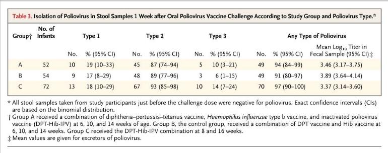
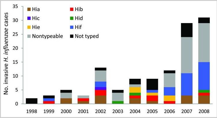
Pathological Blood Coagulation and the Mycotoxic Oxidative Stress Test

 Robert Young PhD

Naturopathic Practitioner – The pH Miracle Ti Sana Detox Medical Spa and Universal Medical Imaging Group

Abstract

Historical analysis suggests that conventional understandings of Disseminated Intravascular Coagulation (DIC) may be misguided; further examination may be necessary.  Here, a theoretical analysis provides an alternative explanation for DIC pathology; it is suggested that the cause and mechanics of DIC are largely due to the proliferation of several intravascular microforms and their associated metabolic toxic acidic waste products — Mycrozymian Acidic Toxins (MAT) and Exotoxic-Mycotoxic-Producing Microorganisms (EMPO).  The Mycotoxic Oxidative Stress Test (MOST) is presented here as an easy, inexpensive and non-invasive alternative to conventional measurements for the detection of intravascular  acidic toxins, DIC  and oxidative stress.

Introduction and Historical Perspective

More than 150 years ago, British physician T. W. Jones asked the question, “Why does the blood circulating in the vessels not coagulate?”[1]  though a general answer to this question is now obvious, the biochemical mechanisms involved in how the blood coagulates (clots) are complex and varied, and all the intricacies have not yet been explained. A. Trousseau, recognized that the blood of cancer patients is in a hyper-coagulable state in the process of coagulation, even while confined in the blood vessels.[2]  The name given to this discovery is still in use today, as “Trousseau’s Syndrome.”[2]  Early in his career, Rudolph Virchow, the Father of Pathology, was interested in thrombosis and embolism.  He speculated that intravascular blood could be altered so it would clot as a result of a stimulus too weak to clot normal blood.[3]  In 1856 Virchow delivered a lecture setting forth this concept.

Although the concept of partial clotting within vessels reaches back to the beginnings of modern medicine, much of the discovery of its biochemical mechanisms – the activation of clotting factors – has been left to chance.  The admission of a patient to the hospital with an unceplained bleeding disorder challenged researchers to discover the cause of hemorrhaging.  Analysis of blood from normal persons helped in the study of the patient with the blood disorder. A new clotting factor was hereby discovered which was missing from the  patient’s blood.  For this reason, several clotting factors have been named after the individuals in which they were missing: e.g., Christmas factor (factor IX)[4], Hageman factor (factor XII)[4].

In this article, the causes of pathological (intravascular) clotting will be described, as will various methods of detecting this condition, especially a blood test I call the Mycotoxin Oxidative Stress Test (MOST).

The Mechanics of Blood Coagulation

Blood clotting is a highly detailed chemical-mechanism involving many distinct components.  The problem for the hematologist hs been to understand it at the biochemical level.  Undoubtedly, efforts to fully understand blood clotting will continue for many more years.

Recalling Antione Bechamp’s[8] and Gunther Enderlein’s[9] research into the sub cellular living elements and combining this with what is known of colloidal flocculation[6], it is suggested that the clotting of blood begins with the end-linking (polymerizing) of the fundamental protein unit called by Bechamp the microzyma[8].  A chain of these living units constitutes fibrinogen, which is still dispersed 9micro-hetergenous0 in the blood, and it may or may not be further processed.  If processing continues, it will be either by continued end-linking or by cross-linking.  End-linked fibrinogen is referred to here as fibrin monomer, which I have suggested is a repair protein also dispersed in the blood. Due to a number of blood clotting factors, the process may continue until the excess fibrin monomer and/or until fibrin becomes excessively end-linked.

Cross-linking the polymerized strands to form a three-dimensional network results in what is called the hard clot (fibrin – the major protein of clotting blood).  Factor XIII, which instigates the forming of these blood networks. is always present but latent in the blood, and must be activated before the formation can occur.  Persons who are producing fibrin monomer or excessively linked fibrinogen are said to be in a hyper-coagulable state, while those having diminished  ability to form clots are in a hypo-coagulated state.  It is the activation of the colloidal clotting factors which is so complex.  Blood clotting may occur through many pathways and be initiated by many different stimuli.  Regardless of initiation factors, the process is a sequence of events in which the activation of one factor triggers another, until, after a series of discrete steps, fibrin is formed.

When blood is clotted prematurely, and the factors involved are consumed (incorporated into) the body recognizes a deficiency of clotting agents and generates more.  Thus, people with a tendency to clot excessively will alternate between a hyper coagulable state and a hypo-coagulatable state.  When in the hypo coagulated state, such people hemorrhage until the deficient clotting factors are replaced.[4]  When only fibrin monomer or excessively linked fibrinogen is formed (no cross-linking), it is quite subtle and may go undetected.  It may be detected by a change in blood viscosity (sedimentation rate), by the Mycotoxic Oxidative Stress Test (described later), or by other more subtle means.  If strands of fibrinogen are cross-linked, however, a suggicient amount of insoluble precipitate of fires may result, and these can be detected microscopically using a phase contrast and dark-field microscopy in prepared slides of fresh tissue or blood.  An excessive formation of fibrin leads to  an impairment in circulation, and eventual organ failure usually results.[5]

With this background, we are in a position to consider a standard medical term: disseminated intravascular coagultion (DIC).[6]  This term encompasses the hyper coagulable state, i refer to as pathological blood coagulation which consists of both insoluble and excess dispersed polymers of colloidal proteins.

Key Ingredients of Pathological Blood Coagulation

Before discussing DIC in more detail, it si necessary to introduce its fur important ingredients according to this view – mycotoxins, endotoxins, exotoxins, and tissue factor.  Any of these elements, or any combination of them, can play a major role in initiating unwanted DIC.[6]  However, mycotoxins or the acids from yeast have been found to be the underlying element which instigates and intensifies the participation of the other three.[6]  Each will now be described in turn and brought into the clotting picture.

(Micrograph 1: left, shows normal hyper-coagulated blood in a healthy blood clot sample and right, hypo coagulated blood in an unhealthy blood clot sample)

Mycotoxins and Metabolism by Fermentation

As discussed in the main text of my published book, Sick and Tired book[7 ]. acidification of blood and body tissues and organs and the accompanying lack of oxygen lead to pathological metabolic fermentation, which is carried out primarily by yeast and mold.  Such pathological microorganisms, or their precursors, ar inherent to the human body and to all higher organisms.  Their precursors according to Bechamp, the microzymas, carry on a nominal and homeostatic fermentation themselves. under healthy conditions.[8]  The primary function of yeast and mold is to decompose the body upon the death of the animal or human organism.  Their premature overgrowth indicates a biochemical environment akin to death.  During pathological metabolic fermentation, high concentrations of several acidic substances called mycotoxins are created.  They are highly damaging, always acidic, metabolic products.  If not immediately buffered by specific antioxidants, such as hydrogen peroxide and the hydroxyl free-radical, mycotoxins can seriously disrupt the physiology by disrupting normal metabolism and by penetrating blood and body cells and poisoning them.  As will be seen, they interact with many of the mechanisms for DIC in various pathological symptomologies.

In my published article called The Finger on the Magic of Life: Antoine Bechamp, 19th Century Genius (1816-1908),  I discuss pleomorphism in some detail.[7] Understanding this phenomenon – the rapid evolution of microorganisms across traditional taxonomic  lines is helpful in getting a complete picture of DIC.  Briefly stated, collodial living microzymas evolve intracellularly into more complex forms (microorganisms), beginning with a healthy primitive stage comprising of repair proteins.  As the disease condition worsens, morbid intermediate forms (filterable bacteria or viruses, cell-wall deficient forms and full bacteria) develop from repair proteins, or directly from microzymas.  A third macrostage comprises the commonly recognized culminate microorganisms which are yeast, fungus to mold.  In terms of pleomorphism, all of these microorganisms represent a single family of variously functioning forms.[8]  The culminate forms produce the lions share of acids, which are mycotoxins and the primary focus of my research.[7][8][9]  For convenience, bacteria, yeast, fungus and mold that produce acidic metabolic wastes and protein cellular fragments called exotoins, endotoxins and mycotoxins will here be referred to collectively ash EMPO, or exotoxic, mycotoxic-producing microorganisms.

What follows is a shortened description or the description and origin of several exotoxins and mycotoxins, referred to collectively microzymian acidic toxins of MAT, which are involved in the processes leading to DIC.  The bio-effects, or the pathology of cellular fermentation, of these toxic metabolites are know as mycotic illness, mycotoxicosis, or mycotoxic stress as seen in the MOST and described and published by Dr. Bolin in the 1940’s.[10]

One such metabolic product is acetyl aldehyde, which is formed by  cellular breakdown of food, especially carbohydrate and the birth of  EMPO.  Acetyl aldehyde can also break down into a secondary substance know as ethyl alcohol.  Although acetyl aldehyde presents an immediate hazard to health and well-being, nature has provided a means of buffering of neutralizing this acidic by-product of cellular digestion and fermentation almost as soon as it is created.[11] The controls of acetyl aldehyde (and ethyl alcohol) are the sulfur amino acids, cysteine, taurine, methionine and the peptide glutathione which is found in red blood cells and almost all cells utilizing oxygen.[12]  In an attempt to buffer or neutralize MAT, the body will also bind or chelate both fats and minerals to them.[12]

Another member of the MAT family is uric acid, which is formed by the digestion of protein and the creation of EMPO.[13]  Uric acid can also break down into secondary substance, on of which is alloxan.[14] This has been shown to damage the insulin-producing pancreatic beta cells leading to diabetes [Refer to Tables 1 and 2]

A shortage of alkalizing nutrients or an excess of MAT initi­ates an immune response in which a special class of free radicals which I call microzymian oxidative buffering species (MOBS) are released.[15] These oxygen metabolites carry unpaired electrons and are intended to disrupt bacteria, yeast, fungus and mold, and buffer exotoxins, endotoxins, and mycotoxins. Current medical savants believe that they can disrupt just about any­thing they contact, including healthy cells and tissue: this is not accurate. The fact is that MOBS carriers a nega­tive surface-charge and repel healthy cells, which also have a negative surface-charge. [16] It is the positively surface-charged bacteria, yeast/fungus, mold, exotoxins, endotoxins, and myco­toxins that MOBS bind too.[17]  This aspect gives some insight into autoimmune phenomena, which are not, as is often maintained, the result of an overburdened immune system. They result either as a side-effect of the immune system’s attempt to remove foreign or toxic ele­ments, or as a direct attempt by the immune system to remove cells or tissue rendered useless or disturb­ing to the body by MAT.

In every degenerative symptomatology I have studied, I have found excessive MAT and MOBS (see Tables 1-3). Some of these degenerative symptoms and their underlying disease conditions, including cancer are described in my recently published paper on a deficiency on alkaline nutrition and cancer. [15] But the fact that myco­toxins cause harm to humans and other animals is purely a secondary effect, since, as noted, the prima­ry function of the microorganism is not to cause illness. We know from the fossil record that pleomorphic microforms existed long before animals.[19] In fact, humans and animals developed in terms of micro­organisms.[20] The reverse, however, is not true. Since micro­organisms appeared first in the developmental sequence, they are not physiologically aware of humans and animals. There is much evidence that human and animal physiologies are highly aware of, and respond to MAT – these acidic compounds signaling the presence of bacteria, yeast, fungi and/or mold or  EMPO.[21].

Endotoxins

Also involved in the process leading to DIC are endotoxins, substances endogenous to symptogenic (i.e., “pathogenic” in orthodox terms) bacteria. Endotoxins are a family of related substances having certain common characteristics, but differing from one bacterial form (or strain) to another. Endotoxins are lipopolysaccharides (LPS). LPS form a widely diversified group because of (1) the number of long- chain fatty acids composing lipids; (2) the number of individual sugars as well as their modes of linkage to one another; (3) the branching of sugar chains; and (4) the number of possible arrangements of these units. Endotoxins also contain proteins, further com­pounding the structural diversity.[22]

One theory on endotoxin states that its purpose is to act as a semi-permeable membrane for the bac­terium, limiting and regulating substances entering the organism.[22] Endotoxin resides solely on or near the interior surface of the cell membrane and is shed into the surrounding medium only upon the death of the bacterium. Thus, as these microforms die off, or are lysed by bodily activity, endotoxin is released. (This fact may well be an explanation for the Herxheimer reaction, in which a patient becomes worse following the administration of toxic drugs or other forms of treatment that drastically alter the associated organ­ism.[23]) Another endotoxin theory states that LPS are a constituent of the membrane, and as the organism grows, endotoxin fragments are repeatedly sloughed off into the medium. This phenomenon has been observed in the digestive tract.[24] Since bacterial translocation into the blood is not only possible but common where epithelial hyperpermeability exists, one can assume that the process will continue there. Both theories may be correct if we think of the first one as true of “adult” forms, and the second as true of newly developed and expanding ones.

Basic to the structure of an endotoxin is the lipid common to all forms, designated lipid A, to which is attached a “core” polysaccharide, identical for large groups of bacteria. To the core polysaccharide is attached the O-antigen, consisting of various lengths of polysaccharide chains which are chemically unique for each type of organism and LPS. These chains pro­vide endotoxin specificity.[25] Experiments conducted over many years indicate that most, if not all, of the toxic effects of an endotoxin may be attributed to the lipid portion, and it is sometimes used per se in experiments rather than the entire molecule.[26] An important additional feature of lipid A is its phos­phate content. Each phosphate group carries a nega­tive charge, and since lipid A is a rather large mole­cule, it provides, essentially, a negatively charged sur­face. The importance of this will be seen shortly.

Exotoxins

These are the metabolic excretions of bacteria. While endotoxin’s ongoing effect is, in a manner of speaking, in the background, exotoxins, like myco­toxins, present a double-edged sword. Not only do they initiate DIC, but they produce, or influence the body to produce, the various and numerous infec­tious symptomatologies, such as typhoid fever, diph­theria, etc. (See “Vaccination Reconsidered” in Section 4 of the Appendix of Sick and Tired for details on the action of diphtheria toxin.)[7] By comparison, mycotoxins not only initiate DIC, but there is much evidence to sug­gest that they produce, or influence the body to pro­duce, degenerative symptomatologies, such as arthri­tis, diabetes, etc., and cancer and AIDS as well.

Tissue Factor

Crucial to the understanding of DIC is recogni­tion of the role of tissue factor (TF), formerly known as thromboplastin. This transmembrane lipoprotein exists on the surface of platelets, vas­cular endothelial cells, leukocytes, monocytes, and most cells producing EMPO.[27] It plays a major role in several biochemical mechanisms leading to DIC.

TF is the primary cell-bound initiator of the blood coagulation cascade. Its gene is activated in wound healing and other conditions. By itself it is capable of initiating clotting, but also becomes active when complexed with factor VII or activated factor VII (Vila).[28] TF has been described as the receptor for factor VII because of the close association between the two proteins and because it causes a shape change (conformational) in factor VII, allowing it to attain activity. Both factor Vila and the TF/VII com­plex activate factors IX and X, which initiate the clotting cascade and the formation of thrombin.[29]

Development of Disseminated
Intravascular Coagulation
(DIC)

DIC Induced by MAT and Tissue Factor

An infusion of toxins into the blood has a direct effect on TF gene expression in leukocytes. Contact of MAT, endotoxins (lipid A), or exotoxins with leukocytes, activates proteins that bind to DNA nucleotide sequences, thereby activating the TF gene.[30] (See Tables 4-6.)

Endothelial cells damaged in culture by exotoxins, endotoxins, or mycotoxins attract polymorphonuclear leukocytes (PMNs), which adhere to the damaged cells. Once the leukocytes are bound, they can still have their TF gene activated if it hasn’t yet occurred, and they may release MOBS in response to toxins and to organisms of disease, possibly creating further dis­turbances. (Cellular disorganization then releases acti­vating proteins into the blood, which is discussed in more detail later.) Research shows that exotoxic and mycotoxic stress resulting in bound PMNs can be blocked by “antioxidants.”[31] These might better be called anti-exotoxins or antimycotoxins. Both observa­tion and study have led the author to conclude that cellular disorganization is initiated and primarily caused by fermentation pathology, not, as is the cur­rent belief, by the MOBS, or free radicals, generated to destroy toxins and microorganisms. MOBS or free radicals, because of their negative charge, are released to chelate or bind EMPO and MAT. It is suggested by current savants that free radical tissue damage is the secondary, “shotgun” effect of intense immune response to EMPO toxification and MAT-damaged cells. This could not be the case since healthy cells or their membranes carry a negative charge and would resist any electromagnetic attraction because of simi­lar charge. The concentration and instability of MAT generated in a compromised terrain, as opposed to the fleeting existence of free radicals, especially exoge­nous ones, also lead to this conclusion.

Endothelial cells grown in culture can be induced to express tissue factor. In one experiment, no procoagulant activity could be detected in the absence of toxins. However, the addition of mycotoxins from Aspergillus niger or Micrococcus neoformas (Mucor racemosus Fresen) resulted in procoagulant activity which reached a maximum in four to six hours and was dose-dependent. The same experiment was applied using E. coli and Salmonella enteritidis endo­toxin with a similar result.[32] A single intravenous injection of a mycotoxin from Aspergillus niger into experimental animals resulted in circulating endothelial cells within five minutes. In other exper­iments with the mycotoxin, detachment of endothe­lial cells from the basement membrane was noted.[33] (See Table 8.)

Removal of endothelial cells has dire conse­quences from two standpoints: First, the surface of these cells is covered with a specific prostaglandin (PGI2) known as prostacyclin. If blood contacts a surface not covered with PGI2, it will clot. For example, surfaces devoid of this prostaglandin are formed whenever a vessel is cut or punctured. An abrasion or other injury may also expose a surface on which PGI2 is lacking. The removal of endothelial cells by exotoxins or mycotoxins creates a surface devoid of PGI2, leading to blood clotting (see Table 7). Secondly, disorganization of endothelial cells cre­ates increased levels of EMPO and MAT which are attracted to an exposed surface (basement mem­brane) which expresses a negative charge. This also leads to clotting.

DIC Induced by Electrostatic Attraction

It was discovered in 1964 that blood will clot sim­ply from contacting a negatively charged surface.[34] Previously it was believed that the clotting process comprised a cascade of enzyme activity in which one activated the next, etc. The discovery that blood could be clotted simply by contacting a negatively charged surface ruled out the purely enzyme hypoth­esis. Only some of the known clotting factors have been shown to be enzymes.[35] As a result of this sur­prising discovery, detailed research was conducted in an attempt to describe the process. In some experi­ments, the negatively charged surfaces of selected, finely divided, inorganic crystals, including alu­minum oxide, barium sulfate, jeweler’s rouge, quartz, and titanium oxide, were considered.[36]

The clotting factor eventually shown to be activat­ed when whole blood contacted negatively charged surfaces was factor XII, also known as the Hageman factor. This is a positively charged protein migrating in an electric field (electrophoresis) toward the anode.[37] It is believed that factor XII is normally in the shape of a hairpin which binds to the negatively charged sur­face at the bend. Electrostatic attraction forces the two arms to lie flat on the surface, thereby exposing the inner faces and activating the molecule.

It was discovered that if the negatively charged particles were smaller than the clotting factor itself, activation was minimal. Or, if the concentration of clotting factor was too great, there was little or no activation.[38] Both of these observations indicated that the process was one of electrostatic attraction between the negatively charged surface and the clot­ting factor, which is a “basic” protein, that is, posi­tively charged.[39]

Activation of factor XII allows the activation of factor XI, which then activates factor IX. Thus, the blood clotting cascade continues to the formation of fibrin in the normal manner.[40] However, due to a series of activations begun by contact of factor XII with a negatively charged surface, trace amounts of factor Xa also show up in the blood. Factor VII is activated to Vila by factor Xa. Factor Vila then acti­vates factors IX and X, leading to the formation of thrombin. Factor Xa, with co-factor Va, continues the clotting cascade until fibrinogen is activated, leading to fibrin formation.[41] (See Table 5.)

As discussed earlier in terms of prostacyclin, beneath endothelial cells is another surface—the basement membrane. Called the extracellular matrix, it is a thin, continuous net of specialized tis­sue between endothelial cells and the underlying connective tissue. It has four or more main con­stituents, including proteoglycans (protein/polysac- charide).[42] The removal of endothelial cells by’MAT exposes this membrane, which is negatively charged by virtue of its sulfonated polysaccharides in the pro­teoglycans. This brings a reduced negatively charged surface into direct contact with the blood, which activates factor XII and the clotting cascade.[43]The positively charged toxic components of MAT also activate factor XII, as do disturbed disorganized cells, yeast/fungus cells, moldy cells, and the phos­phate groups in the lipid A component of endotoxin. (See Tables 2-5.)

To summarize this section, exotoxic, mycotoxic, and oxidative stress resulting from the overgrowth of bacteria, yeast/fungus, and then mold, has multiple actions, all leading to disseminated intravascular coagulation:

MAT activation of tissue factor gene in leukocytes; subsequent activation of factors VII, IX, and X, resulting in the blood clotting cascade.

MAT activation of tissue factor gene in endothelial cells, again leading to the clotting cascade.

MAT damage to endothelial cells, resulting in neu­trophil attraction, with TF gene activation and generation of MOBS, which, in turn, neutralize MAT, protecting healthy endothelial cells or the basement membrane and supporting the janitorial services of the leukocytes.

Removal of negatively charged endothelial cells by positively charged exotoxins, endotoxins, and mycotoxins, creating a surface devoid of PGI2, also exposes the negatively charged basement membrane, leading to the activation of factor XII and initiation of the clotting cascade. Positively charged components of EMPO, exotoxins and mycotoxins, and several other elements, including the lipid A component of bacterial endotoxin, also activate factor XII and the clotting cascade.

Endothelial Cells as Antithrombotics or Procoagulants

Normal, resting (unstimulated) endothelial cells show antithrombotic activity in several ways: (1) by the inhibition of prostacyclin (platelet adhesion and aggregation); (2) the inhibition of thrombin genera­tion; and (3) the activation of the fibrinolytic system, leading to clot lysis.[45] We will take a brief look at the thrombin aspect.

On the surface of endothelial cells is a protein called thrombomodulin, which acts as a receptor for thrombin. When bound to thrombomodulin, throm­bin can activate protein C. Activated protein C then catalyzes the proteolytic cleavage of factors Va and Vila, thereby destroying their participation in blood clotting. Thus thrombin, which normally activates fib­rinogen, plays an opposite role in this case and inhibits the clotting process.[46,47] (See Table 7.)

On the other side of the coin, the endothelial cell becomes a procoagulant agent when acted on by cer­tain lymphokines, such as interleukin-1. Not only can interleukin-1 induce TF gene expression, but it also suppresses transcription of the thrombomodulin gene in endothelial cells. As in other situations, the lymphokine-activated endothelial cell expresses TF on its surface as a result of TF gene activation. This leads to the production of thrombin and the trigger­ing of the blood clotting cascade.[48] (See Table 5.) Many lymphokines also stimulate adhesion of leuko­cytes to endothelial cells damaged by MAT, resulting in recycling of the cells by MOBS, as described later.

DIC Induced by Intracellular Exotoxic, Mycotoxic, Oxidative Stress by Bacteria, Yeast/Fungus and/or Mold

Any cell which has gone from an oxidative to a fer­mentative state can biochemically cause macrophage production of the lymphokine tumor necrosis factor (TNF). This protein has been shown to activate the gene for TF in fermenting cells, which are so behaved due to morbid evolution of bacteria, yeast/fungus, and then mold.[49,50] In the author’s view, a cell having been switched entirely to fermentation metabolism as a result of a physical or emotional disturbance of that cell, is what constitutes cancer (see Tables 5 and 13). (One might argue that this definition does not fit all “forms” of cancer, such as leukemia, for example. This is because leukemia is not cancer, but an immune response to the rise in EMPO and MAT in the body, and a relatively easy compensation to correct.)

The surface of many disorganizing or fermented cells (cancer cells) is characterized by small projec­tions in the plasma membrane which pinch off, becoming free vesicles containing toxins as well as TF complexed with factor VII. These vesicles can aggre­gate and/or lodge anywhere, ultimately releasing their contents. Also, the presence of excessive amounts of TF/factor VII complexes on the surface of fermented cells allows the formation of a fibrin net around the cell and around the entire mass of cells (tumor). This seems to be an attempt by the body to encapsulate and contain the mass. However, fermented cells do escape from the primary fibrin net, perhaps due to some electromagnetic effect, and become free-float­ing in the circulation. They may thus lodge elsewhere and instigate the fermentation of other cells by fungal penetration or by poisoning them and provoking a morbid evolution of their inherent microzymas.

Because of the surrounding fibrin net, these mobi­lized fermenting cells are protected from collection by the immune system while in transit.[51,52] (See Table 4.) The blockage or dissolution of fibrin net forma­tion by an anticoagulant such as heparin allows freed, fermenting (metastasizing) cells to be dismantled by natural killer cells and other immune cells (see Tables 5, 12 and 13).

DIC Induced by MAT/EMPO and Immune System Response (Release of MOBS)

Unsaturated fatty acids are highly susceptible to EMPO as well as MAT. Linoleic acid, a long-chain fatty acid present in white cells, has 18 carbons and 2 unsaturations. Subjected to MAT, linoleic acid binds the exotoxin, endotoxin, or mycotoxin, there­by forming an epoxide at the first unsaturation.[53] Research has revealed that this compound, named leukotoxin, is highly disturbing to other cells. It caus­es platelet lysis, thereby releasing TF and initiating DIC.[54] (See Table 10.) The fact that MAT result in fermented fats lends further credence to the sugges­tion that the initial and primary degenerative damage to structures and substances in the body is caused by exotoxins and/or mycotoxins, and that damage by MOBS, or by other free radicals, is not possible.

Another mechanism leading to DIC is the release of a special glycoprotein, sialic acid, from the terminal ends of cell-membrane polysaccharides, where it is always found. Polysaccharides play a highly significant role in biochemical processes, with both enzymes and membrane receptors recognizing various groupings of specific sugars linked in highly specific ways.

Immediately preceding the release of sialic acid in the polysaccharide chain is the sugar galactose. The sialic acid/galactose arrangement is utilized as a biolog­ical indicator of cellular and molecular aging. As cells age, sialic acid is naturally expressed from the terminal ends of polysaccharides, thereby exposing galactose. A membrane-bound enzyme from the liver, galactose oxi­dase, recognizes galactose and eventually disorganizes it, disrupting cell function integrity and hastening demise. Aged red blood cells, which have expressed a significant amount of sialic acid, are removed from the blood by this process. (I theorize that the biological ter­rain may be at work in normal cell aging. That is, the rate at which sialic acid is expressed is determined by the levels of corrosive acids in the system and the body’s ability to remove them, although there are no doubt intracellular factors at work as well.)

I suggest from my years of  clinical research  that cellular breakdown is compounded by the fermentation of the galactose by the microzyma. This is a process that begins from within and not necessarily from without. Not only does this action create more sialic acid, it creates other toxic waste products such as acetic aldehyde, alcohol, uric acid, oxalic acid, etc. The increase in cellular disturbances and fermenta­tion of the galactose creates biochemical signals for more galactose oxidase. This leads to greater cellular disorganization and developmental morbidity, espe­cially in the red blood cells, and a rise in the level of detrital serum proteins, which encourages clotting. From this perspective, diabetes, arthritis, atheroscle­rosis and other symptomatologies become more clearly “degenerative” (see Tables 2-5, 12 and 13).

Fibrinogen is a rather elaborate protein having the structure of three beads on a string. Expressed on the end beads is sialic acid, which indicates the beginning of disorganization of the fibrinogen and a declining negative charge to the positive. Prior to the declining charge and the expression of sialic acid on the end beads, fibrinogen, which is negatively charged, will not polymerize the healthy blood due to mutual repulsion. However, fibrinogen will poly­merize to damaged cells, EMPO, MAT and other positively charged areas of the body for repair pur­poses. Thus, as more and more sialic acid is expressed, there will be a significant reduction in the charge of the fibrinogen, acting as the primary requirement for the polymerization of fibrinogen (hypercoagulable state). The resulting polymer, fib­rin monomer, is the protein chain used in the repair of cells and clotting of blood.[55] End-linking will take place after the release of sialic acid (positive charge) by whatever means.

With this background, it is interesting to note that blood taken from persons suffering from anxiety is expressing sialic acid from fibrinogen, and is halfway toward clotting. Hormones released during anxiety states are easily fermented, giving more momentum to MAT and thereby resulting in this important change in fibrinogen. It leads to a clotting pattern characteristic of anxiety stress, and is readily identi­fied in the MOST. As can be seen in this picture, the pattern is a “snowstorm” of protein polymeriza­tions measuring from 2 to 10 microns.

allergiesbefore


[Micrograph 2: An Anxiety Profile showing a ‘snowstorm’ of 2 to 10 micron protein polymerizations starting from the center of the clot and moving out towards the edge]

As mentioned earlier, despite the attempt by the body to neutralize EMPO and MAT, an excess will initiate the release of MOBS by immune cells. A major MOBS is superoxide, designated chemically as O 2. It may exist alone or be attached to another ele­ment, such as potassium (KO’2) or sulfur (SO). Again, however, nature has provided a means of pro­tecting healthy cells—their negative charge[1]. Another protection against superoxide is the enzyme superox­ide dismutase (SOD), also found in all healthy cells.

A second member of the MOBS family is hydro­gen peroxide (H202). This molecule is very unstable and tends to react rapidly with other biological mol­ecules, damaging them. The release of hydrogen per­oxide in the body is a response to the overgrowth of decompositional organisms in a declining pH (com­promised biological terrain). The control for healthy cells against hydrogen peroxide is their negative charge and the protective enzyme catalase, one of the most efficient enzymes known.

When leukocytes and other white blood cells are stimulated by the presence of bacteria, yeast/fungus and mold, they treat these organisms as foreign par­ticles to be eliminated. During and prior to phagocy­tosis, the foregoing oxidative cytotoxins, along with the hydroxyl radical (OH’), are generated and released specifically for neutralizing microforms or harmful substances. This release is referred to as an “oxidative burst.” As a result of fermentation and the production of exotoxins and mycotoxins that fer­ment galactose from cells, the immune system is activated. An oxidative burst is released to neutralize the morbid microforms and mycotoxicity.[56] Like other biological processes faced with constantly alarming situations, the continued release of MOBS can get out of control. This may damage endothelial cells, the basement membrane, or other body ele­ments, and this activates fibrinogen to fibrin monomer (repair protein), leading to DIC [see Table 9]. Interestingly, the white blood cells capable of neutralizing MAT through MOBS production are the same ones capable of phagocytosis, the process by which foreign matter, waste products and microor­ganisms are collected and dumped in the liver.[57]

To summarize this section, pathological microforms and their acids create DIC by a number of pathways:

Leukotoxin (linoleic acid bound to mycotoxin) is highly toxic to cells. It causes platelet lysis, there­by releasing TF and initiating DIC.

The expression or release of sialic acid residues from healthy cells that have been disturbed allows for the fermentation of galactose, creating exotox­ins and mycotoxins, biochemically activating galactose oxidase, which further disturbs and dis­organizes healthy cells. This cycle loads the blood with debris.

EMPO and MAT disturb fibrinogen, which releas­es sialic acid and reduces the charge, allowing it to polymerize into fibrin monomer and fibrin nets.

The presence of exotoxins, endotoxins, and myco­toxins and their poisoning of cells activates the immune system. White blood cells generate MOBS (e.g., superoxide [0′2] or hydrogen perox­ide [H202]). These substances bind to and neu­tralize EMPO and MAT. MOBS are repelled by healthy endothelial cells and the basement mem­brane because of their negative charge. Cellular disturbances and disorganization stimulate the generation of fibrin monomer for repair purposes, leading to DIC.

Detection of Disseminated Intravascular Coagulation

The Sonodot Analyzer

The Sonoclot Coagulation Analyzer provides a reaction-rate record of fibrin and clot formation with platelet interaction. An axially vibrating probe is immersed to a controlled depth in a 0.4 ml sample of blood. The viscous drag imposed upon the probe by the fluid is sensed by the transducer. The electronic circuitry quantifies the drag as a change in electrical output. The signal is transmitted to a chart recorder which provides a representation of the entire clot for­mation, clot contraction and clot lysis processes. The analyzer is extremely sensitive to minute changes in visco-elasticity and records fibrin formation at a very early stage. The Sonoclot has been evaluated scientif­ically and shown to provide an accurate measurement of the clotting process.[58,59]

One application of the Analyzer has been the development of a test to distinguish non-advanced breast cancer from tumors that are benign. The ratio­nale for the test is the hypercoagulable state seen in cancer patients (Trousseau’s Syndrome), resulting from the generation of TF by leukocytes (mono­cytes).[60] (See Table 4.)

Fibrin Degradation
Products and Fibrin Monomer

DIC can be seen as a two-step process. First, fib­rinogen, which is always present in the blood, is acti­vated by any of several mechanisms. This activation leads to an automatic polymerization (chain forma­tion) resulting in fibrin monomer. This is not apparent in a microscope unless the blood is allowed to clot, as in the MOST.[61,62] The second step is the precipitation or deposition of fibrin (hard clot) by several other mechanisms. One of these is the formation of cross­links through the action of factor XIII. Another such mechanism may be poor circulation in an organ already blocked by deposited fibrin. The deposition of precipitated fibrin may be detected microscopically in tissue sections and diagnosed as DIC.[62]

Because fibrin monomer is not readily detected, a chemical test for it is of immense value in diagnosing DIC. Research has indicated that its detection may be very useful in the early diagnosis of DIC and MAT.[63] There are three fundamental physiologic areas related to blood clotting: (1) the prevention of blood clotting, (2) the clotting of blood, and (3) the removal of clotted blood once it has formed.

Enzymes are present that are capable of removing (lysing) clotted blood, one of which is plasmin. Another enzyme, plasminogen, is always present in the blood, but is inactive as a proteolytic agent. Plasminogen acti­vator converts plasminogen to plasmin, which can degrade deposited fibrin. This process is not specific for fibrin, however, and other proteins may be affected. When fibrin is degraded (fibrinolysis), fibrin monomer, as well as several other products, are formed. Commercial kits are available for the analysis of fibrin degradation. This test is an indirect measure of the pres­ence of DIC and MAT.[64]

Other tests include:

Protamine Sulfate: Protamine sulfate is a heparin binder sometimes used in surgery for excessive bleed­ing. The test, which indicates fibrin strands and fibrin degradation products, is conducted in a test tube, with fibrin monomer and fibrin forming early and polymer­ization of fibrin degradation products occurring later.[65] Ethanol Gelation: A white precipitate is formed by the addition of ethanol to a solution in a test tube containing fibrin monomer as a degradation product of fibrin, indicating DIC and MAT.[66]

The Mycotoxic Oxidative Stress Test (MOST)

Up to now, blood chemistries have been the prima­ry mode of diagnosis or analysis for the presence of pathology. In the view presented here, the bright-field microscope, is used to easily and inexpensively reveal a disease state as reflected by changes in certain aspects of blood composition and clotting ability. DIC is char­acterized by the abnormal presence in the blood of fib­rin monomer. When allowed to clot, blood containing such an abnormal artifact will exhibit distortions of normal patterns. The presence in the blood of soluble fragments of the extracellular matrix and soluble fibronectin, as well as other factors, will also create abnormal blood clotting patterns as described below.

A small amount of blood from a fingertip is con­tacted with a microscope slide. A series of drops is allowed to dry and clot in a normal manner. Under the compound microscope, the pattern seen in healthy subjects is essentially the same—a dense mat of red areas interconnected by dark, irregular lines, completely filling the area of the drop. The blood of people under mycotoxic/oxidative stress exhibits a variety of characteristic patterns which deviate from nor­mal, but with one striking, common abnormality: “clear” or white areas, in which the fibrin net/red blood cell conglomerate is missing.

BowelCancerLive Blood Dried Blood_0166


[Micrograph 3; An abnormal clot with striking ‘clear’ or white areas or protein polymerization as seen in the hyper coagulated blood of a patient with lower bowel imbalances]

Why the fibrin net is missing may be understood from the following: Two peptides—A and B—in the central protein bead of the fibrinogen structure become bound in the cross-linking process. There are two ways this can be configured: (1) Thrombin is capable of activating peptides A and B, resulting in the formation of a polymer loosely held together only by hydrogen bonds; (2) With peptides A and B acti­vated normally, the resulting hard clot is insoluble, indicating that the peptides are linked by covalent bonds. The difference in bonds results from factor XIII, an enzyme which links the two fibrin strands with a glutamine-lysine peptide bond.

Additional research has shown that the release of sialic acid from fibrinogen inhibits the action of factor XIII, resulting in a soft, white clot. In addition, acetic aldehyde has been shown to inactivate factor XIII directly. The soft clotting, compounded by other polymeric aggregations (described below), results in clear areas in the dry specimens. In the opposite extreme, high serum levels of calcium, for the pur­pose of neutralizing MAT, activates factor XIII, lead­ing to excessive cross-linking of fibrin to form a clot harder than normal. This is reflected in the MOST pattern characteristic of definite hypercalcemia— that of a series of cracks in the clot radiating outward from the center, resembling the spokes of a wheel. High serum calcium is the body’s attempt to com­pensate for the acidity of mycotoxic stress by pulling this alkalizing mineral from bone into the blood. This demand creates endocrine stress in turn, because reabsorption of bone is mediated by parathormone (PTH). Therefore, this clotting pattern indicates cal­cium deficiency and thyroid/parathyroid imbalance.

calciumpattern


[Micrograph 4: A mineral deficiency or more specifically a calcium deficiency pattern associated with an imbalance of they thyroid and/or parathyroid}

Advanced research has shown that there are seven carbohydrate chains in fibrinogen (each terminated by sialic acid). A second action of factor XIII is to ferment a large amount of carbohydrate during clot­ting. Because carbohydrate is most often water solu­ble, the loss of this material undoubtedly adds to the insolubility of a clot, while pathological retention contributes to the softness of the abnormal clot.

Clinical experience demonstrates that the MOST is a reliable indicator of exotoxic and mycotoxic stress and, concurrently, of various disorganizing symptoma­tologies associated with fermentative and oxidative processes. As various cellular degradation occurs, the blood-borne phenomena which accompany such symptoms as diabetes, arthritis, heart attack, stroke, atherosclerosis and cancer show up in the MOST, often with sialic acid beads in the clear areas of poly­merized proteins. (Determination of the liberation of sialic acid from carbohydrate has been approved by the U.S. Food and Drug Administration as an accept­ed indicator for cancer, and is clinically available.)

sialicacid

[Micrograph 5: Sialic acid beads are seen inside the protein
polymerization of the hypocoagulated blood as black dots]

The extent and shape of the clear areas are reflec­tive of particular symptomatologies which have arisen from the way in which the disease condition manifests in a given individual. This observation is borne out by having the patient undergo appropriate alkalizing therapy. With success of treatment based on the patient’s freedom from symptoms, sense of well-being, and live blood exams discussed in the main text of Sick and Tired, Reclaim Your Inner Terrain, Appendix C,[7] repeated analysis with the MOST reveals a progressively improving clotting pattern.

[Micrographs 6 and 7: Medically diagnosed cancer patient with large polymerized protein pools (PPP) in the hypo-coagulated blood above. In the picture below PPP’s have significantly reduced in size and the blood is moving to a more hyper-coagulated state as a result of reducing acid loads with an alkaline lifestyle and diet (7, 70)]

Because of its very nature, the MOST is emi­nently suited to reveal and measure the presence in the blood of abnormal substances, clotting factors, and disorganization of cells due to an inverted way of living, eating, and thinking, which gives rise to MAT. The MOST indicates both the direct and indirect activity of MAT on blood clotting, endothelium, and the extracellular matrix (described next), as well as on biochemical pathways, including hormonal ones. The generation of excessive MOBS in response to EMPO and MAT, the inability that accompanies all degenerative symptoms to neutralize or eradicate EMPO and MAT, and the recognized hyper- and hypocoagulable states seen in various symptomatolo­gies, will beyond doubt be revealed in the MOST.

Aspergillusnigercrystal


[Micrograph 8 and 9: Medically Diagnosed HIV/AIDS micrograph showing above an Aspergullus niger mold crystal using dark field microscopy and below a hypocoagulated blood clot with systemic protein polymerizations measuring in excess of 40 microns using bright field microscopy}

HIV


As mentioned, hormones are easily fermented, and this will show up as a hypocoagulated blood pattern in the MOST. It is my opinion, this hypocoagulated blood appears in the MOST as misty clouds of protein polymerizations throughout the clot, as seen in the accompanying picture.

poorfibrin

[Micrograph 10: Poor fibrin interconnection in the clot associated with endocrine or hormonal imbalance]

The MOST from Solubilized Extracellular Matrix

There is now a clearer picture of the biochemical rationale for correlating abnormal blood clotting patterns with the presence of degenerative symptoms.  A link between symptoms and the distorted clotted blood patterns has been delineated in the MOST.
Another reason for the abnormal clotting patterns accompanying pathological states, in addition to insufficient bonding of fibrinogen peptides as seen in the MOST, is presence in the blood of water-soluble fragments of the extracellular matrix.

Extracellular Matrix Degradation by MAT

The extracellular matrix (EM) is a three-dimen­sional gel, binding cells together and composed of five or more major constituents: collagen (protein), hyaluronic acid (polysaccharide), proteoglycans (pro- tein/polysaccharide), fibronectin and laminin. Also included are glycosaminoglycans and elastin.[67] In every degenerative disease studied by this author, evidence has been found for MAT activity destruc­tive of EM.

One of the proteolytic enzymes activated in response to EMPO and MAT is alpha-1 antitrypsin (capable of neutralizing MAT), normally not active in the presence of the enzyme trypsin. The active por­tion of this anti-exotoxin and antimycotoxin contains the amino acid methionine, which includes a C-S-C linkage. When chelated by the hydroxyl radical (one of the MOBS oxidants), methionine’s central sulfur atom acquires one or two oxygen atoms (forming the sulfone or sulfoxide respectively). The fermentation of methionine is a secondary effect of immune response to an alarming situation, intended to neutral­ize MAT and prevent degradation of the EM. Once alpha-1 antitrypsin is exhausted, MAT will have more access to the EM. If the EM is damaged beyond repair, then the enzyme trypsin is released to disorganize and recycle the cells involved.[68]

A similar scenario holds for the enzymes collage- nase and elastase. Thus, the absence of alpha-1 antitrypsin in the presence of EMPO and MAT activates three enzymes which degrade the extracellular matrix. Degradation of the EM by enzymes and MAT puts into the blood the water-soluble fragments (proteins and glycoproteins) of normally insoluble EM components (see Table 11). The presence of these fragments modifies the normal clotting pattern (described below), as seen in the M/OST, and is therefore an indication of EM degradation, which is always found with degenerative symptoms. (Also present is fibrin monomer, which has been found in the blood of patients suffering from collagen dis­ease.[69] See Table 11.)

Fibronectin is a molecule in EM having several binding sites for various long-chain molecules— heparin (a sulfonated polysaccharide) and collagen, for example. As such, it functions as a cellular glue, bind­ing cells together as well as various components of the EM. A soluble form of fibronectin is normally found free in the blood, and enters into the formation of a blood clot through the action of factor XIII. This form of fibronectin binds to fibrin. Elevated, bound-serum fibronectin results from EM fragmentation by MAT, and accompanies degenerative symptoms such as arthritis and emphysema (collagen diseases).

Water-soluble fragments of the EM bound by fibronectin form a three-dimensional network or gel in the pathologically clotted blood (fibrin and com­ponents of the blood clotting cascade). Since fibronectin binds to both fibrin and collagen, the two polymeric networks are superimposed and intermin­gled, resulting in a modification of the normal clot­ting pattern. Exactly how the pattern is modified depends upon the nature of the collagen abnormally present, the nature and extent of hyaluronate pre­sent, and the degree to which EM fibronectin has been released by MAT.

Conclusion

Thus, it is easily seen that there are many forms which the pattern of clotted blood may take, depending on the individual and the internal terrain that produced the modifying substances. The MOST reveals not only the presence of exotoxic and mycotoxic stress, but indicates as well the nature of the symptom(s) resulting from the stress (see Table 12). Since MAT underlie the entire complex of events which degrade the extracellular matrix, I must conclude that the absence of these exotoxins, endotoxins and mycotoxins would provide substantial improvements in tissue integrity and the overall physiology and functionality of the organism or animal and human.

­

­

References

[1]  Jones, T.W., “Observations on some points in the anatomy, physiology and pathology of the blood.”  British Foreign Medical Review, 1842. 14 : 585.

[2] Trousseau, A., Phlegmasis alba delens. “Clinque Medicale de L’Hotel Dieu de Paris.”, 1865, 3:94

[3]  Virchow, R., “Hypercoagulability: A review of its development, clinical application, and recent progress.”  Gesammelte Abhandlungen our Wussenschaftlichen Medizin, 1856, 26:477.

[4]  Rapaport, S.I., “Blood Coagulation and its Alterations in Hemorrhagic, and Thrombotic Disorders.”  The Western Journal of Medicine, 1993; 158: 153.

[5]  Hamilton, P.J. et al., “Disseminatied Intravascular Coagulation: A Review.”  Journal of Clinical Pathology, 1978, 31: 609

[6] The Harper Collins Illustrated Medical Dictionary, 1994, p.13.

[7] Young, RO, “Sick and Tired, Reclaim Your Inner Terraine,” Woodland Publishing, 1999.

[8] BeChamp, A., “The Blood and Its Third Anatomical Element,”  Hikari Omni Publishing, 1999.

[9]  Schwerdtle, C, Arnoul, F, Enerlein, G, “Introduction to Darkfield Diagnostics”, Semmelweis-Verlag (2006).

[10]  Hawk, BO, Thoma, GE, Inkley, JJ, The Evaluation of the Bolen Test as a Screening Test for Malignancy*, cancerres.aacrjournals.org on December 5, 2015. © 1951 American Association for Cancer Research.

[11]  Uchida, K., “Role of Reactive Aldehyde in Cardiovascular Diseases”,  Labortory of Food and Biodynamics, Nagoya University Graduate School of Bioagricultural Sciences, Nagoya, Japan , Free Radical Biology and MedicineVolume 28, Issue 12, 15 June 2000, Pages 1685–1696

 [12] Chang JCvan der Hoeven LHHaddox CH, “Glutathione reductase in the red blood cells”,  Ann Clin Lab Sci. 1978 Jan-Feb;8(1):23-9.

[13] Kutzing, MK, Firestein, BL, “Altered Uric Acid Levels and Disease States”, Department of Cell Biology and Neuroscience (M.K.K., B.L.F.), Graduate Program in Biomedical Engineering (M.K.K.), Rutgers University, Piscataway, New Jersey. Address correspondence to: Dr. Bonnie L. Firestein, Department of Cell Biology and Neuroscience, Rutgers University, Piscataway, NJ 08854-8082. E-mail: firestein@biology.rutgers.edu

[14] Claudino, M,. Ceolin,,DS, Alberti, S.,  Cestari, TM,  Spadella, CT, Fischer Rubira-Bullen, IR, Gustavo Pompermaier Garlet, Gerson Francisco de Assis, ” Alloxan-Induced Diabetes Triggers the Development of Periodontal Disease in Rats”,  Published: December 19, 2007. DOI: 10.1371/journal.pone.0001320

[15] Young RO (2015), “Alkalizing Nutritional Therapy in the Prevention and Reversal of any Cancerous Condition. Int J Complement Alt Med 2(1): 00046. DOI: 10.15406/ijcam.2015.02.00046

[16] Heloise Pöckel FernandesCarlos Lenz Cesar, and  Maria de Lourdes Barjas-Castro, “Electrical properties of the red blood cell membrane and immunohematological investigation”, Rev Bras Hematol Hemoter. 2011; 33(4): 297–301. doi:  10.5581/1516-8484.20110080 PMCID: PMC3415751

[17] Harris, JO, “The Relationship Between the Surface Charge and the Absorption of Acid Dyes by Bacterial Cells”, Department of Bacteriology, Kansas Agricultural Experiment Station, Manhattan,Kansas, Received for publication March 3, 195.

[18] Young, RO, “Metabolic and Dietary Acids are the Fuel That Lights the Fuse that Ignites Inflammation that Leads to Cancer”. https://www.linkedin.com/pulse/metabolic-dietary-acids-fuse-ignites-inflammation-causes-young. 2015.

[19] Snaders, R, “Did Bacteria Spark Evolution of Multicellular Life?” Berkeley News, Research, Science and Environment,  October 24, 2012.

[20] Wenner, M, “Humans Carry More Bacterial Cells than Human Ones”. Scientific American, November 30th, 2007.

[21} Animals and humans respond to MAT as a poison.

[22]  Morrison, D.C. et al. The effects of bacterial endotox­ins on host mediation systems. American Journal of Pathology, 1978; 93: 526.

[23]  Ibid.

[24]  Ibid.

[25]  Van Deventer, S.J.H. et al. Intestinal Endotoxemia. Gastroenterology, 1988; 94(3): 825-831.

[26]  Morrison, D.C. et al., op. cit.

[27]  Ibid.

[28]  Hu, T. et al. Synthesis of tissue factor messenger RNA and procoagulant activity in breast cancer cells in response to serum stimulation. Thrombosis Research, 1993; 72: 155.

[29]  Rapaport, op. cit. (Ref. 4).

[30]  Ibid.

[31]  Mackman et al. Lipopolysaccharides—mediated tran­scriptional activation of the human tissue factor gene in THP-1 monocytic cells requires both activator protein 1 and nuclear factor kappa B binding sites. Journal of Experimental Medicine, 1991; 174: 1517.

[32]  Yamada, O. et al. Deleterious effects of endotoxins on cultured endothelial cells: An in vitro model of vascular injury. Inflammation, 1981; 5: 115.

[33]  Colucci, M. et al. Cultured human endothelial cells: An in vitro model of vascular injury. Journal of Clinical Investigation, 1983; 71: 1893.

[34]  Cho, T.H. et al. Effects of Escherichia coli toxin on structure and permeability of myocardial capillaries.

[35]  Acta Pathologica Japonica, 1991; 41: 12.

[36]  Rapaport, op. cit. (Ref. 4).

[37]  Ibid.

[38]  Margolis, J. The interrelationship of coagulation of plasma and release of peptides. Annals of the New York Academy of Sciences, 1963; 104: 133.

[39]  23-25. Ibid.

[40]  Morrison, D.C. et al., op. cit.

[41]  Rapaport, op. cit. (Ref. 4).

[42]  Alberts, B. et al, eds. Molecular Biology of the Cell. New York: Garland Publishing, Inc., 1989 (2nd ed.), p. 818.

[43]  Rapaport, op. cit. (Ref. 4).

[44] Bertz, A., et al. Modulation by cytokines of leukocyte endothelial cell interactions. Implications for thrombo­sis. Biorheology, 1990; 27: 455.

[45]  Rapaport, op. cit. (Ref. 4).

[46]  Nachman, R.L. et al. Hypercoagulable states. Annab of Internal Medicine, 1993; 119: 819.

[47]  Ibid.

[48]  Tallman, M.S., et al. New insights into the pathogene­sis of coagulation dysfunction in acute promyelocytic leukemia. Leukemia and Lymphoma, 1993; IT. 27.

[49]  Silberberg, J.M., et al. Identification of tissue factor in two human pancreatic cancer cell lines. Cancer Research, 1989; 49: 5443.

[50]  Grimstad, I.A. et al. Thromboplastin release, but not content, correlates with spontaneous metastasis of can­cer cells. International Journal of Cancer, 1988; 41: 427.

[51]  Gunji, Y. et al. Role of fibrin coagulation in protection of murine tumor cells from destruction by cytotoxic cells. Cancer Research, 1988; 48: 5216.

[52]  Sugiyama, S. et al. The role of leukotoxin (9, 10- epoxy-12-octadecenoate) in the genesis of coagulation abnormalities. Life Sciences, 1988; 43: 221.

[53]  Ibid.

[54]  White, A. et al, eds. Principles of Biochemistry. McGraw-Hill Book Co., New York, 1964, p. 648.

[55]  Mueller, H.E. et al. Increase of microbial neu­raminidase activity by the hydrogen peroxide concen­tration. Experientia, 1972; 23: 397.

[56]  Young, Robert O. Fermentology and oxidology. The study of fungus-produced mycotoxic species and the activation of the immune system and release of microzymian oxidative buffering species (MOBS). Self- published: InnerLight Biological Research Foundation, Alpine, Utah, 1994.

[57]Chandler, WL. et al. Evaluation of a new dynamic vis­cometer for measuring the viscosity of whole blood and plasma. Clinical Chemistry, 1986; 32: 505.

[58]  Saleem, A. et al. Viscoelastic measurement of clot for­mation: A new test of platelet function. Annals of Clinical and Laboratory Science, 1983; 13: 115.

[59]  Spillert, C.R. et al. Altered coagulability: An aid toselective breast biopsy. Journal of the National Medical Association, 1993; 85: 273.

[60]  Bowie, E.J. et al. The clinical pathology of intravascular coagulation. Bibliotheca Haematologica, 1983; 49: 217.

[61]  Muller-Berghaus, G. et al. The role of granulocytes in the activation of intravascular coagulation and the pre­cipitation of soluble fibrin by endotoxin. Blood, 1975; 45: 631.

[62]  Bick, R.L. Disseminated intravascular coagulation. Hematology/Oncology Clinics of North America, 1993; 6: 1259.

[63]  Bredbacka, S. et al. Laboratory methods for detecting disseminated intravascular coagulation (DIC): New aspects. Acta Anaesthesiologica Scandinavica, 1993; 37: 125.

[64]  Sigma Diagnostics, St. Louis, MO 63178; tel: 314- 771-5765.

[65]  Nachman, R.L. et al. Detection of intravascular coag­ulation by a serial-dilution protamine sulfate test. Annals of Internal Medicine, 1971; 75: 895.

[66]  Breen, F.A. et al. Ethanol gelation: A rapid screening test for intravascular coagulation. Annals of Internal Medicine, 1970; 69: 1197.

[67] Hay, E.D., ed. Cell Biology of Extracellular Matrix. New York: Plenum Press, 1981, p. 653.

[68]  Carp, H. et al. In vitro suppression of serum elastase- inhibitory capacity by ROTS generated by phagocytos- ing polymorphonuclear leukocytes. Journal of Clinical Investigation, 1979; 63: 793.

[69]  Wilson, C.L. The alternatively spliced V region con­tributes to the differential incorporation of plasma and cellular fibronectins into fibrin clots. Journal of Cell Biology, 1992; 119: 923.

[70] Young, RO, Young, SR, “The pH Miracle Revised and Updated”, Hachette Publishing, 2010.

Tables

Table 1

Expression of Sialic Acid/Galactose [MAT] from Cell and Protein Degeneration (From All Serum Proteins, RBC/WBC and Other Cell Surfaces)

  1.  Carbohydrate, Proteins, and Fats From Diet, Body Cells or Reserves
  2. As cells breakdown or ferment they give birth to bacteria, yeast, fungus and mold [EMPO] and their associated metabolic acidic waste [MAT]
  3. Exotoxins, Endotoxins, and Mycotoxins [MAT]
  4. Acetyl Aldehyde, Ethyl Alcohol, Uric Acid, Alloxan, Lactic Acid are examples of MAT
  5. MAT  Ferments Other Body Cells and their Extracellular Membranes and Proteins
  6. MAT Modifies Glycoprotein
  7. Binds to liver Galactosidase
  8. Creating an Increase in Cell and Protein Fermentation and Degeneration and Increased Amounts of Exotoxins, Endotoxins and Mycotoxins [MAT]

Table1a

Table 2

Expression of Sialic Acid [MAT] From the Fermentation of Degeneration of Insulin Producing Pancreatic Beta-Cells in Type I, Type II and Type III Diabetes

  1. Pancreatic Insulin producing Beta-Cells with no or minimal Surface Sialic Acid [MAT]A Physical and/or Emotional Disturbance Occurs from Lifestyle and/or Diet
  2. Normal regulation of Insulin Production
  3. A Physical and/or Emotional Disturbance Occurs from Lifestyle and/or Dietary choicesdd
  4. Leads to cellular fermentation and degeneration and the birth of EMPO
  5. This lead to increased abnormal amounts of MAT that the immune system, the alkaline buffering system and the elimination organs has to deal with
  6. Fermenting and degenerating Insulin Producing Beta Cells
  7. Giving Rise to Surface Cell Sialic Acid [MAT}
  8. Increased Amounts of Sialic Acid Activates the Immune Response [MOBS] and Sialidase [AB]
  9. Leads to Lowered or No Insulin Production
  10. Symptoms of Type I, Type II or Type III Expressed
  11. The insulin producing beta cells of the Islets of Langerhans express silica acid on their surface as a break down metabolite.  I have suggested that when insulin producing beta cells are physically disturbed by MAT they begin to disorganize and express sialic acid on the surface of the cell.  This indicates the death of the cell and insulin production will stop.

Table2a

Table 3

HIGH BLOOD PRESSURE, ATHEROSCLEROSIS, HEART ATTACKS, STROKES, and CONGESTIVE HEART FAILURE

  1. A Physical and/or Emotional Disturbance Occurs from Lifestyle and/or Dietary choices
  2. Leads to cellular fermentation and degeneration and the birth of EMPO
  3. This lead to increased abnormal amounts of MAT that activates the immune system to chelate the MAT.
  4. Increased amounts of MAT will cause endothelial breakdown and the expression of Sialic acid.
  5. Increased Amounts of Sialic Acid and damage to the endothelial will cause a reduction in the negative surface-charge leading to the release of Glycoproteins.
  6. The release of Glycoproteins will cause the activation of Factor XII and the blood clotting cascade.
  7. This cause the creation and formation of fibrin monomers and the increase of Platelet Deposition out of the red blood cells for clotting purposes
  8. The immune system will activate and MOBS will be released as well as sodium bicarbonate, calcium, lipids and other alkaline buffers to reduce metabolic acidity.
  9. The build-up of fibrin monomers in the clotting cascade will lead to fibrin nets and clots causing an increase in blood pressure and the risk of blockages potentially causing a Stroke or Heart Attack.

Table3a

Table 4

DISSEMINATED INTRAVASCULAR COAGULATION RESULTING
FROM INTRACELLULAR DISORGANIZATION OR FERMENTATION WHICH GIVES RISE TO MAT
 AND EMPO

  1. A Physical and/or Emotional Disturbance Occurs from Lifestyle and/or Dietary choices
  2. Leads to cellular fermentation and degeneration and the birth of EMPO
  3. This lead to increased abnormal amounts of MAT that activates the Tumor Necrosis Factor (TNF).
  4. Increased amounts of TNF activates the Tissue Factor Gene (TF)
  5. Increased Amounts of TF causes the release of Thromboplastin.
  6. The release of Thromboplastin activates the release of clotting Factors VII (VIIa) and trace amounts of Factor Xa into the blood.
  7. This activates the release of Factors IX and X to IXa and the increase of Factor Xa.
  8. The activation of the blood clotting cascade leads to Disseminated Intravascular coagulation and the clotting or thickening of the blood inside the blood vessels.
  9. The DIC or hyper-coagulation will mask the fermentation of healthy cells to unhealthy cells or cancerous cells.
  10. As the unhealthy cells or cancerous cells increase the body will go into preservation mode and begin forming fibrin nets to encapsulated these unhealthy cells to protect healthy body cells.
  11. As body and blood cells breakdown from MAT this causes an increase of MAT and EMPO leading to systemic latent tissue acidosis and a potential metastatic cancerous condition.

Table4a

 Table 5

DISSEMINATED INTRAVASCULAR COAGULATION RESULTING
IN CELLULAR DISORGANIZATION OR FERMENTATION/OXIDATON AND THE INCREASE OF MAT AND EMPO

  1. A Physical and/or Emotional Disturbance Occurs from Lifestyle and/or Dietary choices.
  2. Leads to cellular fermentation and degeneration and the birth of EMPO
  3. This lead to increased abnormal amounts of MAT that activates the Tumor Necrosis Factor (TNF).
  4. Increased amounts of TNF activates the Tissue Factor Gene (TF)
  5. Increased Amounts of TF causes the release of Thromboplastin.
  6. The release of Thromboplastin activates the release of clotting Factors VII and Factor Xa in the blood.
  7. This activates the release of Factors IX and X to IXa and the increase of Factor Xa.
  8. The activated blood clotting cascade leads to Disseminated Intravascular coagulation and the clotting or thickening of the blood inside the blood vessels.
  9. The DIC or hyper-coagulation will mask the fermentation of healthy cells to unhealthy cells or cancerous cells.
  10. As the unhealthy cells or cancerous cells increase the body will go into preservation mode and begin forming fibrin nets to encapsulated the unhealthy cells.
  11. This leads to tumor formation of the unhealthy or cancerous cells.
  12. As the body and blood cells breakdown this causes an increase of MAT and EMPO leading to an increased risk of  systemic metastatic cancer.

Table5aTable 6

ENDOTHEIAl CELL CONVERSION FROM AN
ANTITHROMBOTIC STATE TO A PROCOAGULANT STATE
CELLULAR DISORGANIZING PATHWAY

  1. A Physical and/or Emotional Disturbance Occurs from Lifestyle and/or Dietary choices
  2. Leads to cellular fermentation and degeneration and the birth of EMPO
  3. This leads to increased abnormal amounts of MAT that damages the protective endothelial cover cells leading to a reduction of PGI2
  4. The absence of PGI2 causes the release of Interleukin-1 and/or Tumor Necrosis Factor (TNF).
  5. In addition the loss of protective endothelial cover cells leads to Tissue Factor Gene Activation and the release of Thrombin causing a pro-coagulate state leading to DIC
  6. Another pathway to DIC would be the loss of protective endothelial cover cells and the absence of PGI2 causes the suppression of Thromomodulin, Protein C leading to procogradulation and DIC.

Talble6

 Table 7

ENDOTHELIAL CELL CONVERSION
FROM AN ANTITHROMBOTIC STATE
(NORMAL PATHWAY)

Table7

Table 8

MECHANISM OF DISSEMINATED INTRAVASCULAR COAGULATION GENERATED BY MAT

Table8Table 9

ACTIVATION OF SIALIDASE AND MICROZYMIAN OXIDATIVE BUFFERING SPECIES (MOBS) BY EMPO AND MAT

Table9

Table 10

DISSEMINATED INTRAVASCULAR COAGULATION RESULTING FROM PHAGOCYTIC OXIDATIVE BURST

Table10

Table 11

MOST BLOOD TEST and DISSEMINATED INTRAVASCULAR COAGULATION WITH SOLUBILIZED EXTRACELLULAR MATRIX

Table11

Table 12

TYPICAL SOURCES OF FERMENTATION INSULT (MAT) IN BIOLOGICAL SYSTEMS INITIATING DIC

Table12

Table 13

POSITIVE CHARGE OF CANCEROUS CELLS AND TUMORS AND THE FORMATION OF FIBRIN NETS AND TREES IN RESPONSE TO MAT

Table13

The 7 Most Acidic and Disease Causing Ingredients Used to Make Vaccines including the MMR Vaccine!

10429808_10153553734673998_8629030119831371097_n
Parents have the right to understand what is being put into their child’s body. The production of vaccines uses many acidic dis-ease causing ingredients. Additionally, the components used during the manufacturing process may violate your personal, religious, or ethical beliefs.
Lastly, remember that whatever colorful language is used by the scientific community (using such words as purified), using such substances can come with an enormous cost.
As you read this list, understand cell lines and vaccines are contaminated with the blood of humans and animals. This is often hidden under the term “adventitious agents.”
Acidic Dis-ease Causing Ingredient #1: Cells From Aborted Fetus

Aborted fetal cells, listed on vaccine package inserts as “Human Fetal Diploid Cells.” Two aborted fetal cell lines, WI-38 and MRC-5, have been grown under laboratory conditions since the 1960s.
The cells are used to grow viruses or acidic waste products used which are then collected from the cell cultures and processed further to produce the acidic vaccine itself.
Terms to Investigate: PERC6, MRC5, WI-38, HEK-293
Which Vaccines? Adenovirus vaccine, DTaP vaccine, Hep A vaccine, Hep B vaccine, MMR vaccine, Rabies vaccine, Varicella (Chickenpox) vaccine
Acidic Dis-ease Causing Ingredient #2: Serum From Aborted Calf Fetus Blood
One of the more toxic methods involved in vaccine manufacturing is the collection of fetal bovine serum. The purpose for serum is providing a nutrient broth of acid for bacteria, yeast and/or mold to evolve in cells.
How is the blood collected?
According to the Humane Research Australia website:
“After slaughter and bleeding of the cow at an abattoir, the mother’s uterus containing the calf fetus is removed during the evisceration process (removal of the mother’s internal organs) and transferred to the blood collection room. A needle is then inserted between the fetus’s ribs directly into its heart and the blood is vacuumed into a sterile collection bag. This process is aimed at minimizing the risk of contamination of the serum with micro-organisms from the fetus and its environment. Only fetuses over the age of three months are used otherwise the heart is considered too small to puncture.
Once collected, the blood is allowed to clot at room temperature and the serum separated through a process known as refrigerated centrifugation.” [1]
Terms to Investigate: Fetal Bovine Serum
Which Vaccines? Adenovirus vaccine, MMR vaccine, Rotavirus vaccine, Varicella (Chickenpox) vaccine
Acidic Dis-ease Causing Ingredient #3: Cells From Armyworms
The FDA approved the Flublok vaccine on January 16, 2013.
This new technology is being touted as the wave of the future. It utilizes an insect cell line (expresSF+®) that is derived from cells of the fall armyworm, Spodoptera frugiperda.
The vaccine package insert for Flublok also mentions:
“Each 0.5 mL dose of Flublok may also contain residual amounts of baculovirus and host cell proteins (≤ 28.5 mcg), baculovirus and cellular DNA (≤ 10 ng) …” [2]
Terms to Investigate: insect cell line (expresSF+)
Which Vaccines? Influenza vaccine
Acidic Dis-ease Causing Ingredient #4: Cells From Monkey Kidneys
As mentioned above, monkey kidney tissue is used to support the production of certain viruses or acidic waste products used in vaccine production. There remains a huge controversy over using these cells and their role contaminating the polio vaccine in the 1950s.
The story is best told in the Congressional papers of a courageous scientist, Bernice Eddy. The Executive Reorganization and Government Research of the Committee on Government Operations United States Senate, Ninety-Second Congress, Second Session [1972] states on page 500:
“The next and only serious vaccine crisis that has occurred since the polio episode was the realization in mid-1961 that a monkey virus later shown to cause tumors in hamsters was contaminating both polio and adenovirus vaccines. The virus, known as SV40, was entering the vaccines and, just as in the polio case were surviving the formalin treatment.
There were several states by which the full extent of the SV40 problem became known. First was the discovery in 1959-1960 by a DBS scientist, once again Bernice Eddy, that an unknown agent in the monkey kidney cells used to produce polio and adenovirus vaccines would cause tumors when the cells were injected into hamsters.”
Page 502:
“In 1954 Eddy, as a polio control officer, found live virus in supposedly killed polio vaccine; in 1955 she was relieved of her duties as polio control officer … After her discoveries concerning the SV40 virus, her staff and animal space were reduced and she was demoted from head of a section to head of a unit.”
Page 505:
“ … even when the contaminating virus was found to be oncogenic [cancer causing] in hamsters, the DBS [Division of Biologics Standards] and its expert advisory committee decided to leave existing stocks on the market rather than risk eroding public confidence by a recall.”
and:
“There has been a tendency on the part of certain higher government circles to play down any open discussion of problems associated with vaccines … ” [3]
Terms to Investigate: Vero (monkey kidney) cell culture, SV40, Bernice Eddy
Which Vaccines? DTaP vaccine,  MMR vaccine, Japanese Encephalitis vaccine, Polio vaccine, Rotavirus vaccine, Vaccinia vaccine 
Acidic Dis-ease Causing Ingredient #5: Cells From Dog Kidneys
On November 20, 2012, the FDA approved the seasonal influenza vaccine, Flucelvax, manufactured by Novartis. [4]
This vaccine is mass-produced using the continuous cell line Madin Darby Canine Kidney (MDCK) as vaccine or acid cell substrate. [5]
Terms to Investigate: Madin Darby Canine Kidney (MDCK)
Which Vaccines? Influenza vaccine
Acidic Dis-ease Causing Ingredient #6: Mouse Brain
Viral or Acid vaccines prepared in tissue culture or mouse brain have been used in many Asian countries. According to the CDC website, the inactivated mouse brain-derived JE vaccine used in the United States since 1992 is no longer available. [6]
Of course, with any dis-ease causing vaccine, the adverse reactions are rarely tracked and downplayed by medical authorities. However, the injuries from vaccines can be quite serious. [7, 8]
Terms to Investigate: inactivated mouse brain (IMB), suckling mouse brain (SMB), JE virus (Beijing-1), acute disseminated encephalomyelitis (ADEM)
Which Vaccines? Japanese encephalitis vaccine, Rabies vaccine
Acidic Dis-ease Causing Ingredient #7: Chicken Embryos
Chickens and their embryos have long been used in the production of acidic vaccines.
These methods were popularized in the 1920s and 1930s by Thomas Rivers and others at the Rockefeller Institute for Medical Research. [9]
Terms to Investigate: Chick embryo
Which Vaccines? Influenza vaccine, Rabies vaccine, Yellow fever vaccine
References
3. Executive Reorganization and Government Research of the Committee on Government Operations United States Senate, Ninety-Second Congress, Second Session. Page 499-505. April 20,21; and May 3,4, 1972.

The Research Validating That The Ingredients in Vaccines Cause Cancer!

SV40, an ingredient found in vaccines is Associated with Causing Different Cancerous Conditions! 

Senior Medical - Vaccination

Below are types of human cancers in which SV40 has been found. By clicking on the name, you will link to some study abstracts that provides additional details. Please note that this is not a comprehensive list of all SV40-associated cancers or all medical/scientific articles written about the detection of SV40 in cancer.

Brain Cancers
Bone Cancers
Chest Cancers
Lymphomas
Thyroid Cancers

Astrocytomas

Huang H., et al. Identification in human brain tumors of DNA sequences specific SV40 large T antigen. Brain Pathol 1999 Jan;9(1):33-42.

Zhen H.N., et al. Expression of the simian virus 40 large tumor antigen (Tag) and formation of Tag-p53 and Tag-pRb complexes in human brain tumors. Cancer 1999 15;86(10):2124-32.

Martini, F., et al. Simian Virus 40 Footprints in Normal Human Tissues, Brain and Bone Tumours of Different Histotypes; 1997. Brown F, Lewis AM (eds): Simian Virus 40 (SV40): A Possible Human Polyomavirus. Dev Biol Stand. Basel, Karger, 1998, vol 94, pp 55-66.

Krieg, P., et al. Episomal simian virus 40 genomes in human brain tumors Proc Natl Acad sci USA 1981 Oct;78(10):6446-50.

Anaplastic Astrocytoma

Huang H., et al. Identification in human brain tumors of DNA sequences specific SV40 large T antigen. Brain Pathol 1999 Jan;9(1):33-42.

Choroid Plexus Papilloma

Huang H., et al. Identification in human brain tumors of DNA sequences specific SV40 large T antigen. Brain Pathol 1999 Jan;9(1):33-42.

Zhen H.N., et al. Expression of the simian virus 40 large tumor antigen (Tag) and formation of Tag-p53 and Tag-pRb complexes in human brain tumors. Cancer 1999 15;86(10):2124-32.

Martini, F., et al. Simian Virus 40 Footprints in Normal Human Tissues, Brain and Bone Tumours of Different Histotypes; 1997. Brown F, Lewis AM (eds): Simian Virus 40 (SV40): A Possible Human Polyomavirus. Dev Biol Stand. Basel, Karger, 1998, vol 94, pp 55-66.

Wang, J and Garcea R.L., et al, Simian virus 40 DNA sequences in human brain and bone tumours. Brown F, Lewis AM (eds): Simian Virus 40 (SV40): A Possible Human Polyomavirus. Dev Biol Stand. Basel, Karger, 1998, vol 94, pp 13-21.

14 of 17 pediatric brain tumors (13 choroid plexus tumors, 3 ependymomas and 1 ganglioneuroma) were positive for SV40 regulatory region sequences. Butel, J.S., et al. Detection of authentic SV40 DNA sequences in human brain and bone tumors Brown F, Lewis AM (eds): Simian Virus 40 (SV40): A Possible Human Polyomavirus. Dev Biol Stand. Basel, Karger, 1998, vol 94, pp 23-32.

Lednicky, et al., Natural simian virus 40 strains are present in human choroid plexus and ependymoma Virology. 1995 Oct 1;212(2):710-7. tumors.

Ependymoma

Huang H., et al. Identification in human brain tumors of DNA sequences specific SV40 large T antigen. Brain Pathol 1999 Jan;9(1):33-42.

Zhen H.N., et al. Expression of the simian virus 40 large tumor antigen (Tag) and formation of Tag-p53 and Tag-pRb complexes in human brain tumors. Cancer 1999 15;86(10):2124-32.

Martini, F., et al. Simian Virus 40 Footprints in Normal Human Tissues, Brain and Bone Tumours of Different Histotypes; 1997. Brown F, Lewis AM (eds): Simian Virus 40 (SV40): A Possible Human Polyomavirus. Dev Biol Stand. Basel, Karger, 1998, vol 94, pp 55-66.

Wang, J and Garcea R.L., et al, Simian virus 40 DNA sequences in human brain and bone tumours. Brown F, Lewis AM (eds): Simian Virus 40 (SV40): A Possible Human Polyomavirus. Dev Biol Stand. Basel, Karger, 1998, vol 94, pp 13-21.

14 of 17 pediatric brain tumors (13 choroid plexus tumors, 3 ependymomas and 1 ganglioneuroma) were positive for SV40 regulatory region sequences. Butel, J.S., et al. Detection of authentic SV40 DNA sequences in human brain and bone tumors Brown F, Lewis AM (eds): Simian Virus 40 (SV40): A Possible Human Polyomavirus. Dev Biol Stand. Basel, Karger, 1998, vol 94, pp 23-32.

Lednicky, et al., Natural simian virus 40 strains are present in human choroid plexus and ependymoma Virology. 1995 Oct 1;212(2):710-7. tumors.

Gemistocytic Astrocytoma

Huang H., et al. Identification in human brain tumors of DNA sequences specific SV40 large T antigen. Brain Pathol 1999 Jan;9(1):33-42.

Glioblastoma

Huang H., et al. Identification in human brain tumors of DNA sequences specific SV40 large T antigen. Brain Pathol 1999 Jan;9(1):33-42.

Zhen H.N., et al. Expression of the simian virus 40 large tumor antigen (Tag) and formation of Tag-p53 and Tag-pRb complexes in human brain tumors. Cancer 1999 15;86(10):2124-32.

Martini, F., et al. Simian Virus 40 Footprints in Normal Human Tissues, Brain and Bone Tumours of Different Histotypes; 1997. Brown F, Lewis AM (eds): Simian Virus 40 (SV40): A Possible Human Polyomavirus. Dev Biol Stand. Basel, Karger, 1998, vol 94, pp 55-66.

Krieg, P., et al. Episomal simian virus 40 genomes in human brain tumors Proc Natl Acad sci USA 1981 Oct;78(10):6446-50.

Gliosarcoma

Huang H., et al. Identification in human brain tumors of DNA sequences specific SV40 large T antigen. Brain Pathol 1999 Jan;9(1):33-42.

Medulloblastoma

Huang H., et al. Identification in human brain tumors of DNA sequences specific SV40 large T antigen. Brain Pathol 1999 Jan;9(1):33-42.

Zhen H.N., et al. Expression of the simian virus 40 large tumor antigen (Tag) and formation of Tag-p53 and Tag-pRb complexes in human brain tumors. Cancer 1999 15;86(10):2124-32.

Krieg, P., et al. Episomal simian virus 40 genomes in human brain tumors Proc Natl Acad sci USA 1981 Oct;78(10):6446-50.

Meningioma

Zhen H.N., et al. Expression of the simian virus 40 large tumor antigen (Tag) and formation of Tag-p53 and Tag-pRb complexes in human brain tumors. Cancer 1999 15;86(10):2124-32.

Krieg, P., et al. Episomal simian virus 40 genomes in human brain tumors Proc Natl Acad sci USA 1981 Oct;78(10):6446-50.

Weiss A.F., et al. Simian virus 40-related antigens in three human meningiomas defined chromosome loss Proc Natl Acad Sci USA 1975 Feb;72(2):609-13.

Oligodendroglioma

Huang H., et al. Identification in human brain tumors of DNA sequences specific SV40 large T antigen. Brain Pathol 1999 Jan;9(1):33-42.

Krieg, P., et al. Episomal simian virus 40 genomes in human brain tumors Proc Natl Acad sci USA 1981 Oct;78(10):6446-50.

Pituitary Adenoma

Zhen H.N., et al. Expression of the simian virus 40 large tumor antigen (Tag) and formation of Tag-p53 and Tag-pRb complexes in human brain tumors. Cancer 1999 15;86(10):2124-32.

Osteosarcoma

Martini, F., et al. Simian Virus 40 Footprints in Normal Human Tissues, Brain and Bone Tumours of Different Histotypes; 1997. Brown F, Lewis AM (eds): Simian Virus 40 (SV40): A Possible Human Polyomavirus. Dev Biol Stand. Basel, Karger, 1998, vol 94, pp 55-66.

Wang, J and Garcea R.L., et al, Simian virus 40 DNA sequences in human brain and bone tumours. Brown F, Lewis AM (eds): Simian Virus 40 (SV40): A Possible Human Polyomavirus. Dev Biol Stand. Basel, Karger, 1998, vol 94, pp 13-21.

Ewing’s Tumors

Martini, F., et al. Simian Virus 40 Footprints in Normal Human Tissues, Brain and Bone Tumours of Different Histotypes; 1997. Brown F, Lewis AM (eds): Simian Virus 40 (SV40): A Possible Human Polyomavirus. Dev Biol Stand. Basel, Karger, 1998, vol 94, pp 55-66.

Mesothelioma

Cristaudo A, et al., SV40 enhances the risk of malignant mesothelioma among people exposed to asbestos: a molecular epidemiologic case-control study. Cancer Res. 2005 Apr 15;65(8):3049-52.

Pass HI, et al., Evidence of an important role for SV40 in mesothelioma. Thorac Surg Clin. 2004 Nov;14(4):489-95.

Carbone M, et al., New developments about the association of SV40 with human mesothelioma. Oncogene. 2003 Aug 11;22(33):5173-80.

Non-Hodgkins Lymphoma

Vilchez RA, et al., Simian virus 40 tumor antigen expression and immunophenotypic profile of AIDS-related non-Hodgkin’s lymphoma. Virology. 2005 Nov 10;342(1):38-46.

Butel JS, et al., Association between SV40 and non-Hodgkin’s lymphoma. Leuk Lymphoma. 2003;44 Suppl 3:S33-9.

Papillary Thyroid Carcinomas

Vivaldi, et al., Simian virus 40-like sequences from early and late regions in human thyroid tumors of different histotypes. J Clin Endocrinol Metab. 2003 Feb;88(2):892-9.

Anaplastic Thyroid Carcinomas (ATC)

Vivaldi, et al., Simian virus 40-like sequences from early and late regions in human thyroid tumors of different histotypes. J Clin Endocrinol Metab. 2003 Feb;88(2):892-9.

OPPOSE Senate Bill 792 – Mandatory Vaccines for Adults in California!